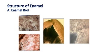
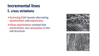
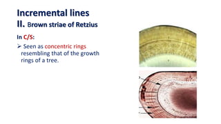
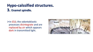
This document discusses the structure and properties of enamel. It begins by defining enamel as the outermost layer of tooth covering made of highly mineralized tissue. The structure of enamel is described including enamel rods, interrod substance, and rod sheaths. Physical properties like hardness, thickness and chemical composition consisting mainly of hydroxyapatite are covered. Incremental growth lines including cross striations, striae of Retzius and neonatal line are also summarized. Hypo-mineralized enamel structures such as enamel spindles, tufts and lamellae are defined. Finally, the surface structure of enamel including outer structureless enamel and perikymata grooves are described.
It is a presentation in detail about the strongest structure of the oral cavity "ENAMEL". It is a simple topic but people find it difficult to learn about it. I hope my presentation is a simple method to learn about it. I would like to thank my professors for assign me this project and i learn't a lot from it and still learning my basics daily.
Amelogenesis is the formation of enamel. During amelogenesis, the ameloblast (enamel-forming cells) undergo various stages i.e the life cycle of ameloblast.
For more content check out my blog: www.rkharitha.wordpress.com "a little about everything dental"
A Complete presentation explaining the complete morphology of Maxillary first molar, for the benefit of people like me who tried and failed to find everything in one package
The presentation discusses about tooth enamel in detail including its formation, characteristics, structure and histological features along with its clinical considerations. It is well supported with diagrams for better understanding of the text.
Suggestions and feedback will be well appreciated.
I prepared this presentation during the first year of my MDS. This will give you a basic idea and necessary information about the pulp of the teeth and its histology. Hope you guys find it useful.
PHYSICAL PROPERTIES
CHEMICAL PROPERTIES
STRUCTURE OF ENAMEL
DEVELOPMENT OF ENAMEL
EPITHELIAL ENAMEL ORGAN
AMELOGENESIS
LIFE CYCLE OF AMELOBLASTS
AGE CHANGES IN ENAMEL
DEFECTS OF AMELOGENESIS
CLINICAL IMPLICATIONS
Indian Dental Academy: will be one of the most relevant and exciting training center with best faculty and flexible training programs for dental professionals who wish to advance in their dental practice,Offers certified courses in Dental implants,Orthodontics,Endodontics,Cosmetic Dentistry, Prosthetic Dentistry, Periodontics and General Dentistry.
Upload By : Ahmed Ali Abbas
Babylon University College of Dentistry
download this file from Website on google theoptimalsmile.wix.com/dentistry
Oral histology
This Slide, gives a Brief introduction to the Anatomy of the tooth specifically the outer shell, the enamel, including the structures, development and abnormalities.
Created by Dr. Mohsen S. Mohamed
For Ozident.com
Enamel is the hardest tissue and outer covering of tooth. The presentation consists of physical ,chemical properties , structure , developmental stages of enamel, age changes , clinical implications, and defects in enamel. Learning about enamel will enhance the basic knowledge of new dental aspirants about dentistry.
The Indian Dental Academy is the Leader in continuing dental education , training dentists in all aspects of dentistry and
offering a wide range of dental certified courses in different formats.
Enamel significance in operative dentistry /certified fixed orthodontic cour...Indian dental academy
Welcome to Indian Dental Academy
The Indian Dental Academy is the Leader in continuing dental education , training dentists in all aspects of dentistry and offering a wide range of dental certified courses in different formats.
Indian dental academy has a unique training program & curriculum that provides students with exceptional clinical skills and enabling them to return to their office with high level confidence and start treating patients
State of the art comprehensive training-Faculty of world wide repute &Very affordable.
These simplified slides by Dr. Sidra Arshad present an overview of the non-respiratory functions of the respiratory tract.
Learning objectives:
1. Enlist the non-respiratory functions of the respiratory tract
2. Briefly explain how these functions are carried out
3. Discuss the significance of dead space
4. Differentiate between minute ventilation and alveolar ventilation
5. Describe the cough and sneeze reflexes
Study Resources:
1. Chapter 39, Guyton and Hall Textbook of Medical Physiology, 14th edition
2. Chapter 34, Ganong’s Review of Medical Physiology, 26th edition
3. Chapter 17, Human Physiology by Lauralee Sherwood, 9th edition
4. Non-respiratory functions of the lungs https://academic.oup.com/bjaed/article/13/3/98/278874
Ozempic: Preoperative Management of Patients on GLP-1 Receptor Agonists Saeid Safari
Preoperative Management of Patients on GLP-1 Receptor Agonists like Ozempic and Semiglutide
ASA GUIDELINE
NYSORA Guideline
2 Case Reports of Gastric Ultrasound
Flu Vaccine Alert in Bangalore Karnatakaaddon Scans
As flu season approaches, health officials in Bangalore, Karnataka, are urging residents to get their flu vaccinations. The seasonal flu, while common, can lead to severe health complications, particularly for vulnerable populations such as young children, the elderly, and those with underlying health conditions.
Dr. Vidisha Kumari, a leading epidemiologist in Bangalore, emphasizes the importance of getting vaccinated. "The flu vaccine is our best defense against the influenza virus. It not only protects individuals but also helps prevent the spread of the virus in our communities," he says.
This year, the flu season is expected to coincide with a potential increase in other respiratory illnesses. The Karnataka Health Department has launched an awareness campaign highlighting the significance of flu vaccinations. They have set up multiple vaccination centers across Bangalore, making it convenient for residents to receive their shots.
To encourage widespread vaccination, the government is also collaborating with local schools, workplaces, and community centers to facilitate vaccination drives. Special attention is being given to ensuring that the vaccine is accessible to all, including marginalized communities who may have limited access to healthcare.
Residents are reminded that the flu vaccine is safe and effective. Common side effects are mild and may include soreness at the injection site, mild fever, or muscle aches. These side effects are generally short-lived and far less severe than the flu itself.
Healthcare providers are also stressing the importance of continuing COVID-19 precautions. Wearing masks, practicing good hand hygiene, and maintaining social distancing are still crucial, especially in crowded places.
Protect yourself and your loved ones by getting vaccinated. Together, we can help keep Bangalore healthy and safe this flu season. For more information on vaccination centers and schedules, residents can visit the Karnataka Health Department’s official website or follow their social media pages.
Stay informed, stay safe, and get your flu shot today!
Explore natural remedies for syphilis treatment in Singapore. Discover alternative therapies, herbal remedies, and lifestyle changes that may complement conventional treatments. Learn about holistic approaches to managing syphilis symptoms and supporting overall health.
Prix Galien International 2024 Forum ProgramLevi Shapiro
June 20, 2024, Prix Galien International and Jerusalem Ethics Forum in ROME. Detailed agenda including panels:
- ADVANCES IN CARDIOLOGY: A NEW PARADIGM IS COMING
- WOMEN’S HEALTH: FERTILITY PRESERVATION
- WHAT’S NEW IN THE TREATMENT OF INFECTIOUS,
ONCOLOGICAL AND INFLAMMATORY SKIN DISEASES?
- ARTIFICIAL INTELLIGENCE AND ETHICS
- GENE THERAPY
- BEYOND BORDERS: GLOBAL INITIATIVES FOR DEMOCRATIZING LIFE SCIENCE TECHNOLOGIES AND PROMOTING ACCESS TO HEALTHCARE
- ETHICAL CHALLENGES IN LIFE SCIENCES
- Prix Galien International Awards Ceremony
Title: Sense of Taste
Presenter: Dr. Faiza, Assistant Professor of Physiology
Qualifications:
MBBS (Best Graduate, AIMC Lahore)
FCPS Physiology
ICMT, CHPE, DHPE (STMU)
MPH (GC University, Faisalabad)
MBA (Virtual University of Pakistan)
Learning Objectives:
Describe the structure and function of taste buds.
Describe the relationship between the taste threshold and taste index of common substances.
Explain the chemical basis and signal transduction of taste perception for each type of primary taste sensation.
Recognize different abnormalities of taste perception and their causes.
Key Topics:
Significance of Taste Sensation:
Differentiation between pleasant and harmful food
Influence on behavior
Selection of food based on metabolic needs
Receptors of Taste:
Taste buds on the tongue
Influence of sense of smell, texture of food, and pain stimulation (e.g., by pepper)
Primary and Secondary Taste Sensations:
Primary taste sensations: Sweet, Sour, Salty, Bitter, Umami
Chemical basis and signal transduction mechanisms for each taste
Taste Threshold and Index:
Taste threshold values for Sweet (sucrose), Salty (NaCl), Sour (HCl), and Bitter (Quinine)
Taste index relationship: Inversely proportional to taste threshold
Taste Blindness:
Inability to taste certain substances, particularly thiourea compounds
Example: Phenylthiocarbamide
Structure and Function of Taste Buds:
Composition: Epithelial cells, Sustentacular/Supporting cells, Taste cells, Basal cells
Features: Taste pores, Taste hairs/microvilli, and Taste nerve fibers
Location of Taste Buds:
Found in papillae of the tongue (Fungiform, Circumvallate, Foliate)
Also present on the palate, tonsillar pillars, epiglottis, and proximal esophagus
Mechanism of Taste Stimulation:
Interaction of taste substances with receptors on microvilli
Signal transduction pathways for Umami, Sweet, Bitter, Sour, and Salty tastes
Taste Sensitivity and Adaptation:
Decrease in sensitivity with age
Rapid adaptation of taste sensation
Role of Saliva in Taste:
Dissolution of tastants to reach receptors
Washing away the stimulus
Taste Preferences and Aversions:
Mechanisms behind taste preference and aversion
Influence of receptors and neural pathways
Impact of Sensory Nerve Damage:
Degeneration of taste buds if the sensory nerve fiber is cut
Abnormalities of Taste Detection:
Conditions: Ageusia, Hypogeusia, Dysgeusia (parageusia)
Causes: Nerve damage, neurological disorders, infections, poor oral hygiene, adverse drug effects, deficiencies, aging, tobacco use, altered neurotransmitter levels
Neurotransmitters and Taste Threshold:
Effects of serotonin (5-HT) and norepinephrine (NE) on taste sensitivity
Supertasters:
25% of the population with heightened sensitivity to taste, especially bitterness
Increased number of fungiform papillae
Tom Selleck Health: A Comprehensive Look at the Iconic Actor’s Wellness Journeygreendigital
Tom Selleck, an enduring figure in Hollywood. has captivated audiences for decades with his rugged charm, iconic moustache. and memorable roles in television and film. From his breakout role as Thomas Magnum in Magnum P.I. to his current portrayal of Frank Reagan in Blue Bloods. Selleck's career has spanned over 50 years. But beyond his professional achievements. fans have often been curious about Tom Selleck Health. especially as he has aged in the public eye.
Follow us on: Pinterest
Introduction
Many have been interested in Tom Selleck health. not only because of his enduring presence on screen but also because of the challenges. and lifestyle choices he has faced and made over the years. This article delves into the various aspects of Tom Selleck health. exploring his fitness regimen, diet, mental health. and the challenges he has encountered as he ages. We'll look at how he maintains his well-being. the health issues he has faced, and his approach to ageing .
Early Life and Career
Childhood and Athletic Beginnings
Tom Selleck was born on January 29, 1945, in Detroit, Michigan, and grew up in Sherman Oaks, California. From an early age, he was involved in sports, particularly basketball. which played a significant role in his physical development. His athletic pursuits continued into college. where he attended the University of Southern California (USC) on a basketball scholarship. This early involvement in sports laid a strong foundation for his physical health and disciplined lifestyle.
Transition to Acting
Selleck's transition from an athlete to an actor came with its physical demands. His first significant role in "Magnum P.I." required him to perform various stunts and maintain a fit appearance. This role, which he played from 1980 to 1988. necessitated a rigorous fitness routine to meet the show's demands. setting the stage for his long-term commitment to health and wellness.
Fitness Regimen
Workout Routine
Tom Selleck health and fitness regimen has evolved. adapting to his changing roles and age. During his "Magnum, P.I." days. Selleck's workouts were intense and focused on building and maintaining muscle mass. His routine included weightlifting, cardiovascular exercises. and specific training for the stunts he performed on the show.
Selleck adjusted his fitness routine as he aged to suit his body's needs. Today, his workouts focus on maintaining flexibility, strength, and cardiovascular health. He incorporates low-impact exercises such as swimming, walking, and light weightlifting. This balanced approach helps him stay fit without putting undue strain on his joints and muscles.
Importance of Flexibility and Mobility
In recent years, Selleck has emphasized the importance of flexibility and mobility in his fitness regimen. Understanding the natural decline in muscle mass and joint flexibility with age. he includes stretching and yoga in his routine. These practices help prevent injuries, improve posture, and maintain mobilit
Title: Sense of Smell
Presenter: Dr. Faiza, Assistant Professor of Physiology
Qualifications:
MBBS (Best Graduate, AIMC Lahore)
FCPS Physiology
ICMT, CHPE, DHPE (STMU)
MPH (GC University, Faisalabad)
MBA (Virtual University of Pakistan)
Learning Objectives:
Describe the primary categories of smells and the concept of odor blindness.
Explain the structure and location of the olfactory membrane and mucosa, including the types and roles of cells involved in olfaction.
Describe the pathway and mechanisms of olfactory signal transmission from the olfactory receptors to the brain.
Illustrate the biochemical cascade triggered by odorant binding to olfactory receptors, including the role of G-proteins and second messengers in generating an action potential.
Identify different types of olfactory disorders such as anosmia, hyposmia, hyperosmia, and dysosmia, including their potential causes.
Key Topics:
Olfactory Genes:
3% of the human genome accounts for olfactory genes.
400 genes for odorant receptors.
Olfactory Membrane:
Located in the superior part of the nasal cavity.
Medially: Folds downward along the superior septum.
Laterally: Folds over the superior turbinate and upper surface of the middle turbinate.
Total surface area: 5-10 square centimeters.
Olfactory Mucosa:
Olfactory Cells: Bipolar nerve cells derived from the CNS (100 million), with 4-25 olfactory cilia per cell.
Sustentacular Cells: Produce mucus and maintain ionic and molecular environment.
Basal Cells: Replace worn-out olfactory cells with an average lifespan of 1-2 months.
Bowman’s Gland: Secretes mucus.
Stimulation of Olfactory Cells:
Odorant dissolves in mucus and attaches to receptors on olfactory cilia.
Involves a cascade effect through G-proteins and second messengers, leading to depolarization and action potential generation in the olfactory nerve.
Quality of a Good Odorant:
Small (3-20 Carbon atoms), volatile, water-soluble, and lipid-soluble.
Facilitated by odorant-binding proteins in mucus.
Membrane Potential and Action Potential:
Resting membrane potential: -55mV.
Action potential frequency in the olfactory nerve increases with odorant strength.
Adaptation Towards the Sense of Smell:
Rapid adaptation within the first second, with further slow adaptation.
Psychological adaptation greater than receptor adaptation, involving feedback inhibition from the central nervous system.
Primary Sensations of Smell:
Camphoraceous, Musky, Floral, Pepperminty, Ethereal, Pungent, Putrid.
Odor Detection Threshold:
Examples: Hydrogen sulfide (0.0005 ppm), Methyl-mercaptan (0.002 ppm).
Some toxic substances are odorless at lethal concentrations.
Characteristics of Smell:
Odor blindness for single substances due to lack of appropriate receptor protein.
Behavioral and emotional influences of smell.
Transmission of Olfactory Signals:
From olfactory cells to glomeruli in the olfactory bulb, involving lateral inhibition.
Primitive, less old, and new olfactory systems with different path
TEST BANK for Operations Management, 14th Edition by William J. Stevenson, Ve...kevinkariuki227
TEST BANK for Operations Management, 14th Edition by William J. Stevenson, Verified Chapters 1 - 19, Complete Newest Version.pdf
These lecture slides, by Dr Sidra Arshad, offer a quick overview of physiological basis of a normal electrocardiogram.
Learning objectives:
1. Define an electrocardiogram (ECG) and electrocardiography
2. Describe how dipoles generated by the heart produce the waveforms of the ECG
3. Describe the components of a normal electrocardiogram of a typical bipolar leads (limb II)
4. Differentiate between intervals and segments
5. Enlist some common indications for obtaining an ECG
Study Resources:
1. Chapter 11, Guyton and Hall Textbook of Medical Physiology, 14th edition
2. Chapter 9, Human Physiology - From Cells to Systems, Lauralee Sherwood, 9th edition
3. Chapter 29, Ganong’s Review of Medical Physiology, 26th edition
4. Electrocardiogram, StatPearls - https://www.ncbi.nlm.nih.gov/books/NBK549803/
5. ECG in Medical Practice by ABM Abdullah, 4th edition
6. ECG Basics, http://www.nataliescasebook.com/tag/e-c-g-basics
3. Enamel
Definition.
Properties
A. Physical
B. Chemical.
structure of enamel
A. Rod.
B. Inter rod
C. Rod sheath.
incremental lines
I. Cross striations.
II. Brown striae of Retzius.
III. Neonatal line.
4. Enamel
Hypo-calssified structures.
1. Incremental lines.
2. ADJ
3. Enamel spindle.
4. Enamel tufts.
5. Enamel lamellae.
surface structure.
a. Outer structureless enamel.
b. perikymata.
c. Rod end.
d. Cracks
e. Afibrillar cementum .
5. Definition
A material like colored glass that is
heated until it is liquid and then
used to decorate the surface of
metal, glass, or pottery.
6. Definition
Ectodermal non-collagenous tissue
covering the anatomical crown of
teeth forming a protective
converging against masticatory
forces. It is the most highly
mineralized tissue in the body.
Acellular, inert, non vital and
insensitive tissue, when destroyed
by any means usually wear or caries
it can not be replaced or
regenerated.
7. Enamel
Definition.
Properties
A. Physical
B. Chemical.
structure of enamel
A. Rod.
B. Inter rod
C. Rod sheath.
incremental lines
I. Cross striations.
II. Brown striae of Retzius.
III. Neonatal line.
9. Properties
Physical properties
1. Color
Depending on degree of translucency which depends on
a. Degree of calcification.
b. Homogenisity of enamel.
Yellowish white Grayish white
More translucent More opaque
10. 2. Thickness
Varies from 2.5 mm over the working surfaces to a feather edge at the
cervical line.
Properties
Physical properties
11. 3. Hardness
The hardest calcified tissue due to
a. High mineral content.
b. Crystalline arrangement.
Micro-hardness is grater at surface and cusp tip than ADJ and cervical
line.
Enamel of permanent teeth is harder than that of deciduous.
Properties
Physical properties
12. 4. Brittleness
Brittle therefore an underlying layer of resilient dentin is necessary to
maintain its integrity.
Properties
Physical properties
13. 5. Permeability
It acts as a semi permeable membrane for some ions and dyestuffs
of small molecular size through pores between the crystals.
Permeability is mainly from saliva to the outer layer of enamel, but
less from the pulp to the inner enamel layer across the dentin.
Properties
Physical properties
14. Properties
chemical properties
By weight
96% inorganic
crystalline calcium phosphate “hydroxyapatite”
Ca10 (PO4)6 (OH)2
(OH) can be replaced by F
4% organic
1-2% non-collagenous proteins “ E Prs.”
and 2-3% water. Fine lacy network, forms
an envelop surrounding each crystal.
By volume both are equal
15. Enamel
Definition.
Properties
A. Physical
B. Chemical.
Structure of enamel
A. Rod.
B. Inter rod
C. Rod sheath.
Incremental lines
I. Cross striations.
II. Brown striae of Retzius.
III. Neonatal line.
17. Structure of enamel
Unite structure of enamel is enamel
rod enveloped with rod sheath and
surrounded by inter rod substance.
Enamel
Rod
Rod
SheathInter rod
substance
18. When rod is cut transversely, different
patterns are revealed rounded, oval,
fish scales & key hole.
When rod is cut longitudinally it
appears cylindrical separated by less
calcified dark lines “cross striations”.
Structure of enamel
A. Enamel Rod
19. I. By light microscope (LM)
Either TS or LS it appear clear and structure less because
of the tightly packed crystals to allow light to pass through.
The use of electron microscope with thinner sections and
grater resolving power.
Structure of Enamel
A. Enamel Rod
20. II. By electron microscope (EM)
LS.. Cylindrical separated by less calcified dark lines.
TS .. Oval, fish scales, rounded, hexagonal or Key hole
with the head directed occlusally and the tail points
cervically.
Crystals.. at the central part parallel to the long axis of
the rod. “0-40 degrees” as they approach the rod
boundary, they flare laterally to an increasing degree.
The difference in the angulation causes difference in
the reflective index.
Structure of Enamel
A. Enamel Rod
21. III. Number
Varies 5 millions at lower central and up to 12 millions at the upper 1st
permanent molar.
IV. Diameter
Ratio between inner and outer E. surface is 1:2.
i.e. 3-4 microns at ADJ…. 8 microns at outer surface.
Structure of Enamel
A. Enamel Rod
22. V. Direction
Structure of Enamel
A. Enamel Rod
Vertical
Oblique
Vertical
Oblique
Horizontal
Knife edge
23. VI. Course
From ADJ outward … wavy course.
BUT just before they reach the outer surface
they become straight.
Under cusp tip or incisal edge; rods become
twisted to give maximum strength.. This is
known as Gnarled Enamel.
Structure of Enamel
A. Enamel Rod
25. Bands of Hunter and
Schreger
An optical phenomenon produced
solely by changes in rod direction.
They are seen most clearly in
longitudinal ground sections
by reflected light.
Found in the inner two thirds of
enamel.
Structure of Enamel
A. Enamel Rod
26. Bands of Hunter and
Schreger
These bands appear as
alternating dark (diazones)
light (parazones).
Scanning electron microscopy
clearly reveals the difference
orientation of groups of rods
within these zones.
Structure of Enamel
A. Enamel Rod
27. Bands of Hunter and
Schreger
It may be due to:
1. Change in the direction of enamel
rods.
2. Variation in calcification of the
enamel.
3. Alternate zones having different
permeability and organic material.
Structure of Enamel
A. Enamel Rod
29. It separates enamel rods from each
other.
It is as highly calcified as the enamel
rods.
It has a different refractive index.
In human teeth it appears to be
minimum or even absent in certain
areas.
Structure of Enamel
B. Inter rod Substance
32. The peripheral part of the enamel
rod is called rod sheath.
It forms an incomplete envelope
around the rod.
It is less calcified than the rod
itself i.e. it has a higher organic
content, thus it resists
decalcification more than the rod.
Structure of Enamel
C. Rod Sheath
33. It is formed along the interface
between groups of crystals with
different angulation in the rods and
inter rod regions, due to the abrupt
change in crystal orientation.
For this reason the crystals in the rod
sheath are not so tightly packed,
allowing more spaces between them for
more organic material to be present.
Structure of Enamel
C. Rod Sheath
36. Enamel
Definition.
Properties
A. Physical
B. Chemical.
Structure of enamel
A. Rod.
B. Inter rod
C. Rod sheath.
Incremental lines
I. Cross striations.
II. Brown striae of Retzius.
III. Neonatal line.
37. Incremental lines
Incremental means increasing or adding on, especially in a
regular series.
Incremental lines of enamel means growth lines or bands
seen in tooth enamel.
It includes :
I. Cross striations.
II. Brown striae of Ratzius.
III. Neonatal line.
39. Periodic bands of 3-4 µm intervals across
the rod.
These represent daily rhythm of the
enamel deposition.
More visible when GLS of enamel is
treated with mild etching solution, the
cylindrical clear crystalline enamel rod
appears to be divided horizontally into
equal segments by less calcified dark
lines.
Also known as short increments.
Incremental lines
I. cross striations
40. Scanning EM reveals alternating
constrictions and expansions.
Close examination reveled that
constrictions are concavities in the
rod structure.
Incremental lines
I. cross striations
41. Brownish bands appear in the GS of
enamel.
It represents the weekly rhythm of enamel
formation “periods of activity alternating
with periods of rest”.
Metabolic disturbance leads to prolonged
periods of rest and broadening of the
incremental lines.
Incremental lines
II. Brown striae of Retzius
42. In L/S:
At the middle and cervical parts: they
run obliquely and deviate occlusally
reach the enamel surface and become
represented as a series of transverse
depressions (perikymata).
At cusp tips and incisal ridges: these
bands form semicircle as they do not
reach the surface (surround the tip of
dentin).
Incremental lines
II. Brown striae of Retzius
43. In C/S:
Seen as concentric rings
resembling that of the growth
rings of a tree.
Incremental lines
II. Brown striae of Retzius
44. The darkest stria of Retzius that occurs at the
time of birth. It is due to the stress of birth.
Present only in deciduous teeth and first
permanent molars (enamel develops partly
before and partly after birth).
Separates prenatal enamel from postnatal
enamel.
The quality of prenatal enamel is better than
the postnatal enamel (more protected
condition and constant nutrition of the fetus).
Incremental lines
III.Neonatal Line
45. Enamel
Definition.
Properties
A. Physical
B. Chemical.
Structure of enamel
A. Rod.
B. Inter rod
C. Rod sheath.
Incremental lines
I. Cross striations.
II. Brown striae of Retzius.
III. Neonatal line.
46. Enamel
Hypo-calssified structures.
I. Incremental lines.
II. ADJ
III. Enamel spindle.
IV. Enamel tufts.
V. Enamel lamellae.
surface structure.
a) Outer structureless enamel.
b) perikymata.
c) Rod end.
d) Cracks
e) Afibrillar cementum .
47. Hypo-calssified structures.
2. Amelo-Dentinal Junction “ADJ”
The junction between enamel and dentin is
seen as a scalloped profile in ground section
with the convexities directed towards the
dentin.
These scallops provide undercuts that was
thought at one time to be the cause of the firm
attachment between enamel and dentin.
However these scallops occasionally seem to
be absent and in this case the ADJ appears
smooth and still enamel and dentin are firmly
attached!!!!
48. The cause of the firm attachment is due to
the interdigitating at the ADJ between the
fibrils of the first formed layer of dentin and
the fibrils of the organic matrix of the first
formed layer of enamel.
Consequently the interdigitating between
the hydroxyapatite crystals that are
deposited during the mineralization of both
structures.
Hypo-calssified structures.
2. Amelo-Dentinal Junction “ADJ”
49. It is a short, straight, thin, dark
structures which extend for only short
distances into enamel.
They are residual tubules formed when
odontoblastic processes extend across
ADJ during odontogenesis before
enamel forms.
Hypo-calssified structures.
3. Enamel spindle.
50. In GS; the odontoblastic
processes disintegrate and are
replaced by air which appears
dark in transmitted light.
Hypo-calssified structures.
3. Enamel spindle.
51. Ribbon like structure; project from ADJ for a
short distance into enamel (1/5-1/3).
Occur developmentally because of abrupt
changes in the direction of groups of rods that
arise from different regions of the scalloped
ADJ.
They appear to be branched and contain
greater concentration of enamel proteins.
They resemble tufts of grass.
Hypo-calssified structures.
4. Enamel Tufts.
52. Best seen in transverse thick sections of
enamel with low magnification.
Usually best demonstrated in G.S but
can also seen in carefully demineralized
sections of enamel.
Hypo-calssified structures.
4. Enamel Tufts.
53. Fissure or leaf like defects and extend from
enamel surface for varying depth in enamel.
This defect is filled with organic material.
Could help in spread of caries.
Types
I. Type A “ true lamella”
II. Type B
III. Type C
Hypo-calssified structures.
5. Enamel Lamellae.
56. Enamel
Hypo-calssified structures.
1. Incremental lines.
2. ADJ
3. Enamel spindle.
4. Enamel tufts.
5. Enamel lamellae.
surface structure.
a. Outer structureless enamel.
b. perikymata.
c. Rod end.
d. Cracks
e. Afibrillar cementum .
57. surface structure
a. Outer structure less enamel
The outer most 30 microns of enamel.
Found in all deciduous teeth an 70% of
permanent teeth.
Most common in cervical area and least often
in cusp tip or incisal edge.
No rod outline is visible; crystals are arranged
parallel to each other and perpendicular to the
surface.
it is highly mineralized “hyper-mineralized”
58. Tome’s process is absent during formation of
this layer; which insure its role in rod and inter
rod substance and rod sheath. “responsible for
crystal arrangement”
Similar to it; there is inner structure less
enamel, which is formed before Tome’s process
formation.
In inner structure less enamel, crystals are
arranged parallel to each other and
perpendicular to dentine surface.
surface structure
a. Outer structure less enamel
Outer structure less
Inner structure less
ADJ
59. Transverse wave like groves believed
to be the external manifestation of
striae of Retzius.
Continuous around the tooth and
parallel to the CEJ.
There are 30mm in cervical area and
decrease toward the occlusal surface to
about 10mm.
surface structure
b. perikymata
60. They are concave depression vary in
depth and shape.
They are shallow at the cervical region
and deepen near the incisal or occlusal
surface.
surface structure
c. Rod End.
61. They are narrow fissure like structure
found on almost all enamel surfaces and
extend for varying distance.
They are actually the outer edge of enamel
lamellae. Completely disappear by careful
decalcification.
surface structure
d. Cracks.
62. It is coronal cementum is formed on the cervical
portion of the crown; it serves no anchoring
function.
Its presence is restricted to localized areas of REE
degeneration.
The exposed enamel provides a surface on which
cementoblasts from the dental follicle are able to
deposit cementum.
surface structure
e. Afibrillar cementum.
63. In histological sections it may appear as
an "island or a "spur" of cementum
continuous with radicular cementum
and overlapping the cervical enamel.
Generally, coronal cementum in
humans is acellular and afibrillar.
surface structure
e. Afibrillar cementum.
64. Enamel
Definition.
Properties
A. Physical
B. Chemical.
Structure of enamel
A. Rod.
B. Inter rod
C. Rod sheath.
Incremental lines
I. Cross striations.
II. Brown striae of Retzius.
III. Neonatal line.
65. Enamel
Hypo-calssified structures.
1. Incremental lines.
2. ADJ
3. Enamel spindle.
4. Enamel tufts.
5. Enamel lamellae.
surface structure.
a. Outer structureless enamel.
b. perikymata.
c. Rod end.
d. Cracks
e. Afibrillar cementum .