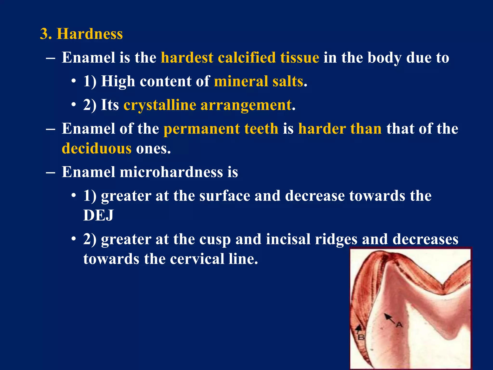
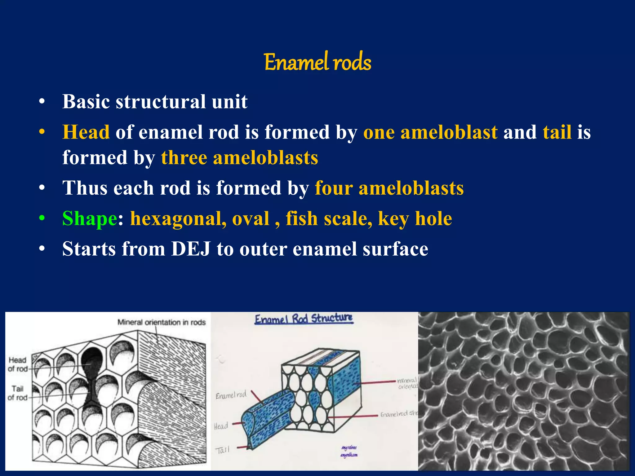
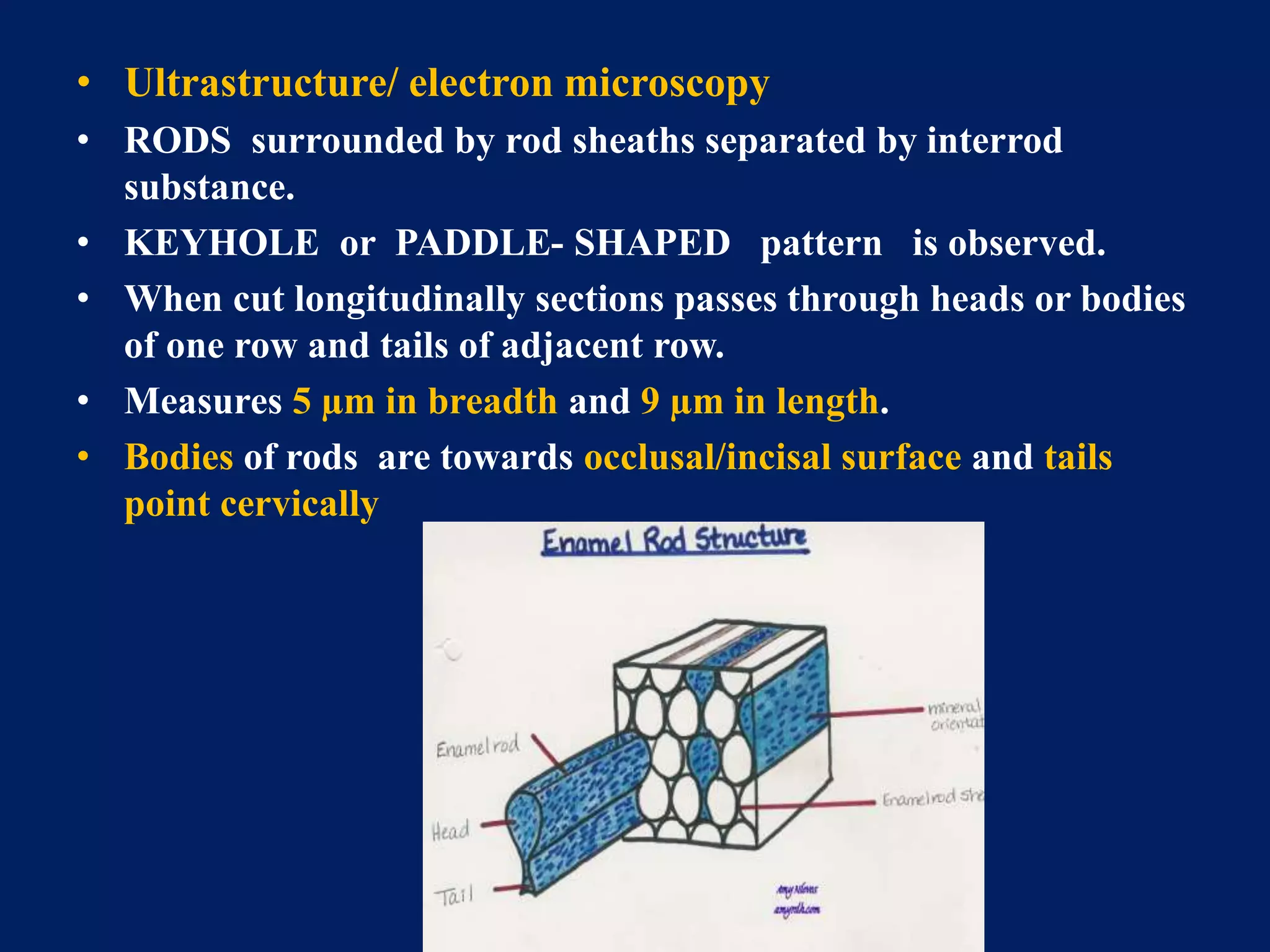
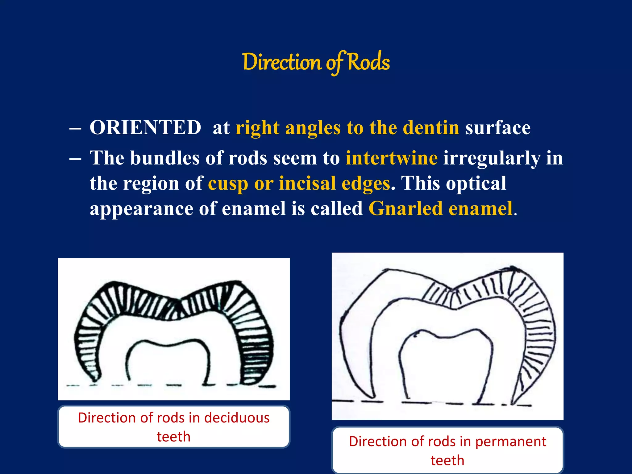
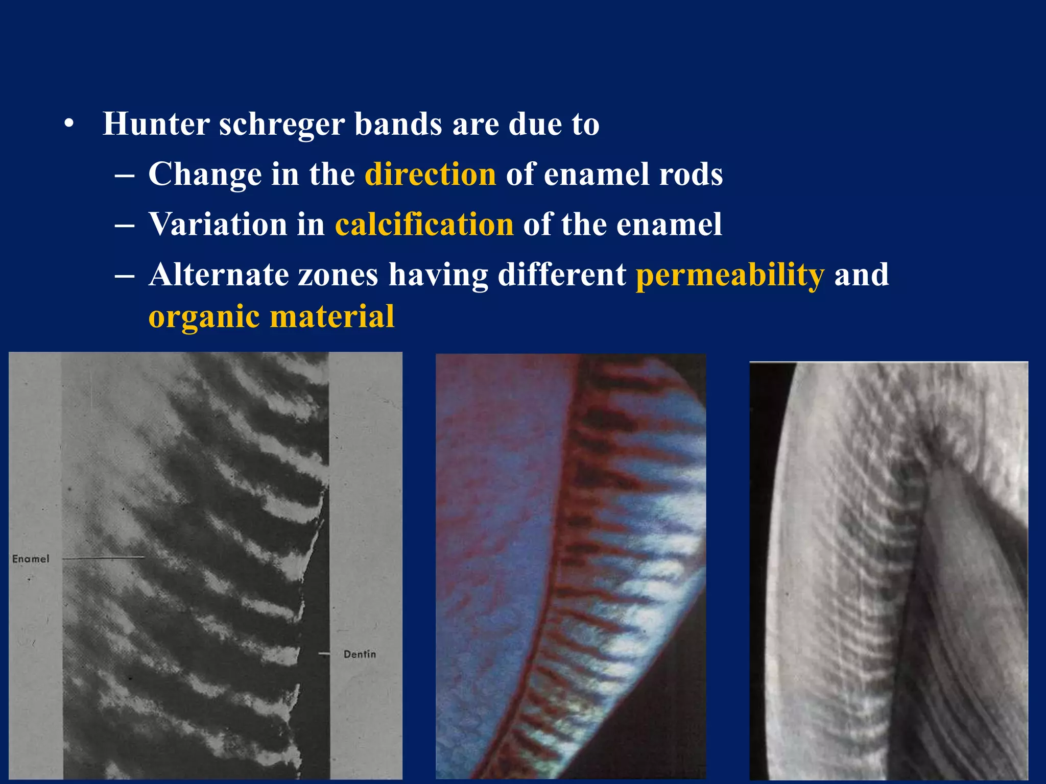
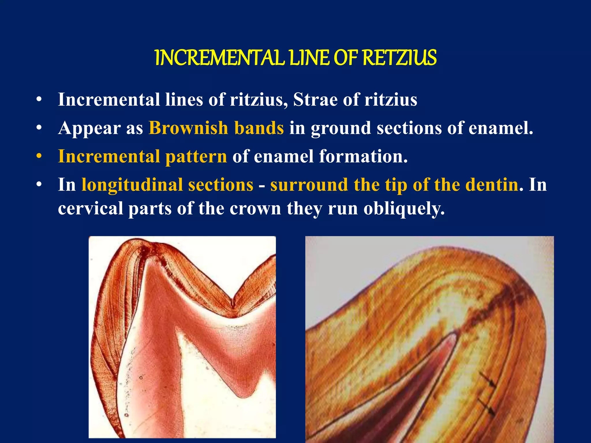
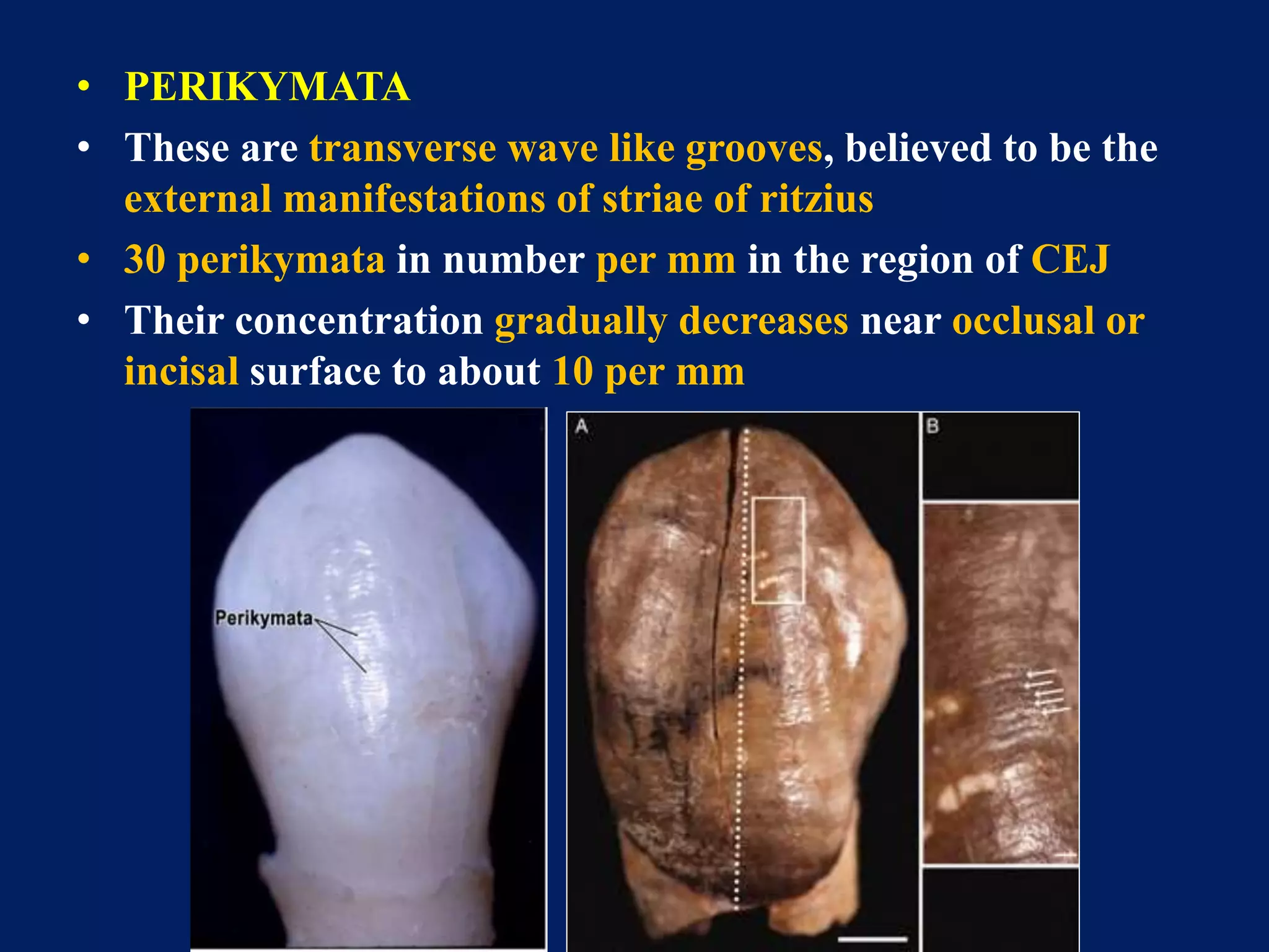
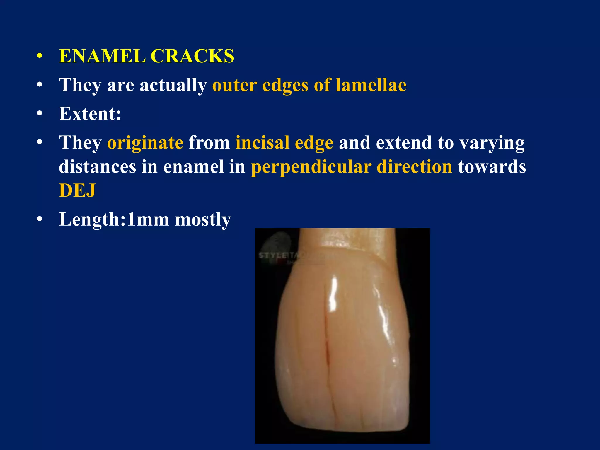
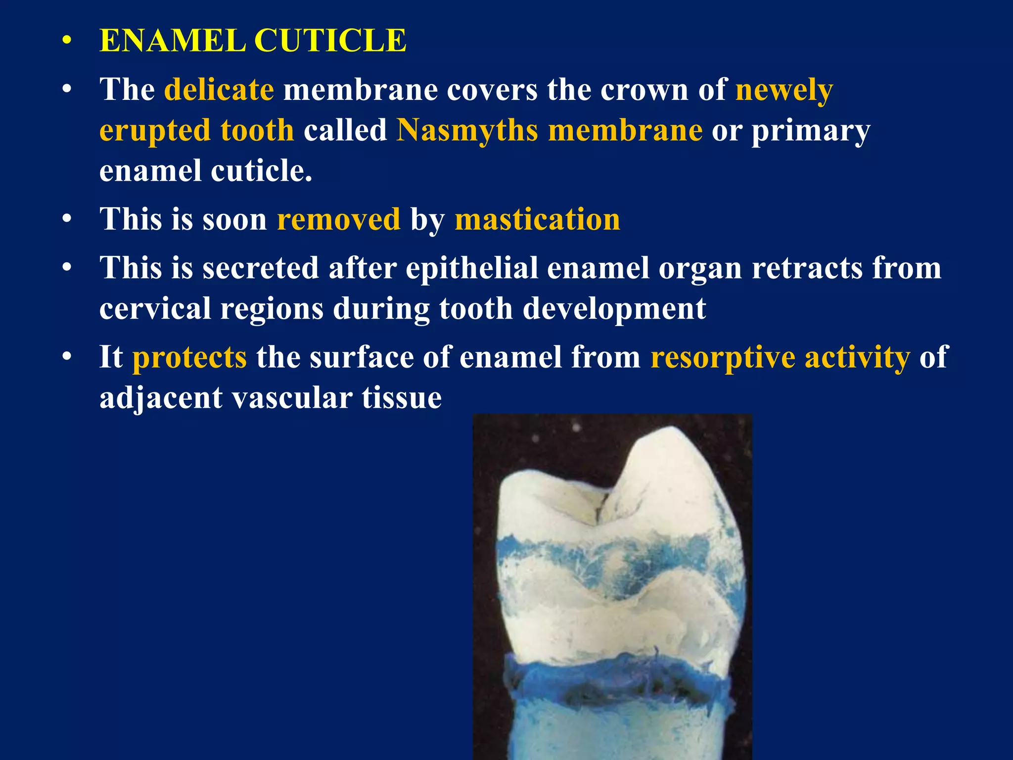
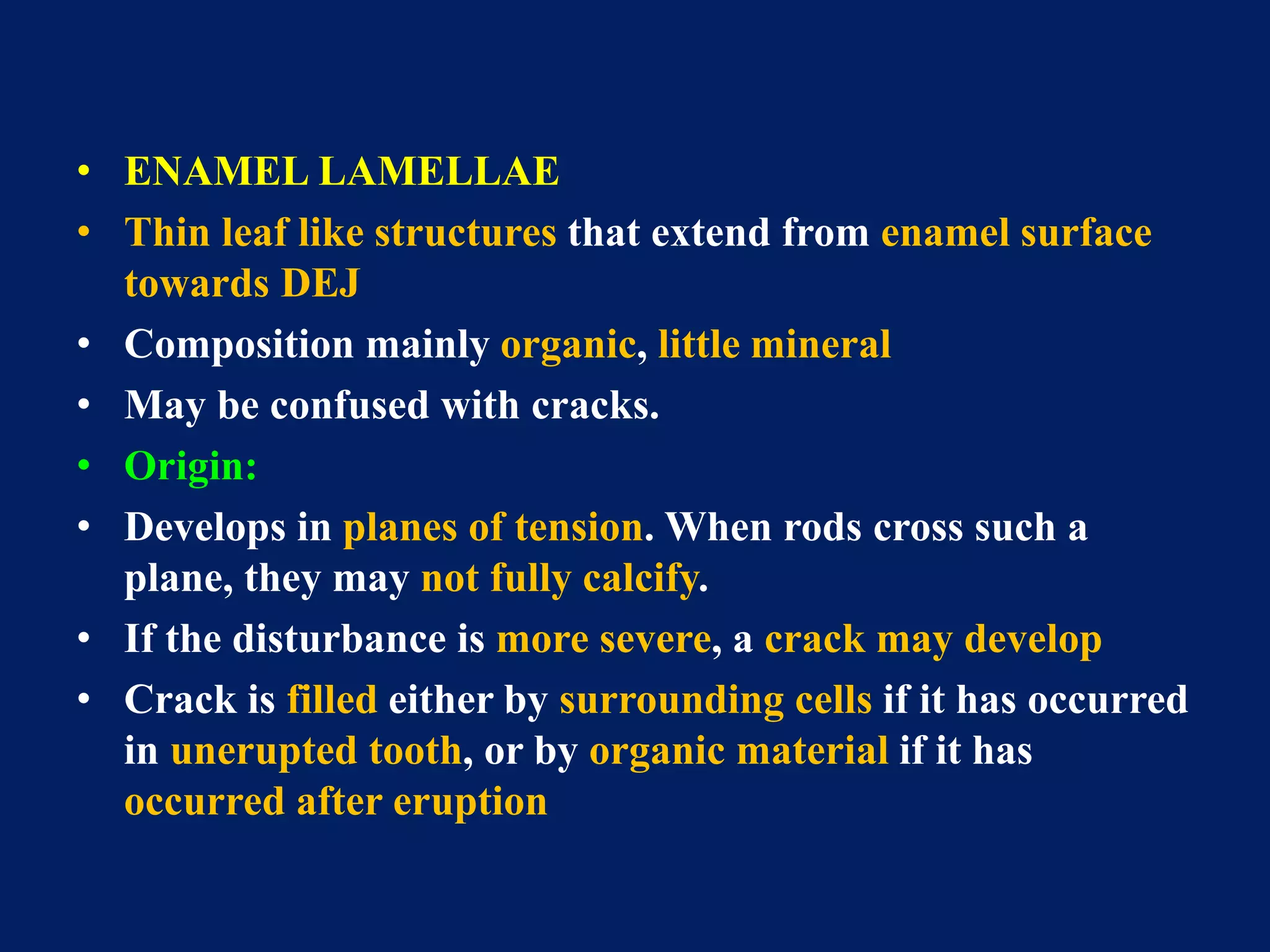
It is a presentation in detail about the strongest structure of the oral cavity "ENAMEL". It is a simple topic but people find it difficult to learn about it. I hope my presentation is a simple method to learn about it. I would like to thank my professors for assign me this project and i learn't a lot from it and still learning my basics daily.
Amelogenesis is the formation of enamel. During amelogenesis, the ameloblast (enamel-forming cells) undergo various stages i.e the life cycle of ameloblast.
For more content check out my blog: www.rkharitha.wordpress.com "a little about everything dental"
PHYSICAL PROPERTIES
CHEMICAL PROPERTIES
STRUCTURE OF ENAMEL
DEVELOPMENT OF ENAMEL
EPITHELIAL ENAMEL ORGAN
AMELOGENESIS
LIFE CYCLE OF AMELOBLASTS
AGE CHANGES IN ENAMEL
DEFECTS OF AMELOGENESIS
CLINICAL IMPLICATIONS
I prepared this presentation during the first year of my MDS. This will give you a basic idea and necessary information about the pulp of the teeth and its histology. Hope you guys find it useful.
A Complete presentation explaining the complete morphology of Maxillary first molar, for the benefit of people like me who tried and failed to find everything in one package
Ameloblast are the enamel forming cells. Understanding of life cycle of ameloblast aids in the understanding of various developmetal anomalies in particular and various other oral pathologies.
The Indian Dental Academy is the Leader in continuing dental education , training dentists in all aspects of dentistry and
offering a wide range of dental certified courses in different formats.
Upload By : Ahmed Ali Abbas
Babylon University College of Dentistry
download this file from Website on google theoptimalsmile.wix.com/dentistry
Oral histology
Enamel significance in operative dentistry /certified fixed orthodontic cour...Indian dental academy
Welcome to Indian Dental Academy
The Indian Dental Academy is the Leader in continuing dental education , training dentists in all aspects of dentistry and offering a wide range of dental certified courses in different formats.
Indian dental academy has a unique training program & curriculum that provides students with exceptional clinical skills and enabling them to return to their office with high level confidence and start treating patients
State of the art comprehensive training-Faculty of world wide repute &Very affordable.
Indian Dental Academy: will be one of the most relevant and exciting training center with best faculty and flexible training programs for dental professionals who wish to advance in their dental practice,Offers certified courses in Dental implants,Orthodontics,Endodontics,Cosmetic Dentistry, Prosthetic Dentistry, Periodontics and General Dentistry.
1. Done By : Dr.Mohamad Ghazi Kassem
2. • Enamel is an Epithelially derived hard, protective covering of teeth • Fully formed enamel is the most highly mineralized extracellular matrix known • It is highly brittle yet exhibits certain degree of resistance to withstand fracture
3. • Enamel is the hardest substance of the body, its hardness is comparable to mild steel. • Average knoop hardness number for enamel is approximately 343 • Surface of enamel is more mineralized and hard than deeper enamel. • Unlike other calcified structures in the body enamel is unique as it is totally acellular.
4. Composition of enamel Enamel consists of approximately 96% of inorganic material and 4% of organic material and water by weight. The organic component forms the matrix and the inorganic component comprises of various minerals. Percentage of dental tissue components by weight
5. The organic matrix of enamel is made from non-collagenous proteins and enzymes. Of the enamel proteins 90% are amelogenins and 10% are nonamelogenins. The different types of nonamelogenins associated with formation of enamel are ameloblastin, enamelin and tuftelin. The primary function of the organic material is to direct the growth of enamel crystals.
6. The inorganic component hydroxyapetite crystals. of enamel is comprised almost entirely of Enamel hydroxyapetite crystals are the largest hydroxyapetite crystals of all the calcified tissues in the body. In addition to hydroxyapetite crystals enamel also contains carbonates and trace elements. These crystals are susceptible to dissolution by acids and hence provides the basis for dental caries. SEM
7. Enamel is translucent and varies in colour from light yellow to whitish It varies in thickness, with maximum over cusps (2.5 mm) to a feather edge at the cervical line Thickness of enamel in primary teeth is nearly half than that in permanent teeth
8. Although enamel is an extremely hard tissue it is partially permeable to some fluids, bacteria and other products of the oral cavity The permeability of enamel is due to the presence of cracks and microscopic spaces on the surface of enamel which allows penetration of fluids The permeability of enamel decreases and hardness increases with age
9. Structure of enamel Rod and interrod enamel The fundamental units of enamel are rods and interrod enamel. The rod and interrod enamel is built from closely packed and long ribbon like hydroxyapetite crystals. The rod is shaped like a cylinder with a wide head portion, a neck and a thinner tail portion . Each rod is formed by four ameloblasts. SEM
10. ameloblasts SEM
11. Rods are formed nearly perpendicular to DEJ and curve slightly towards the cusp tip The follow a wavy course as the traverse from the DEJ to the surface of the crown The length of most rods is much longer than the thickness of enamel SEM
mandibular premolars, common triats and differences between mandibular first and second premolar. buccal aspect, lingual aspect, mesial aspect, distal aspect, occlusal aspect of mandibular premolars
Lung Cancer: Artificial Intelligence, Synergetics, Complex System Analysis, S...Oleg Kshivets
RESULTS: Overall life span (LS) was 2252.1±1742.5 days and cumulative 5-year survival (5YS) reached 73.2%, 10 years – 64.8%, 20 years – 42.5%. 513 LCP lived more than 5 years (LS=3124.6±1525.6 days), 148 LCP – more than 10 years (LS=5054.4±1504.1 days).199 LCP died because of LC (LS=562.7±374.5 days). 5YS of LCP after bi/lobectomies was significantly superior in comparison with LCP after pneumonectomies (78.1% vs.63.7%, P=0.00001 by log-rank test). AT significantly improved 5YS (66.3% vs. 34.8%) (P=0.00000 by log-rank test) only for LCP with N1-2. Cox modeling displayed that 5YS of LCP significantly depended on: phase transition (PT) early-invasive LC in terms of synergetics, PT N0—N12, cell ratio factors (ratio between cancer cells- CC and blood cells subpopulations), G1-3, histology, glucose, AT, blood cell circuit, prothrombin index, heparin tolerance, recalcification time (P=0.000-0.038). Neural networks, genetic algorithm selection and bootstrap simulation revealed relationships between 5YS and PT early-invasive LC (rank=1), PT N0—N12 (rank=2), thrombocytes/CC (3), erythrocytes/CC (4), eosinophils/CC (5), healthy cells/CC (6), lymphocytes/CC (7), segmented neutrophils/CC (8), stick neutrophils/CC (9), monocytes/CC (10); leucocytes/CC (11). Correct prediction of 5YS was 100% by neural networks computing (area under ROC curve=1.0; error=0.0).
CONCLUSIONS: 5YS of LCP after radical procedures significantly depended on: 1) PT early-invasive cancer; 2) PT N0--N12; 3) cell ratio factors; 4) blood cell circuit; 5) biochemical factors; 6) hemostasis system; 7) AT; 8) LC characteristics; 9) LC cell dynamics; 10) surgery type: lobectomy/pneumonectomy; 11) anthropometric data. Optimal diagnosis and treatment strategies for LC are: 1) screening and early detection of LC; 2) availability of experienced thoracic surgeons because of complexity of radical procedures; 3) aggressive en block surgery and adequate lymph node dissection for completeness; 4) precise prediction; 5) adjuvant chemoimmunoradiotherapy for LCP with unfavorable prognosis.
TEST BANK for Operations Management, 14th Edition by William J. Stevenson, Ve...kevinkariuki227
TEST BANK for Operations Management, 14th Edition by William J. Stevenson, Verified Chapters 1 - 19, Complete Newest Version.pdf
Couples presenting to the infertility clinic- Do they really have infertility...Sujoy Dasgupta
Dr Sujoy Dasgupta presented the study on "Couples presenting to the infertility clinic- Do they really have infertility? – The unexplored stories of non-consummation" in the 13th Congress of the Asia Pacific Initiative on Reproduction (ASPIRE 2024) at Manila on 24 May, 2024.
Pulmonary Thromboembolism - etilogy, types, medical- Surgical and nursing man...VarunMahajani
Disruption of blood supply to lung alveoli due to blockage of one or more pulmonary blood vessels is called as Pulmonary thromboembolism. In this presentation we will discuss its causes, types and its management in depth.
- Video recording of this lecture in English language: https://youtu.be/lK81BzxMqdo
- Video recording of this lecture in Arabic language: https://youtu.be/Ve4P0COk9OI
- Link to download the book free: https://nephrotube.blogspot.com/p/nephrotube-nephrology-books.html
- Link to NephroTube website: www.NephroTube.com
- Link to NephroTube social media accounts: https://nephrotube.blogspot.com/p/join-nephrotube-on-social-media.html
Ozempic: Preoperative Management of Patients on GLP-1 Receptor Agonists Saeid Safari
Preoperative Management of Patients on GLP-1 Receptor Agonists like Ozempic and Semiglutide
ASA GUIDELINE
NYSORA Guideline
2 Case Reports of Gastric Ultrasound
Explore natural remedies for syphilis treatment in Singapore. Discover alternative therapies, herbal remedies, and lifestyle changes that may complement conventional treatments. Learn about holistic approaches to managing syphilis symptoms and supporting overall health.
3. Contents
• Introduction
• Physical characteristics
• Chemical characteristics
• Structure
• Development
• Life cycle of ameloblast
• Amelogenesis
• Clinical considerations
4.
5. Introduction
• Enamel is an epithelially derived protective covering of
variable thickness over the entire surface of the crown
• It is the hardest biologic tissue in the body
• It attains the maximum thickness of about 2-2.5mm on the
cusps of molars and premolars.
• It thins down to almost knife edge at the neck of the tooth
7. Physical characteristics
1. Thickness
– 2.5 mm (thickest) over cusp of molar
– 2.0 mm over incisal edge
– Knife edge thickness at the cervical region
– Thick at maxillary lingual surfaces of molars and
mandibular buccal surface
8. 2. Color
– Depends on thickness and translucency of enamel
– Grayish white to yellowish white
– Yellowish – at thin areas underlying dentin
– Grayish – thick opaque enamel
– Bluish – incisal area
9. 3. Hardness
– Enamel is the hardest calcified tissue in the body due to
• 1) High content of mineral salts.
• 2) Its crystalline arrangement.
– Enamel of the permanent teeth is harder than that of the
deciduous ones.
– Enamel microhardness is
• 1) greater at the surface and decrease towards the
DEJ
• 2) greater at the cusp and incisal ridges and decreases
towards the cervical line.
10. 4. Brittleness
– Its structure and hardness render it brittle, specially
when it looses its elastic foundation of healthy dentin
5. Solubility: dissolves in acidic media
6. Permeability:
– It acts has a semi permeable membrane for certain ions
and dye stuffs of small molecular size through pores
between the crystals.
– Permeability is mainly from saliva to outer layers of
enamel. But less from the pulp to the inner enamel
layers across the dentin.
7. Specific gravity: 2.8
8. Translucency: semi translucent
12. • Organic content : mainly proteins (Amelogenenins, Non
Amelogenins)
• Amelogenins
• 90%
• Low molecular weight
protein
• Rich in proline, histidin,
glutamin, leucine.
• Non amelogenins
• 10%
• High molecular weight
• Proteins – ameloblastin,
tuftelin, enamelin.
• Rich in glycine, aspartic
acid and serine.
13. Structure of enamel
• Rods
• Rod sheath
• Inter-prismatic substance
• Striations
• Directions of rods
• Hunter-schreger bands
• Incremental lines of ritzius
• Surface structures
• Enamel cuticle
• Enamel lamellae
• Enamel tufts
• Dentinoenamel junction
• Odontoblast processes and enamel spindles
14. • Hypocalcified areas of enamel
– Rod sheath
– Incremental lines of ritzius
– Enamel lamellae
– Enamel tuft
– Enamel cracks
– Enamel spindles
– Neonatal lines
15. Enamel rods
• Basic structural unit
• Head of enamel rod is formed by one ameloblast and tail is
formed by three ameloblasts
• Thus each rod is formed by four ameloblasts
• Shape: hexagonal, oval , fish scale, key hole
• Starts from DEJ to outer enamel surface
16. • Number:
• Lower lateral incisor - 5 millions
• Upper 1st molar - 12 millions
• Course: Tortous from DEJ
• Length: greater than thickness of enamel
• Diameter: 4 µm in light microscopy.
• Increases from DEJ to outer enamel surface by a ratio of
1:2 since outer surface of enamel is greater than inner
surface
• Appearance : clear crystalline.
17. • Light microscopy:
• Rods appear hexagonal
• In cross section: fish scale appearance
• Recently – arcade outline near DEJ and keyhole outline at
enamel surface.
18. • Ultrastructure/ electron microscopy
• RODS surrounded by rod sheaths separated by interrod
substance.
• KEYHOLE or PADDLE- SHAPED pattern is observed.
• When cut longitudinally sections passes through heads or bodies
of one row and tails of adjacent row.
• Measures 5 µm in breadth and 9 µm in length.
• Bodies of rods are towards occlusal/incisal surface and tails
point cervically
19. • Hydroxyapatite crystals
• Arranged approximately parallel to the long axis of the
rods
• Length 0.05 to 1µm
• Width 90 µm
• Pyramid shape
20. The rod sheath
• A thin peripheral layer
• Darker than rod
• Relatively acid resistant
• Less calcified and contains more organic matter than the
rod itself
21. Inter-prismatic substance
• Cementing enamel rods together
• More calcified than the rod sheath
• Less calcified than the rod itself
• Appears to be minimum in human teeth
22. STRIATIONS
• Enamel rods is built up of segments of uniform length of
about 4µm, seperated by dark lines that gives it a straited
appearance
• More visible by action of mild acids
• Appearance is because of formation of enamel matrix in
rhythmic manner.
• More pronounced in hypocalcified areas
Cross-striation within a ground, longitudinal section of enamel
23. Direction of Rods
– ORIENTED at right angles to the dentin surface
– The bundles of rods seem to intertwine irregularly in
the region of cusp or incisal edges. This optical
appearance of enamel is called Gnarled enamel.
Direction of rods in deciduous
teeth Direction of rods in permanent
teeth
24. • In deciduous teeth, direction of rods is horizontal in
cervical and central parts of the crown. Near incisal edge
or tip of cusp they gradually increase in oblique direction
and almost vertical in the cusp tip region
• In permanent teeth, in occlusal two third of the crown
direction of rods is oblique.
• In cervical direction rods deviate from the horizontal in
apical direction
25.
26. • GNARLED ENAMEL
• Near the dentin in the region of cusps or incisal edge,
bundles of rods seem to interwine more irregular,
especially in section cut obliquely. This optical appearance
of enamel is called gnarled enamel.
27. HUNTER- SCHREGER BANDS.
• Optical phenomenon seen in reflected light
• Seen in longitudinal ground section
• Due to abrupt change in direction of enamel rod
• Alternating light (diazones) and dark (parazones) strips of
varying widths best seen in longitudinal ground section
under oblique reflected light.
• Originate at the DEJ and pass outward, ending at some
distance from the outer enamel surface.
28. • Hunter schreger bands are due to
– Change in the direction of enamel rods
– Variation in calcification of the enamel
– Alternate zones having different permeability and
organic material
29.
30. INCREMENTAL LINE OF RETZIUS
• Incremental lines of ritzius, Strae of ritzius
• Appear as Brownish bands in ground sections of enamel.
• Incremental pattern of enamel formation.
• In longitudinal sections - surround the tip of the dentin. In
cervical parts of the crown they run obliquely.
31.
32.
33. • In transverse section - appears as concentric circles.
• DEJ to outer surface of enamel
• Reflects variations in structure and mineralization that
occur during growth of enamel.
• Broadening of incremental lines may reflect metabolic
disturbance at the time of matrix formation
• NEONATAL LINE or NEONATAL RING: Accentuated
incremental line of Retzius.
• Where they end as shallow furrows known as perikymata
34.
35. Neonatal line
• The enamel of the deciduous teeth and 1st permanent
molar (it is incremental line that is boundary between the
enamel formed before and after the birth)
• The neonatal line is usually the darkest and thickest striae
of ritzius
• Etiology
– Due to sudden change in the environment and nutrition
– The antenatal enamel is better calcified than the
postnatal enamel
38. • PRISMLESS ENAMEL
• About 30µm thick
• Present in 70% permanent teeth and all deciduous teeth
• Found least often over the cusp tips
• Found commonly in the cervical areas
• No enamel prisms visible.
• All the apatite crystals are parallel to one another and
perpendicular to the striae of ritzius
• More mineralized than the bulk of enamel beneath it,
39. • PERIKYMATA
• These are transverse wave like grooves, believed to be the
external manifestations of striae of ritzius
• 30 perikymata in number per mm in the region of CEJ
• Their concentration gradually decreases near occlusal or
incisal surface to about 10 per mm
40.
41. • ENAMEL ROD ENDS
• These are concave and vary in depth
• They are shallow cervically and deep occlusally/incisally
• Pits - 1-1.5µm in diameter
• Enamel caps - 10-15µm elevations
• Enamel brochs - Larger enamel elevations
42. • ENAMEL CRACKS
• They are actually outer edges of lamellae
• Extent:
• They originate from incisal edge and extend to varying
distances in enamel in perpendicular direction towards
DEJ
• Length:1mm mostly
43. • ENAMEL CUTICLE
• The delicate membrane covers the crown of newely
erupted tooth called Nasmyths membrane or primary
enamel cuticle.
• This is soon removed by mastication
• This is secreted after epithelial enamel organ retracts from
cervical regions during tooth development
• It protects the surface of enamel from resorptive activity of
adjacent vascular tissue
44. • PELLICLE
• Erupted enamel is covered by a precipitate of salivary
proteins called pellicle
• This pellicle reforms within hours after mechanical
cleaning.
• It becomes colonized by microorganisms within a day or
two after formation which forms bacterial plaque
45. • ENAMEL LAMELLAE
• Thin leaf like structures that extend from enamel surface
towards DEJ
• Composition mainly organic, little mineral
• May be confused with cracks.
• Origin:
• Develops in planes of tension. When rods cross such a
plane, they may not fully calcify.
• If the disturbance is more severe, a crack may develop
• Crack is filled either by surrounding cells if it has occurred
in unerupted tooth, or by organic material if it has
occurred after eruption
46. • Types
• Type A : consists of poorly calcified rod segments
• Resticted to enamel
• Type B:
• Consists of degenerating cells. May reach into dentin
• Type C: containing organic material, presumably from
saliva.
• If connective tissue invades the cracks in enamel,
cementum may be formed
47. • Significance:
• It has been suggested that lamellae may be a site of
weakness in a tooth and may form a road of entry for
bacteria that initiate caries
48.
49. `
• ENAMEL TUFTS
• Arises from DEJ
• Thin ribbon like structure, resembling tufts of grass which
is created by examining such area under low magnification
in thick ground section
• Tufts consists of hypo calcified enamel rods and
interprismatic substance.
50. • They arise at DEJ and reach to enamel to about 1/5 to 1/3rd
of its thickness
• Their presence and their development are consequence of
an adaptation to spatial condition of enamel
• Significance
• Enamel tuft prevents enamel fractures
51.
52. • DENTINOENAMEL JUNCTION
• Scalloped structure: the surface of the dentin at DEJ is
pitted, in shallow depression of dentin, fir rounded
projection of enamel.
• It appears scalloped due to the mixing of crystals of dentin
and enamel with each other
53. • Significance of scalloping:
• It ensures firm hold of the enamel cap to dentin
54. • ENAMEL SPINDLES
• Occasionally odontoblastic process passes across DEJ into
enamel, since many are thickened at their end, they have
been termed enamel spindles.
• Directions of spindles and rods are divergent as rods are
formed at right angle to ameloblast and spindles are
parallel to ameloblasts.
58. Age changes
• Most common change s are – attrition or wear of occlusal
and proximal surfaces
• Loss of verticle dimension of crown and by flattening of
proximal contour.
59. • Perikymata and rod ends
• At eruption these are prominent
• With age they reduce
• Generalized loss of rod ends
• Flattening of perikymata.
• The rate at which structures are lost depends upon
– Location of surface of tooth location of tooth in mouth
60. • WITH AGE
• Teeth darken
– Increase in organic content
– Deepening of dentin colour
• Decrease in permeability
• Fluoride ions increase with age
• Nitrogen increases with age
• Their resistant to decay may be increased
• Reduced permeability of older teeth to fluid
• Enamel may become harder with age
61. Clinical considerations
• Grooves and fissures on the occlusal surfaces of molars
and premolars – weak spots for the action of caries as the
maintenance is difficult – use of pit fissure sealants
• Lamellae, tufs and spindles may facilitate spread of caries.
• Striae of ritzius are the areas of hypomineralization thus
fecilitate the lateral spread of caries.
• Fluoridation decreases caries.