Downloaded 148 times

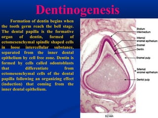
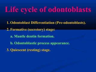
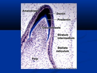
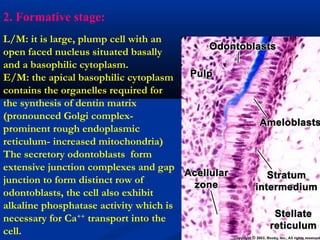
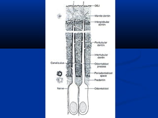
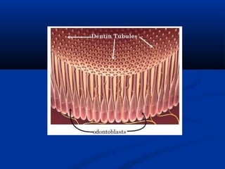
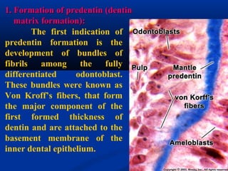
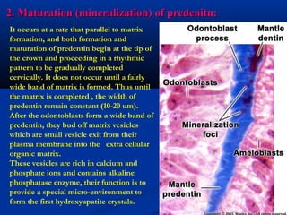
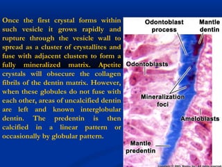


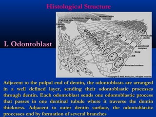
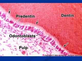


Dentin is the mineralized hard tissue that forms the bulk of the tooth beneath enamel and cementum. It has two main properties that distinguish it from enamel: it is sensitive and forms throughout life at the expense of the dental pulp. Dentinogenesis, or dentin formation, begins when the tooth germ reaches the bell stage. Odontoblasts differentiate from ectomesenchymal cells of the dental papilla and secrete dentin matrix, which then undergoes mineralization to form the bulk of dentin, including mantle dentin and circumpulpal dentin. This process of matrix formation and mineralization by odontoblasts is ongoing throughout life.


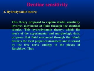
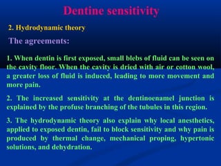
![]Dental Occlusion part 1](https://cdn.slidesharecdn.com/ss_thumbnails/occlusionpart1-160420073612-thumbnail.jpg?width=640&height=640&fit=bounds)


