
More Related Content
What's hot
Similar to ENAMEL
Similar to ENAMEL (20)
More from CmenonMenon
Recently uploaded
Recently uploaded (20)
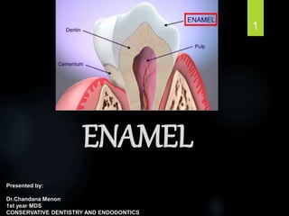
ENAMEL
- 1. ENAMEL Presented by: Dr.Chandana Menon 1st year MDS CONSERVATIVE DENTISTRY AND ENDODONTICS 1
- 2. TABLE OF CONTENTS: Definition Introduction Physical Properties Chemical properties Structure of enamel Surface structures of enamel Development of enamel Age changes of enamel 2
- 3. Clinical considerations Developmental defects of enamel Caries and enamel Non carious lesions of enamel Effects of flouride Effects of laser Bleaching Adhesion to enamel Formulation of enamel walls Effects of burs Conclusion 3
- 4. Enamel is an eccentric hard tissue because of its origin, chemically distinct nature of the various noncollagenous matrix proteins expressed by ameloblasts, and its large mineral crystals Ectodermal , Non – collagenous, Non vital 4 tencate
- 5. INTRODUCTION Hardest calcified tissue in human body. Protective and resistant covering over the entire surface of the crown. Ameloblast – cells responsible for formation of enamel, are lost as the tooth erupts into the oral cavity, and hence enamel cannot renew itself. 5
- 6. PHYSICAL PROPERTIES THICKNESS: Increase in thickness viewed as adaptation to functional demands. • Maximum thickness of 2-2.5mm at cusps of molars and premolars. • Knife edge at the neck of the tooth • Thicker at lingual surface of maxillary molar and buccal surface of mandibular molars. 6
- 7. HARDNESS: Hardest calcified tissue due to high content of mineral salts and their crystalline arrangement. Brittle, which is particularly apparent when the enamel loses its foundation of sound dentin. SPECIFIC GRAVITY : 2.8 7
- 8. PERMEABILITY: Semipermeable , permitting complete or partial passage of certain molecules COLOR: Color ranges from yellowish white to grayish white. •Translucent and thin enamel, visible dentin more opaque enamel 8
- 9. CHEMICAL PROPERTIES: The enamel consist mainly of Inorganic material (96%) Organic material and water (4%) The inorganic material of the enamel is hydroxyapatite. 9
- 10. The organic material consists of Unique proteins (found exclusively in the enamel) Lipids The proteins found in the enamel are of two main types: Amelogenins Nonamelogenins 10
- 11. AMELOGENINS: 11 Heterogenous group of low molecular weight protein. Accounts 90% of the enamel proteins. Hydrophobic. Rich in proline, histidine, glutamine and leucine
- 12. NONAMELOGENIN: 12 High molecular weight proteins. Constitute about 10% of enamel matrix protein. Enamelin, ameloblastin and tuftelin are the important proteins of this group. They are rich in glycine, aspartic acid and serine.
- 13. HYDROXYAPATITE CRYSTALS: The inorganic material of enamel Chemical formula-- (Ca10(PO4)6(OH)2 ) Crystals are Hexagonal in cross section. 13
- 14. Has a central core or C axis of hydroxyl ion around which Calcium and phosphorous ions are arranged in triangles. During the formation, magnesium can replace calcium and carbonate can replace hydroxyl ion which destabilize the lattice. 14
- 15. Fluoride may substitute hydroxyl ions. Conferring greater stability and resistance to acidic dissolution. Cores of the crystallites are richer in magnesium and carbonate. So greater solubility in acids than the peripheral portions. Carbonate rich crystals are more attacked by acids in caries 15
- 16. DRY WEIGHT % OF ENAMEL OXYGEN 43.4 % CALCIUM 36.6 % PHOSPHORUS 17.7 % SODIUM 0.67 % CARBON 0.64 % MAGNESIUM 0.35 % 16
- 17. WATER: Water is present as a part of the crystal, between crystals and between rods and surrounding the rods. Pores are present between the crystals, especially at the boundaries of the rods and these are filled with water. 17
- 18. STRUCTURE OF ENAMEL: The enamel is composed of 18 Enamel rods or prisms Rod sheath Interprismatic substance
- 19. ENAMEL RODS / PRISMS HA crystals are arranged parallel to each other to form the enamel rods/prisms. Number of rods Diameter of rods – Avg 4 micro m - increase from DEJ to the surface (1:2) Runs a tortuous course from DEJ to the surface - length of rods > thickness of enamel 19 - 5 million in lower lateral incisor - 12 million in upper first molars
- 20. In electron microscopic study - key shaped or paddle shaped prisms In decalcified section of teeth - hexagonal round or oval, resembles fish scales DEJ : Arcade outline Enamel surface :keyhole shaped outline 20
- 21. ROD SHEATHS Organic matrix forms an envelope surrounding each apatite crystals Less calcified than rods More organic matter than the rod 21
- 22. INTER PRISMATIC SUBSTANCE Cements the rods Less calcified areas than the rod More calcified than rod sheath 22
- 23. STRIATIONS Cross striations demarcate the rod segments More pronounced in insufficiently calcified enamel Length – 4 micro m Due to diurnal rhythmic deposition of matrix 23
- 24. Direction of rods Starts perpendicular to the surface of dentin 24
- 25. Has a wavy course from the dentin to enamel surface It’s a functional adaptation to resist cleavage in an axial direction 25
- 26. During cavity preparation the direction of enamel rods should be followed Enamel margins should be well supported by dentin Unsupported enamel margins are brittle Can Break & produce leakage leading to secondary caries 26
- 27. Gnarled enamel If disks are cut in an oblique direction, near the dentin in the region of cusps - bundles of rods seem to intertwine This enamel does not yield readily to the pressure of hand instruments 27
- 28. Hunter – Schreger bands In longitudinal ground sections under reflected light -As alternating dark and light strips of varying widths Result of change in the direction of rods Composed of alternating zones - slightly different permeability - different content of organic material 28
- 29. Starts from DEJ and ends at some distance from the outer surface HSB patterns have evolved to optimise resistance to attrition, abrasion and tooth fracture. Certain aspects of HSB packing densities and distributions have beneficial roles in enamel bonding. Hunter–Schreger Band patterns and their implications for clinical dentistry, journal of oral rehabilitation 29
- 30. Incremental lines of retzius In ground sections - Brownish bands In transverse section - As concentric circles In longitudinal sections - suround the tip of dentin 30
- 31. Due to successive apposition of layers of enamel during formation Mean daily formation is 3.5 microns Increases from inner to outer enamel Metabolic disturbance may alter this rhythm 31
- 32. Neonatal lines Accentuated incremental lines In deciduous teeth and 1st permanent molar Forms the boundary of enamel formed before birth and after birth Prenatal enamel is more calcified. 32
- 33. Surface structures of enamel 1. Prismless enamel No prisms are visible (30 micro m) In 70% permanent & all deciduous teeth Heavily mineralized with more inorganic content 33
- 34. Surface is denser and less permeable when compared to prismatic enamel Less at the cusp tips & more cervically 34
- 35. 2. Perikymata External manifestations of striae of Retzius, parallel to each other and CEJ. As transverse wave like grooves About 30/mm at CEJ - reduces to 10/mm at occlusal and incisal third May contribute to the accumulation of plaque 35
- 36. 3. Enamel Cuticle Primary enamel cuticle - Nasmyth’s memberane covers entire crown of newly erupted tooth. Wavy course, protects enamel from resorptive activity prior to tooth eruption 36
- 37. Primary Enamel Cuticle Delicate membrane that covers the entire surface of newly erupted tooth Is the basal lamina secreted by ameloblasts Soon removed after eruption, from the incisal and occlusal thirds 37
- 38. Secondary Enamel Cuticle Covered the cervical area of enamel Continuous with the cementum Its probably of mesodermal origin 38
- 39. Pellicle It is the precipitate of salivary proteins Covers the crown Reforms with in hours after mechanical cleaning It colonizes micro organism to form a bacterial plaque 39
- 40. 4. Enamel lamellae Leaf like structures that extend from surface of enamel towards the dentinoenamel junction. Hypomineralized structure It may be a site of weakness and forms the road for entry of bacteria 40
- 41. Types of Enamel lamellae Type A Type B Type C Consist Poorly calcified rod segments Degenerated cells Organic matter from saliva Tooth unerupted unerupted erupted Location Restricted to enamel Reach into dentin Reach into dentin Occurrence Less common Less common More common 41
- 42. 5. Enamel tufts Arise at DEJ & reach into enamel to about 1/5th to 1/3rd of enamel thickness. Consists of hypocalcified enamel rods & interprismatic substance. 42
- 43. Ribbon-like, resemble tuft of grass Extend in the direction long axis of crown Abundantly seen in longitudinal sections. 43
- 44. Dentino Enamel Junction(DEJ) Surface of dentin is pitted at DEJ Into the shallow depression of dentin, the rounded end of enamel is fitted DEJ appears scalloped, convexity directed towards dentin. 44
- 45. Ridges of DEJ are more pronounced in occlussal area. Provide better adhesion of enamel and dentin. 45
- 46. Cemento Enamel junction The relation between enamel and cementum at the cervical region of the tooth is variable. In 60% -cementum overlaps the enamel In 30% -cementum meets enamel in a relatively sharp line. In 10% -enamel and cementum do not meet. 46
- 47. 6.Enamel spindles Extension of odontoblastic processes across the DEJ into the enamel. Appears dark in transmitted light. Mainly found in the cuspal region Hypomineralized structure 47
- 48. Development of Enamel Some special features of enamel Enamel is the only hard tissue, which does not have collagen in its organic matrix The enamel present in the fully formed crown has no viable cells All the enamel is formed before eruption of teeth 48
- 49. Only ectodermal derivative of the tooth Derived from the enamel organ Enamel organ is differentiated from the primitive oral epithelium lining the stomodeum They also lacks the vessels and nerves 49
- 50. Dental lamina Serve as the primordium for the ectodermal portion of the deciduous teeth. - enamel organ First evidence of tooth development begins in the sixth week in utero or three weeks after the rupture of the bucco pharyngeal membrane. 50
- 51. 51 Developmental stages of teeth – Bud stage Cap stage Bell stage
- 52. Enamel organ of cap stage 52 Outer enamel epithelium Inner enamel epithelium
- 53. Life cycle of Ameloblasts (1) Morphogenic stage (2) Organizing stage (3) Formative stage (4) Maturative stage (5) Protective stage (6) Desmolytic stage 53
- 54. Before formation of enamel, the cells of inner enamel epithelium differentiate in to AMELOBLASTS 54
- 55. 1. Morphogenic stage Early bell stage Ameloblasts interact with the adjacent mesenchymal cells Determination of shape of the dentino enamel junction and the crown Cells are short and columnar with large oval nuclei. 55
- 56. 2. Organizing stage Late bell stage The inner enamel epithelium interacts with the adjacent connective tissue cells, which differentiate into odontoblasts. Formation of dentin begins Cells become longer 56
- 57. Migration of centrioles and Golgi regions from proximal ends to distal end- reversal of polarity. 57
- 58. 3. Formative stage The ameloblasts enter their formative stage after the first layer of dentin has been formed. Enamel formation begins. First layer of enamel is formed and the ameloblasts migrate to form the Tome’s process. 58
- 59. 4. Maturative stage Enamel maturation (full mineralization) occurs after most of the thickness of the enamel matrix has been formed in the occlusal or incisal area. Matrix formation is still in progress at the cervical area 59
- 60. Ameloblasts are sightly reduced in length, spindle shaped cells in stratum intermedium. Distal extremities of ameloblast display microvilli and cytoplasmic vacuoles containing material resembling enamel matrix indicating- absorptive function. 60
- 61. 5. Protective stage Enamel has completely developed and has fully calcified The ameloblasts cease to be differentiated These cell layers then form a stratified epithelial covering of the enamel -Reduced Enamel Epithelium(REE) 61
- 62. Reduced Enamel Epithelium(REE) REE protects the enamel till eruption. In case of premature breakdown, CT comes in contact with the enamel surface and deposits cementum 62
- 63. 6. Desmolytic stage REE proliferates and induces atrophy of connective tissue separating it from oral epithelium. Fusion of both epithelium takes place. 63
- 64. 64
- 65. 65 FIGURE 4.45 The life cycle of an ameloblast. The cells of the internal enamel epithelium (1) start to differentiate, beginning at the future enamel–dentine junction of the cusp tip. The differentiating cell (2) is characterized by a reversed polarity; the cell becomes columnar and the nucleus moves to that part of the furthest from the dentine. Secreting organelles are formed and the end of the cell adjacent to the dentine becomes the site for secretion. At the next stage (3), the cell secretes the initial enamel component of the enamel–dentine junction. This thin layer will be continuos with the inter-rod enamel of the later formed tissue. As the cell retreats, the secreting pole becomes morphologically distinct as a pyramidal Tomes process (4a). Crystallites are formed at both surfaces of the process. The proximal region between two processes, deep in the junctional regions, always secretes ahead of the more distal region so that pits surrounded by inter-rod enamel are formed. These are then filled, giving the prism configuration to the tissue. Simultaneous secretion of both organic material and mineral continues until the full thickness of the tissue is formed. In this secreting phase, two appearances of ameloblasts can be distinguished by the position of the nuclei within the cell: high (4a) and low (4b). At the beginning of secretion, half the cells are in each form. Towards the end of secretion, most of the high nuclei have moved to a low position, effectively increasing the areas of the ameloblast cells as the surface of forming enamel increases. When the full thickness of enamel has formed, ameloblasts lose the secretory extension, the Tomes process (5a). Up to 50% of them die and are phagocytosed by others in the layer. The maturation phase lasts two to three times longer than the secretory phase. During the maturation phase there is a regular, repetitive modulation of cell morphology between a ruffled (5a) and a smooth (5b) surface apposed to the enamel. Once the maturation changes are complete, the cells regress in height (6). At this stage, they serve to protect the enamel surface during eruption and later will contribute to form the junctional epithelium.
- 66. 66 AMELOGENESIS
- 67. Two processes are in involved, 1.Organic matrix formation - Development of tomes process - Distal terminal bars 2.Matrix mineralization and maturation 67
- 68. 1. Organic matrix formation Ameloblasts begin secretory activity when a small amount of dentin has been laid down Deposition of islands of matrix proteins along the predentin Amelogenin is the major component of enamel matrix proteins 68
- 69. Tomes’ process Surfaces of the ameloblasts facing the developing enamel are not smooth. There is an interdigitation of the cells and the enamel rods that they produce The projections of the ameloblasts into the enamel matrix - Tomes’ process 69
- 70. “Picket fence” arrangement of Tome’s processes. 70
- 71. 4 ameloblast results in the synthesis of 1 enamel rod Head of rod by 1 ameloblast Tail of rod by 3 ameloblasts Inter rod enamel is formed by junctional complexes and from adjacent ameloblast 71
- 72. 72 Drawing illustrating one interpretation of relationships between enamel rods and ameloblasts. Cross-sections of ameloblasts are indicated by thin lines arranged in regular hexagonal array. Enamel rods are indicated by thicker curved black lines, outlining keyhole- or paddle-shaped rods. Gray lines indicate approximate orientation of enamel crystals, which are parallel to long axes of rods in their “bodies” and approach a position perpendicular to long axes in “tails.” One can see that each rod is formed by four ameloblasts and that each ameloblast contributes to four different rods. (Source: Modified from Boyde A: In Stack MV and Fearnhead RW, editors: Tooth enamel, Bristol, 1965, John Wright & Sons Ltd).
- 73. Distal terminal bars Junctional complexes encircle the distal and the proximal end of ameloblast Junctional complexes at distal end – distal terminal bar It separates Tome’s process from the cell proper 73
- 74. 2. Matrix mineralization Nucleation is initiated by the apatite crystallites of dentin on which enamel is laid Occurs in matrix segments and interprismatic substances are laid down. Initial mineral is octacalcium phosphate. It is unstable and one unit of it is converted to 2 units of Hydroxyapatite. 74
- 75. Maturation(second stage) Gradualcompletion of mineralization. Its begins before matrix has reached its full thickness Rods mature from - depth to surface The crystals increase in size from 1.5 – 25 micro m Loss of volume of organic matrix by withdrawal of proteins & water 75
- 76. 76
- 77. AGE CHANGES IN ENAMEL Most apparent change - Attrition, wear of occlusal & proximal surface - loss of vertical dimension Anterior teeth lose rapidly than do posterior 77
- 78. Changes in the organic portions - teeth may become darker - resistance to decay increases Increase in the size of the crystals - decrease the pores between them - greatly reduce the permeability 78
- 79. Some clinical considerations Grooves & fissures They are formed at the junction of the developmental lobes of the enamel. Sound coalescence of the lobes results in grooves, faulty coalescence results in fissures. Deep enamel fissures acts as a niche for acidogenic bacteria - Easily penetrate the floor - spreads along the DEJ - undermining of enamel 79
- 80. Surface of enamel in the cervical region should be kept smooth & well polished. - otherwise food debris & bacterial plaque accumulate on the roughened surface. 80
- 81. Developmental Defects of Enamel Hypoplasia Hypocalcification 81
- 82. Hypoplasia Incomplete or defective formation of enamel matrix These are the principal expressions of pathologic amelogenesis Manifested as pitting, furrowing, even total absence 82
- 83. Hypocalcification Maturation is lacking or incomplete A deficiency of mineral content is found Opaque or chalky white areas on normally contoured enamel surface 83
- 84. Systemic hypocalcification Major cause is the increased fluoride content of water (>1.5 ppm). If the injury is during the maturation stage deficiency of calcification will occur 84
- 85. Hereditary hypocalcification Normal amount of enamel produced,but hypomineralized. It is soon discolored, abraded by mastication, or peeled off in layers. Affected teeth may have areas of coronal discoloration, or they may have actual pits and irregularities 85
- 86. AMELOGENESIS IMPERFECTA It represents a group of hereditary defects that cause disruption to the structure and clinical appearance of enamel. It is entirely an ectodermal disturbance and the mesodermal components of the tooth are normal. 86
- 87. According to the 3 stages in the development of normal enamel, 3 basic types are recognized. 1.Hypoplastic type There is defective matrix formation. Enamel has not formed to full normal thickness on newly erupted teeth The teeth are small and may be white, yellow, or brown, 87
- 88. Defective mineralization of formed matrix. The enamel may have a normal thickness, but it’s too soft. The teeth may be white, yellow, or brown, and the enamel may be rough. 88
- 89. 3. Hypo maturation type The teeth are opaque to yellow or brown with sensitivity. The enamel has a normal thickness, but it’s too soft, so the teeth appear mottled and may wear away and break. 89
- 90. Caries and Enamel Is an irreversible microbial disease of the calcified tissue of the teeth, characterized by demineralization of inorganic part and destruction of organic substance of tooth,which often leads to cavitation. 90
- 91. Clinical characteristics On clean dry tooth the earliest evidence of caries is a white spot which are chalky white and opaque They are revealed only when the tooth surface is dry. The surface texture is unaltered and these areas of enamel lose their translucency because of the extensive subsurface porosity caused by demineralization. 91
- 92. Zones of enamel caries Zone1: translucent zone. Zone2: dark zone. Zone3: body of lesion. Zone4: surface zone 92
- 93. 1. Translucent zone Deepest zone Represent advancing front of lesion Pore volume is 1% 93
- 94. 2. Dark zone Lies adjacent and superficial to translucent zone Does not transmit polarized light Total pore volume 2- 4% 94
- 95. 3. Body of the lesion Largest portion Area of gretest demineralization Pore volume 5 – 25 % Striae of retzius is more prominent 4. Surface zone Relatively stable layer and unaffected by caries Radiopacity similar to normal enamel Pore volume less than 5% of the spaces. 95
- 96. Non carious lesions of enamel Attrition Abrasion Erosion Localized nonhereditary enamel hypoplasia Localized non hereditary enamel hypocalcification Discoloration Amelogenesis imperfecta 96
- 97. Discoloration Can occur due to: Extrinsic factors: 1. Tobacco/tea stains 2. Poor oral hygiene 3. Food colors 4. Existing restorations 5. Chromogenic bacteria 97
- 98. Intrinsic factors: 1. Caries. 2. Fluorosis. 3. Tetracycline and other drugs. 4. Age changes. 5. Non vital teeth 6. Internal resorption. 7. Hereditary disorders. 98
- 99. Effects of fluoride on enamel 1.Anti cariogenic property Increased enamel resistance or reduction in enamel solubility. Calcium hydroxy apatite fluroapatite 99
- 100. 2.Increased rate of post eruptive maturation 3.Remineralisation of incipient lesion 4.Inhibition of demineralisation 5.Interference with microorganisms 6.Modification of tooth morphology 10 0
- 101. Flourosis ► Caused by excessive systemic flouride during enamel matrix formation and calcification. ► Mild intermittent white spotting ► Chalky or opaque areas ► Surface pitting ► Marked wear of enamel surface ► Brown stains ► Severe cases-corroded appearance 10 1
- 102. Flourosis 10 2
- 103. Bleaching The lightening of the color of a tooth through the application of a chemical agent to oxidize the organic pigmentation of the tooth is referred to as bleaching. Hydrogen peroxide is most commonly used. 10 3
- 104. Types of Bleaching A) Non-Vital bleaching In-Office /Thermocatalytic technique Out of the office technique/Walking Bleach B)Vital Bleaching In Office technique /Power bleaching. Dentist prescribed home applied technique (Night guard vital bleaching) 10 4
- 105. Enamel Microabrasion It is not a bleaching technique. A selective erosion process that removes stained enamel. Currently microabrasion is recommended for the removal of stains that are superficial and localized in enamel. 10 5
- 106. Uses of 18% hydrochloric acid and pumice . Only one commercially developed system currently exists for enamel microabrasion. The PREMA system (Premier enamel micro abrasion) 10 6
- 107. ADHESION TO ENAMEL ACID ETCHING Achieved through acid etching, which enlarges its surface area for bonding. Buonocore in 1955. Underlying mechanism of the bond suggested that resin tags were formed and micromechanically interlocked with the enamel micro porosities created by etching 107
- 108. As there are 30,000 to 40,000 enamel rods/sq mm and the etch penetration increases the bondable surface area 10 to 20 fold Etching transforms the smooth enamel surface into an irregular surface with a high surface free energy 108
- 109. Acid etching removes about 10µm of the enamel surface and creates a micro porous layer from 5 to 50 µm deep. When doing etching for bonding composites - Prismless enamel should not provide mechanical retention - So etching should go beyond to the prismatic enamel 109
- 110. GENERAL PRINCIPLES FOR FORMULATION OF ENAMEL WALLS The enamel portion of a wall should be the smoothest portion of the preparation anatomy. Junction between different enamel walls should be rounded -decreasing stress concentration there. Enamel walls should be well supported by dentin 110
- 111. cavosurface angle Amalgam restoration -90 degree (butt joint). Cast inlay -130-140 degree (lap joint) For Amalgam class 2 – bevelling the gingival margin with GMT : 15-20 degree. 111
- 112. Effect of burs Higher speed results in more rougher surface Straight cut provides smoother finish than cross cut design. Tungsten carbide provide smoother finish than stainless or diamond burs. 112
- 113. Conclusion Much of the art of restorative dentistry comes from efforts to simulate the color, texture, translucency and contours of enamel with synthetic dental materials such as resin composite or porcelain Lifelong preservation of the patient’s own enamel is one of the defining goals of the dentist. 113
- 114. References Oral history - Ten Cate Orbans histology and embryology Operative dentistry - Clifford M. Sturdevant Operative dentistry - Marzouk. Philips’ science of dental materials- Anusavice Shafer’s Textbook of Oral Pathology 114
- 115. 11 5