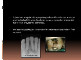
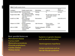
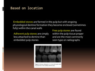
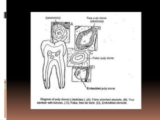
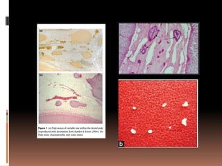
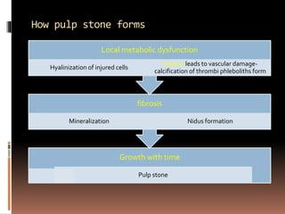
Pulp calcification
 Pulp stones are primarily a physiological manifestation (as are most
other pulpal calcifications) and may increase in number and/or size
due to local or systemic pathology.
 The aetiological factors involved in their formation are still not fully
apparent.
AGE CHANGES IN PULP
 Pulp spaces of teeth decrease in size through the deposition of
secondary and tertiary dentine.
 Increase in the number of collagenous bundles in old coronal
pulps occur
 As a result of calcification of the blood vessels and nerves in
the pulp, their numbers decrease
 As part of the pulp ageing process there is also a considerable
decrease in the number of cells -fibroblasts, odontoblasts and
mesenchymal cells
 Fat deposits occur in the pulp with age
ETIOLOGY
 Etiological factors for pulp stone formation are not well
understood
Pulp degeneration
Inductive interactions between epithelium and pulp tissue
Age
Circulatory disturbances in pulp
Orthodontic tooth movement
Idiopathic factors
Genetic predisposition
Most possible factors are
•Periodontal disease
•Carious teeth
•Restorative procedures
•Fluoride prophylaxsis
•Cardiac disease
•Kidney stones
Systemic or genetic diseases
such as dentine dysplasia,
Dentinogenesis imperfecta
Certain syndromes such as
Van derWoude syndrome
Types of calcified bodies
• Central cavity filled with
epithelial remnants and
surrounded peripherally by
odontoblasts
DENTICLES
• Compact degenerative masses
of calcified tissuesPULP
STONES
Diffuse calcifications- amorphous and un organized linear strands
of calcificationThey appear as amorphous irregular calcific
deposits in the pulp tissue, usually following collagen fiber
bundles or blood vessels.They are usually found in root canals
and less often in the coronal area.
Based on morphology
 TRUE DENTICLES
 Localized masses of calcified
tissue that resembles dentin
 Resembles more of secondary
dentin
 More common in pulp chamber
than in root canal
 Seldom larger than a fraction of
millimeter
 usually located near the apical
foramen.
 FALSE DENTICLES
 Do not exhibit dentinal tubules
 Appear as lamellae deposited
around a central nidus
 Larger than true denticles
 May fill entire pulp chamber
Based on location
Embedded stones are formed in the pulp but with ongoing
physiological dentine formation they become enclosed (sometimes
fully) within the canal walls
Adherent pulp stones are simply
less attached to dentine than
embedded pulp stones
Free pulp stones are found
within the pulp tissue proper
and are the most commonly
seen type on radiographs
How pulp stone forms
Growth with time
Pulp stone
fibrosis
Mineralization Nidus formation
Local metabolic dysfunction
Hyalinization of injured cells
Trauma leads to vascular damage-
calcification of thrombi phleboliths form
What does stone contain
 The stones were composed of two major elements:
calcium and phosphorus. The average concentrations
were 32.1% and 14.7%, respectively, resulting in a
calcium/phosphorus weight ratio of 2.19
 Other elements included fluorine (0.88%), sodium
(0.75%) and magnesium (0.51%). Potassium, chlorine,
manganese, zinc and iron in trace concentrations.
Clinical implications
 May block access to canal orifices and alter the internal anatomy
 Attached stones may deflect or engage the tip of exploring
instruments
 Ultrasonic instrumentation with the use of special tips makes their
removal far easier
 Should a stone be attached to the canal wall and a file can be passed
alongside the stone, it may be removed by careful instrumentation
 Pulp stones present little clinical difficulty during root canal
treatment when magnification, good access and appropriate
instruments are employed.

Pulp calcification

  • 1.
  • 2.
     Pulp stonesare primarily a physiological manifestation (as are most other pulpal calcifications) and may increase in number and/or size due to local or systemic pathology.  The aetiological factors involved in their formation are still not fully apparent.
  • 3.
    AGE CHANGES INPULP  Pulp spaces of teeth decrease in size through the deposition of secondary and tertiary dentine.  Increase in the number of collagenous bundles in old coronal pulps occur  As a result of calcification of the blood vessels and nerves in the pulp, their numbers decrease  As part of the pulp ageing process there is also a considerable decrease in the number of cells -fibroblasts, odontoblasts and mesenchymal cells  Fat deposits occur in the pulp with age
  • 4.
    ETIOLOGY  Etiological factorsfor pulp stone formation are not well understood Pulp degeneration Inductive interactions between epithelium and pulp tissue Age Circulatory disturbances in pulp Orthodontic tooth movement Idiopathic factors Genetic predisposition
  • 5.
    Most possible factorsare •Periodontal disease •Carious teeth •Restorative procedures •Fluoride prophylaxsis •Cardiac disease •Kidney stones Systemic or genetic diseases such as dentine dysplasia, Dentinogenesis imperfecta Certain syndromes such as Van derWoude syndrome
  • 6.
    Types of calcifiedbodies • Central cavity filled with epithelial remnants and surrounded peripherally by odontoblasts DENTICLES • Compact degenerative masses of calcified tissuesPULP STONES Diffuse calcifications- amorphous and un organized linear strands of calcificationThey appear as amorphous irregular calcific deposits in the pulp tissue, usually following collagen fiber bundles or blood vessels.They are usually found in root canals and less often in the coronal area.
  • 8.
    Based on morphology TRUE DENTICLES  Localized masses of calcified tissue that resembles dentin  Resembles more of secondary dentin  More common in pulp chamber than in root canal  Seldom larger than a fraction of millimeter  usually located near the apical foramen.  FALSE DENTICLES  Do not exhibit dentinal tubules  Appear as lamellae deposited around a central nidus  Larger than true denticles  May fill entire pulp chamber
  • 9.
    Based on location Embeddedstones are formed in the pulp but with ongoing physiological dentine formation they become enclosed (sometimes fully) within the canal walls Adherent pulp stones are simply less attached to dentine than embedded pulp stones Free pulp stones are found within the pulp tissue proper and are the most commonly seen type on radiographs
  • 12.
    How pulp stoneforms Growth with time Pulp stone fibrosis Mineralization Nidus formation Local metabolic dysfunction Hyalinization of injured cells Trauma leads to vascular damage- calcification of thrombi phleboliths form
  • 13.
    What does stonecontain  The stones were composed of two major elements: calcium and phosphorus. The average concentrations were 32.1% and 14.7%, respectively, resulting in a calcium/phosphorus weight ratio of 2.19  Other elements included fluorine (0.88%), sodium (0.75%) and magnesium (0.51%). Potassium, chlorine, manganese, zinc and iron in trace concentrations.
  • 14.
    Clinical implications  Mayblock access to canal orifices and alter the internal anatomy  Attached stones may deflect or engage the tip of exploring instruments  Ultrasonic instrumentation with the use of special tips makes their removal far easier  Should a stone be attached to the canal wall and a file can be passed alongside the stone, it may be removed by careful instrumentation  Pulp stones present little clinical difficulty during root canal treatment when magnification, good access and appropriate instruments are employed.