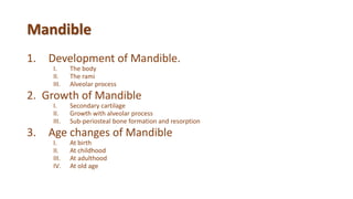
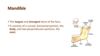
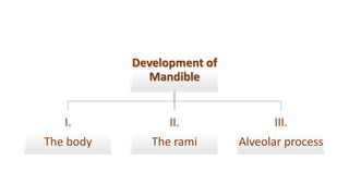
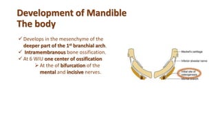
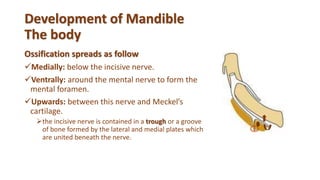
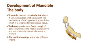
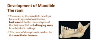
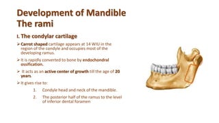
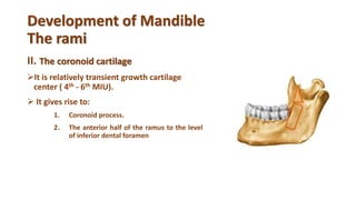
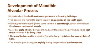
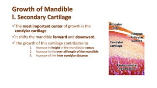
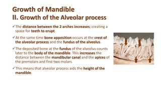
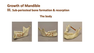
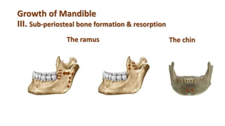
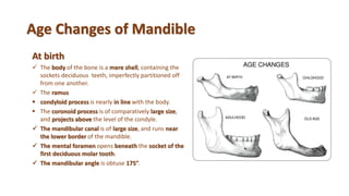
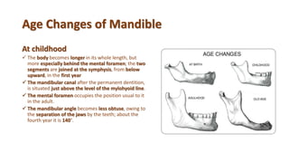
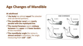
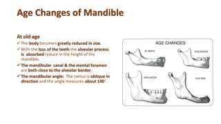
Menatalla M. Elhindawy
Development and Growth of
Mandible
Mandible
1. Development of Mandible.
I. The body
II. The rami
III. Alveolar process
2. Growth of Mandible
I. Secondary cartilage
II. Growth with alveolar process
III. Sub-periosteal bone formation and resorption
3. Age changes of Mandible
I. At birth
II. At childhood
III. At adulthood
IV. At old age
Mandible
The largest and strongest bone of the face.
It consists of a curved, horizontal portion, the
body, and two perpendicular portions, the
rami.
Development of
Mandible
I.
The body
II.
The rami
III.
Alveolar process
Meckel's cartilage
 It is cartilages of the 1st. branchial arch.
 It extends as an unbroken rod of cartilage.
 Their proximal ends are connected with the
ear capsules.
Their distal extremities are joined to one
another at the symphysis by mesodermal
tissue.
It has a close relation to the mandibular nerve.
Meckel's Cartilage & Mandibular Nerve
 At the junction between poximal and
middle thirds the mandibular nerve divides
into the lingual and inferior alevolar nerve.
 The lingual nerve passes forward in the
medial side of the cartilage.
 Inferior alveolar nerve lies lateral to its
upper margin and runs forward parallel to
it.
 At the junction between middle and distal
thirds, the inferior alveolar nerve
terminates by dividing into the mental and
incisive branches.
Meckel’s cartilage
IncisiveMandibular Inferior alv.
Meckel's cartilage
Fate and Derivatives
The proximal portion gives the Malleus and Incus, two of the bones
of the middle ear.
The middle portion is replaced by fibrous tissue, which persists to
form the sphenomandibular ligament & the perichondrium of the
cartilage persist as sphenomallular ligament.
The distal portion gives rise to the cartilaginous remnants in the
midline of the mandible.
 Develops in the mesenchyme of the
deeper part of the 1st branchial arch.
 Intramembranous bone ossification.
 At 6 WIU one center of ossification
At the of bifurcation of the
mental and incisive nerves.
Development of Mandible
The body
Development of Mandible
The body
Ossification spreads as follow
Medially: below the incisive nerve.
Ventrally: around the mental nerve to form the
mental foramen.
Upwards: between this nerve and Meckel’s
cartilage.
the incisive nerve is contained in a trough or a groove
of bone formed by the lateral and medial plates which
are united beneath the nerve.
Development of Mandible
The body
Forwards: towards the middle line where
it comes into close relationship with the
similar bone of the opposite side, but from
which it is separated by connective tissue.
Backward: produces at first a trough of
bone in which lies the inferior dental nerve
and much later the mandibular canal is
formed.
The ossification stops at the site of future
lingula.
Development of Mandible
The rami
The ramus of the mandible develops
by a rapid spread of ossification
backwards into the mesenchyme of
the first branchial arch diverging away
from Meckel’s cartilage.
This point of divergence is marked by
the mandibular foramen.
Development of Mandible
The rami
I. The condylar cartilage
Carrot shaped cartilage appears at 14 WIU in the
region of the condyle and occupies most of the
developing ramus.
It is rapidly converted to bone by endochondral
ossification.
 It acts as an active center of growth till the age of 20
years.
It gives rise to:
1. Condyle head and neck of the mandible.
2. The posterior half of the ramus to the level
of inferior dental foramen
Development of Mandible
The rami
II. The coronoid cartilage
It is relatively transient growth cartilage
center ( 4th - 6th MIU).
 It gives rise to:
1. Coronoid process.
2. The anterior half of the ramus to the level
of inferior dental foramen
Development of Mandible
Alveolar Process
It starts when the deciduous tooth germs reach the early bell stage.
The bone of the mandible begins to grow on each side of the tooth germ.
By this growth the tooth germs come to be in a bony trough, which also includes
the alveolar nerves and vessels.
Later on, septa of bone between the adjacent tooth germs develop, keeping each
tooth separate in its bony crept.
 The mandibular canal is separated from the bony crypts by a horizontal plate of
bone.
The alveolar processes grow rapidly during the periods of tooth eruption.
Any questions??
Growth of
Mandible
I.
Secondary cartilage
II.
Growth of the
Alveolar Process
III.
Sub-periosteal bone
formation &resorption
Growth of Mandible
I. Secondary Cartilage
The most important center of growth is the
condylar cartilage.
It shifts the mandible forward and downward.
 the growth of this cartilage contributes to
1. Increase in height of the mandibular ramus
2. Increase in the over all length of the mandible
3. Increase of the inter condylar distance
Growth of Mandible
II. Growth of the Alveolar process
The distance between the 2 arches increases; creating a
space for teeth to erupt.
At the same time bone apposition occurs at the crest of
the alveolar process and the fundus of the alveolus.
The deposited bone at the fundus of the alveolus counts
later to the body of the mandible. This increases the
distance between the mandibular canal and the apices of
the premolars and first two molars.
This means that alveolar process aids the height of the
mandible.
Growth of Mandible
III. Sub-periosteal bone formation & resorption
The body
Growth of Mandible
III. Sub-periosteal bone formation & resorption
The ramus The chin
Any questions??
Age Changes of Mandible
At birth
 The body of the bone is a mere shell, containing the
sockets deciduous teeth, imperfectly partitioned off
from one another.
 The ramus
 condyloid process is nearly in line with the body.
 The coronoid process is of comparatively large size,
and projects above the level of the condyle.
 The mandibular canal is of large size, and runs near
the lower border of the mandible.
 The mental foramen opens beneath the socket of the
first deciduous molar tooth.
 The mandibular angle is obtuse 175°.
Age Changes of Mandible
At childhood
 The body becomes longer in its whole length, but
more especially behind the mental foramen; the two
segments are joined at the symphysis, from below
upward, in the first year
 The mandibular canal after the permanent dentition,
is situated just above the level of the mylohyoid line.
 The mental foramen occupies the position usual to it
in the adult.
 The mandibular angle becomes less obtuse, owing to
the separation of the jaws by the teeth; about the
fourth year it is 140°.
Age Changes of Mandible
At adulthood
 the body has almost equal The alveolar
and sub-dental portions.
The mandibular canal runs nearly
parallel with the mylohyoid line.
The mental foramen: opens midway
between the upper and lower borders
of the mandible.
The mandibular angle the ramus is
almost vertical in direction, the angle
measuring from 110° - 120°.
Age Changes of Mandible
At old age
The body becomes greatly reduced in size.
With the loss of the teeth the alveolar process
is absorbed reduce in the height of the
mandible.
The mandibular canal & the mental foramen
are both close to the alveolar border.
The mandibular angle: The ramus is oblique in
direction and the angle measures about 140°.
Any questions??
Thank you..

Development of Mandible

  • 1.
  • 2.
  • 3.
    Mandible 1. Development ofMandible. I. The body II. The rami III. Alveolar process 2. Growth of Mandible I. Secondary cartilage II. Growth with alveolar process III. Sub-periosteal bone formation and resorption 3. Age changes of Mandible I. At birth II. At childhood III. At adulthood IV. At old age
  • 4.
    Mandible The largest andstrongest bone of the face. It consists of a curved, horizontal portion, the body, and two perpendicular portions, the rami.
  • 5.
  • 6.
    Meckel's cartilage  Itis cartilages of the 1st. branchial arch.  It extends as an unbroken rod of cartilage.  Their proximal ends are connected with the ear capsules. Their distal extremities are joined to one another at the symphysis by mesodermal tissue. It has a close relation to the mandibular nerve.
  • 7.
    Meckel's Cartilage &Mandibular Nerve  At the junction between poximal and middle thirds the mandibular nerve divides into the lingual and inferior alevolar nerve.  The lingual nerve passes forward in the medial side of the cartilage.  Inferior alveolar nerve lies lateral to its upper margin and runs forward parallel to it.  At the junction between middle and distal thirds, the inferior alveolar nerve terminates by dividing into the mental and incisive branches. Meckel’s cartilage IncisiveMandibular Inferior alv.
  • 8.
    Meckel's cartilage Fate andDerivatives The proximal portion gives the Malleus and Incus, two of the bones of the middle ear. The middle portion is replaced by fibrous tissue, which persists to form the sphenomandibular ligament & the perichondrium of the cartilage persist as sphenomallular ligament. The distal portion gives rise to the cartilaginous remnants in the midline of the mandible.
  • 9.
     Develops inthe mesenchyme of the deeper part of the 1st branchial arch.  Intramembranous bone ossification.  At 6 WIU one center of ossification At the of bifurcation of the mental and incisive nerves. Development of Mandible The body
  • 10.
    Development of Mandible Thebody Ossification spreads as follow Medially: below the incisive nerve. Ventrally: around the mental nerve to form the mental foramen. Upwards: between this nerve and Meckel’s cartilage. the incisive nerve is contained in a trough or a groove of bone formed by the lateral and medial plates which are united beneath the nerve.
  • 11.
    Development of Mandible Thebody Forwards: towards the middle line where it comes into close relationship with the similar bone of the opposite side, but from which it is separated by connective tissue. Backward: produces at first a trough of bone in which lies the inferior dental nerve and much later the mandibular canal is formed. The ossification stops at the site of future lingula.
  • 12.
    Development of Mandible Therami The ramus of the mandible develops by a rapid spread of ossification backwards into the mesenchyme of the first branchial arch diverging away from Meckel’s cartilage. This point of divergence is marked by the mandibular foramen.
  • 13.
    Development of Mandible Therami I. The condylar cartilage Carrot shaped cartilage appears at 14 WIU in the region of the condyle and occupies most of the developing ramus. It is rapidly converted to bone by endochondral ossification.  It acts as an active center of growth till the age of 20 years. It gives rise to: 1. Condyle head and neck of the mandible. 2. The posterior half of the ramus to the level of inferior dental foramen
  • 14.
    Development of Mandible Therami II. The coronoid cartilage It is relatively transient growth cartilage center ( 4th - 6th MIU).  It gives rise to: 1. Coronoid process. 2. The anterior half of the ramus to the level of inferior dental foramen
  • 15.
    Development of Mandible AlveolarProcess It starts when the deciduous tooth germs reach the early bell stage. The bone of the mandible begins to grow on each side of the tooth germ. By this growth the tooth germs come to be in a bony trough, which also includes the alveolar nerves and vessels. Later on, septa of bone between the adjacent tooth germs develop, keeping each tooth separate in its bony crept.  The mandibular canal is separated from the bony crypts by a horizontal plate of bone. The alveolar processes grow rapidly during the periods of tooth eruption.
  • 16.
  • 17.
    Growth of Mandible I. Secondary cartilage II. Growthof the Alveolar Process III. Sub-periosteal bone formation &resorption
  • 18.
    Growth of Mandible I.Secondary Cartilage The most important center of growth is the condylar cartilage. It shifts the mandible forward and downward.  the growth of this cartilage contributes to 1. Increase in height of the mandibular ramus 2. Increase in the over all length of the mandible 3. Increase of the inter condylar distance
  • 19.
    Growth of Mandible II.Growth of the Alveolar process The distance between the 2 arches increases; creating a space for teeth to erupt. At the same time bone apposition occurs at the crest of the alveolar process and the fundus of the alveolus. The deposited bone at the fundus of the alveolus counts later to the body of the mandible. This increases the distance between the mandibular canal and the apices of the premolars and first two molars. This means that alveolar process aids the height of the mandible.
  • 20.
    Growth of Mandible III.Sub-periosteal bone formation & resorption The body
  • 21.
    Growth of Mandible III.Sub-periosteal bone formation & resorption The ramus The chin
  • 22.
  • 23.
    Age Changes ofMandible At birth  The body of the bone is a mere shell, containing the sockets deciduous teeth, imperfectly partitioned off from one another.  The ramus  condyloid process is nearly in line with the body.  The coronoid process is of comparatively large size, and projects above the level of the condyle.  The mandibular canal is of large size, and runs near the lower border of the mandible.  The mental foramen opens beneath the socket of the first deciduous molar tooth.  The mandibular angle is obtuse 175°.
  • 24.
    Age Changes ofMandible At childhood  The body becomes longer in its whole length, but more especially behind the mental foramen; the two segments are joined at the symphysis, from below upward, in the first year  The mandibular canal after the permanent dentition, is situated just above the level of the mylohyoid line.  The mental foramen occupies the position usual to it in the adult.  The mandibular angle becomes less obtuse, owing to the separation of the jaws by the teeth; about the fourth year it is 140°.
  • 25.
    Age Changes ofMandible At adulthood  the body has almost equal The alveolar and sub-dental portions. The mandibular canal runs nearly parallel with the mylohyoid line. The mental foramen: opens midway between the upper and lower borders of the mandible. The mandibular angle the ramus is almost vertical in direction, the angle measuring from 110° - 120°.
  • 26.
    Age Changes ofMandible At old age The body becomes greatly reduced in size. With the loss of the teeth the alveolar process is absorbed reduce in the height of the mandible. The mandibular canal & the mental foramen are both close to the alveolar border. The mandibular angle: The ramus is oblique in direction and the angle measures about 140°.
  • 27.
  • 28.