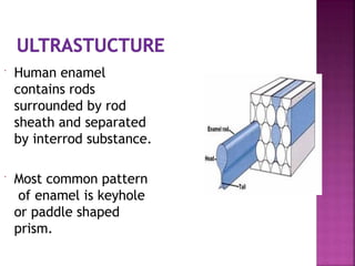
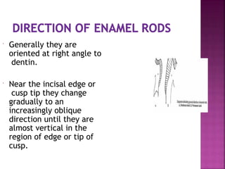
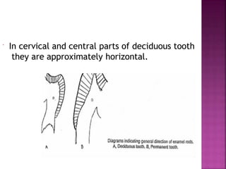
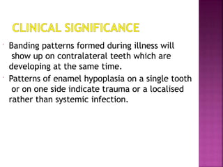
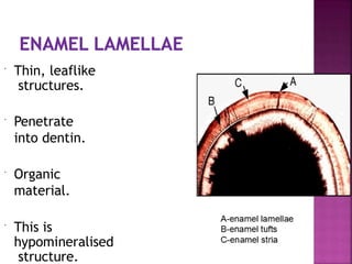
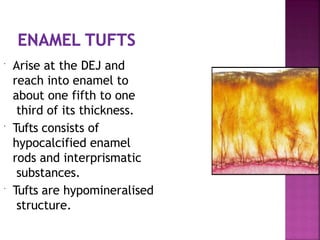
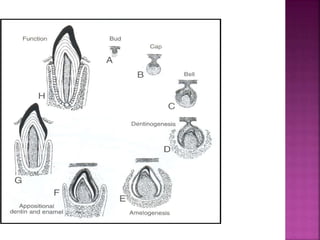
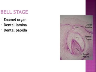
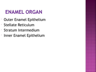
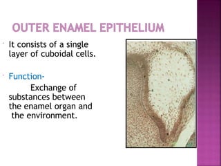
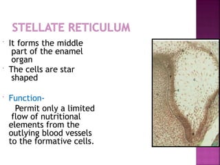
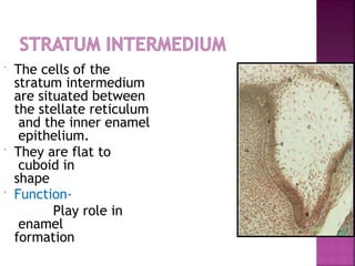
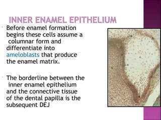
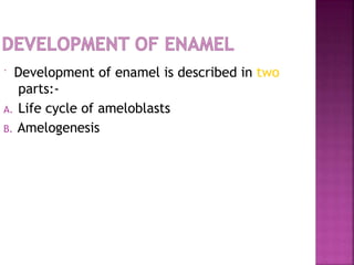
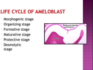
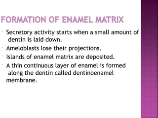
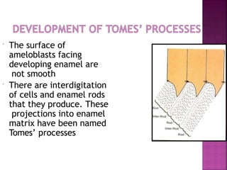
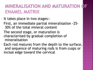
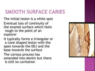
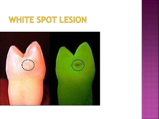
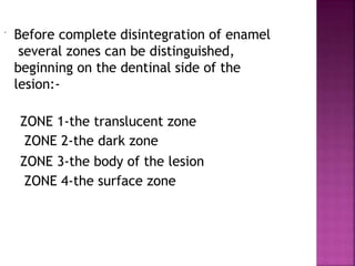
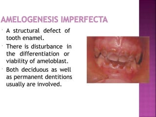
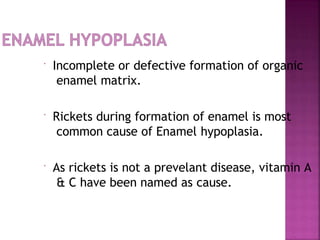
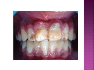
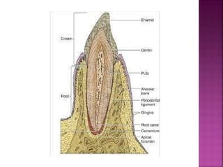
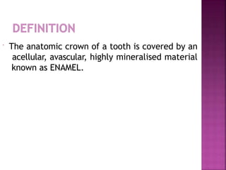
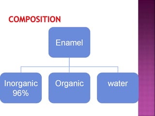
Enamel
 The anatomic crown of a tooth is covered by an
acellular, avascular, highly mineralised material
known as ENAMEL.
 It is the hardest calcified tissue in the human
body.
 It is the only calcified tissue arising from
ectoderm.
 It contains the largest crystals among the
mineralized tissues.
 It protects the less mineralised underlying
dentin of the tooth.
 It serves as a surface for chewing, grinding and
crushing of food.
1) Hardness
 Its high mineral content makes it the hardest
substance in the human body
 Surface enamel vs subsurface enamel
 Hardness and density also decrease from the
cuspal/incisal tip towards the cervical margin
and from the surface towards the DEJ
2) Brittleness
 The hardness which is comparable to mild steel
makes enamel brittle.
 Compensated by the cushioning effect of
underlying resilient dentin.
 Enamel is stiffer and more brittle than
dentin.
3) Permeability
 Enamel can act in a sense like a semipermeable
membrane, permitting complete or partial
passage of certain molecules.
4) Thickness
 It varies with shape of the tooth and location
 Reaching a maximum of 2.5mm in the incisal or
occlusal areas and thinning down to almost a
knife-edge at the CEJ
5) Color
 Enamel is naturally transparent.
 Ranges from yellowish white to grayish white.
 Young enamel has a low translucency and whiter
in colour.
 The translucency increases with age and the
yellow colour of underlying dentin becomes
darker and more apparent with age.
6) Specific gravity- 2.8
7) Compressive strength- 384 Mpa
8) Modulus of elasticity- 84 Gpa
9) Knoop hardness number- 350-430 KHN
10) Tensile strength- 10 MPa
10) Co-efficient of thermal expansion- 11.4
11) Density- 2.97
12) Refractive index-
Average refractive index of 1.62
13) Solubility-
 It dissolves in acid media.
 It is influenced by certain ions and molecules like
fluoride, carbonates, organic matrix etc.
 Surface enamel is less soluble than deeper
enamel
14) Abrasion resistance-
 Is high, allowing it to wear down slowly
 Calcium
hydroxyapatite
[Ca10(PO4)6(OH)2].
 The mineral content
increases from the DEJ
to the surface.
 Most crystallites are
regularly hexagonal in
cross-section.
 A fine lacy network of organic material appears
between the crystal.
 According to frank(1979), in the mature state,
the matrix constitutes of:-
-only 0.3 %
-58%
-42%
-
trace





Enamel
Protein
s Lipids
Lactates, ions,
sugars, citrates
 The proteins present in enamel are:-
1.Amelogenins
 2. non ameligenins
• Ameloblastin .
Amelin
• Enamelin .
Tuftelin
d
i
r
e
c
t
 90%.
 Important in crystal growth & organization.
 Nanospheres between which enamel crystals forms.
 Absence leads to hypoplastic.
 Also found in formation of acellular cementum.
Structure
 This is structureless layer of enamel.
 30µm
 Present in 70% permanent teeth & all
deciduous teeth.
 More heavily mineralized than bulk
beneath it.
9/16/2014
 Enamel rods near
dentin at the incisal
edge or cusps
forms more
complicated, this
optical
arrangement of
enamel is called
Gnarled enamel.
9/16/2014
 Basic structural unit is
the enamel prism or
rod.
 It consists of a tightly
packed mass of
millions of small,
elongated
hydroxyapatite
crystals in an
organised pattern.
 Many show fish scales
appearance in cross section.
 Width is 4µm and length.
 Number of rods estimated as ranging from 5 million
in lower lateral incisor to 12 million in upper first
molars.
 Diameter of enamel increases from DEJ to outer
surface at a ratio about 1:2.
 The cross-sectional--
the keyhole
arrangement of enamel
prisms with the heads
pointing occlusally and
the tails pointing
cervically.
 Head of each rod is
made up of 1
ameloblast and tail is
made up of 3
ameloblasts.
 Human enamel
contains rods
surrounded by rod
sheath and separated
by interrod substance.
 Most common pattern
of enamel is keyhole
or paddle shaped
prism.
 In a longitudinal section, appearance of
rods separate by interrod substance.
 Polarized light and roentgen-ray study
indicated that apatite crystals are
arranged approximately parallel to long
axis of prisms.
9/16/2014
 It confers strength to the enamel.
 Their direction is an important consideration in
the cavity preparation for restorations
 Enamel rods that are supported by hard
restorative material rather than more pliant
dentin are more likely to fracture
 Fracturing of unsupported rods in poorly
designed restorative preparations causes loss of
enamel around the margins of the filling
material resulting in marginal leakage and
makes the tooth more susceptible to carious
attack.
 Additionally, it is also important to note that
the inclination of rods differs in permanent and
primary teeth and must be accounted for during
cavity preparation.
 Each enamel rod is built up of segments
separated by dark lines that give it a
striated appearance.
 The striations are more pronounced in
enamel that is insufficiently
calcified.
9/16/2014
 Generally they are
oriented at right angle to
dentin.
 Near the incisal edge or
cusp tip they change
gradually to an
increasingly oblique
direction until they are
almost vertical in the
region of edge or tip of
cusp.
 In cervical and central parts of deciduous tooth
they are approximately horizontal.
 More or less changes
in the direction of
rods may be regarded
as functional
adaptation minimizing
the risk of cleavage
due to occlusal
loading forces. This
changes in direction of
rods is responsible for
appearance the
Hunter-Schreger
bands.
 Careful decalcification and staining gives
evidence that these are not solely optical
phenomenon.
 These are composed of zones different
permeability and organic content.
 Some books suggest that this is an optical
phenomenon produced merely by changes in
direction of lights.
9/16/2014
 They appears as brownish bands
in ground section of the enamel.
 They illustrate the incrimental
pattern of enamel, during
formation of crown.
 They reflect variation in
structure and mineralisation,
and are either hypomineralised
or hypermineralised.
 Banding patterns formed during illness will
show up on contralateral teeth which are
developing at the same time.
 Patterns of enamel hypoplasia on a single tooth
or on one side indicate trauma or a localised
rather than systemic infection.
 A delicate membrane that covers entire portion
of newly erupted crown is enamel cuticle or
Nasmyth’s membrane.
 It soon get removed by mastication.
 This is secreted by ameloblast when enamel
formation is complete.
 This is hypomineralised structure.
 Thin, leaflike
structures.
 Penetrate
into dentin.
 Organic
material.
 This is
hypomineralised
structure.
 Lamellae may develop in planes of tension.
Where rods cross such a plane, a short segment
of the rod may not fully calcify.
 This leads to formation of three types of
lamellae:-
type A:- poorly calcified rods
type B:- degenerated cells
type C:- arising in erupted teeth where the cracks are
filled organic matter, presumably originating from saliva.
9/16/2014
 Represents a significant weakness in the
structure of enamel and is susceptible to
cracking and and form a road for the entry of
bacteria that initiate caries.
 Arise at the DEJ and
reach into enamel to
about one fifth to one
third of its thickness.
 Tufts consists of
hypocalcified enamel
rods and interprismatic
substances.
 Tufts are hypomineralised
structure.
 No major clinical significance, but represent
areas of enamel weakness.
 The surface of dentin
at DEJ is pitted which
fit rounded
projections of enamel.
 Scalloped appearance.
 The DEJ is more
prominent in the
occlusal area.
 It is hypomineralised
structure.
 Occasionally odontoblast
processes pass across
the DEJ into enamel,
many of them are
thickened at there end,
they are termed as
enamel spindles.
 This is hypomineralised
structure.
 No major clinical significance but may confer
additional permeability to the deeper layers of
enamel.
 Striae of Retzius often
extends from DEJ to
outer surface, when
they end in shallow
furrows known as
Perikymata
1) Prismless enamel- Primary teeth are more
likely to have a prismless surface zone than
are permanent teeth. A difference in the
reaction to conditioning agents is suspected
because less etching occurs on primary tooth
than on permanent tooth enamel during
acid conditioning.
2)Thickness- enamel is twice as thick on
permanent teeth as in primary teeth.
 3) Neonatal line-it is the
most prominent
incremental line in
primary teeth.it is due
to the metabolic trauma
to the developing tooth
at or near the time of
birth. Prenatal enamel
is less pigmented and
more free of defects
than postnatal enamel.
4)Enamel of primary teeth is whiter than that of
permanent teeth. This is believed to be because
much of primary tooth enamel is formed
prenatally and is not subject to some
enviornmental factors.
5)Direction of enamel rods-in the cervical area
the enamel rods in primary teeth are oriented
horizontally while in permanent teeth they are
inclined apically.
 As the development of tooth progresses through
various stages of tooth development, the actual
formation of starts once ameloblasts are formed.
 Ameloblasts are formed when tooth
development progresses to formation of bell
stage.
 Enamel organ
 Dental lamina
 Dental papilla
 Outer Enamel Epithelium
 Stellate Reticulum
 Stratum Intermedium
 Inner Enamel Epithelium
 It consists of a single
layer of cuboidal cells.
 Function-
Exchange of
substances between
the enamel organ and
the environment.
 It forms the middle
part of the enamel
organ
 The cells are star
shaped
 Function-
Permit only a limited
flow of nutritional
elements from the
outlying blood vessels
to the formative cells.
 The cells of the
stratum intermedium
are situated between
the stellate reticulum
and the inner enamel
epithelium.
 They are flat to
cuboid in
shape
 Function-
Play role in
enamel
formation
 Before enamel formation
begins these cells assume a
columnar form and
differentiate into
ameloblasts that produce
the enamel matrix.
 The borderline between the
inner enamel epithelium
and the connective tissue
of the dental papilla is the
subsequent DEJ
 Development of enamel is described in two
parts:-
A. Life cycle of ameloblasts
B. Amelogenesis
 Morphogenic stage
 Organizing stage
 Formative stage
 Maturative stage
 Protective stage
 Desmolytic
stage
 Before ameloblast differentiate and produce
enamel, they interact with adjacent
mesenchymal cells, determining shape of DEJ &
crown.
 During this stage cells are short columnar with
oval nuclei that almost fill cell body.
• This is characterized by presence of cells in inner
el
epithelium.
 During this stage there is changes in
organization and number of cytoplasmic
organelles related to initiation of
enamel matrix.
• Enamel maturation begins after most of thickness of
enamel matrix has been laid down.
 Ameloblast after maturation of enamel matrix
forms a protective layer i.e. reduced enamel
epithelium which is protective to enamel until
tooth erupt in oral cavity.
 By desmolysis the cells of reduced enamel
epithelium help in eruption of tooth.
 There are two processes involved in
development of enamel
Formation of enamel matrix
Maturation
 Secretory activity starts when a small amount of
dentin is laid down.
 Ameloblasts lose their projections.
 Islands of enamel matrix are deposited.
 A thin continuous layer of enamel is formed
along the dentin called dentinoenamel
membrane.
 The surface of
ameloblasts facing
developing enamel are
not smooth
 There are interdigitation
of cells and enamel rods
that they produce. These
projections into enamel
matrix have been named
Tomes’ processes
 The head of each rod is formed by one
ameloblast where as 3 others contribute to
tail of each rod. That is each rod is
formed by four ameloblasts and each
ameloblast contributes to four different
rods.
 Ameloblasts are shorter.
 They have a villous surface near the enamel and
ends of cells are packed with mitochondria-
typical of absorptive cells.
 Organic components and water are lost during
mineralization.
 Over 90% of initially secreted protein is lost.
 It takes place in two stages:-
First, an immediate partial mineralisation -25-
30% of the total mineral content
The second stage, or maturation is
characterised by gradual completion of
mineralisation
 Each rod matures from the depth to the surface,
and sequence of maturing rods is from cusps or
incisal edge toward the cervical
DESTRUCTION OF ENAMEL-
Bacterial
i.e. Dental caries
Non Bacterial
i.e.
attrition,abrasion,erosion,abfraction
Dental Caries
The high mineral content of enamel which
makes this tissue the hardest in the human
body , also makes it susceptible to a
demineralisation process which often occurs as
dental caries.
Enamel caries is of two types:-
1)Smooth surface caries
2)Pit and fissure caries
 The initial lesion is a white spot
 Eventual loss of continuity of
the enamel surface which feels
rough to the point of an
explorer
 It typically forms a triangular or
a cone shaped lesion with the
apex towards the DEJ and the
base towards the surface
 The carious process has
extended into dentin but there
is still no cavitation
 Before complete disintegration of enamel
several zones can be distinguished,
beginning on the dentinal side of the
lesion:-
ZONE 1-the translucent zone
ZONE 2-the dark zone
ZONE 3-the body of the lesion
ZONE 4-the surface zone
 Caries beginning in a
fissure with
decalcification extending
from its sides and bottom.
 It forms a cone shaped
lesion with the base at
the DEJ and apex at
towards the
surface
 It reaches the dentin and
spreads laterally.
 There is separation of
enamel and dentin and
fracture of the enamel
roof.
 Attrition
 Abrasion
 Erosion
 Abfraction
 The physiologic
wearing away of a
tooth as a result of
tooth-tooth contact.
 This phenomenon is
physiologic rather
than pathologic
 Pathologic wearing
away of tooth
substance through
some abnormal
mechanical process.
 Generally occurs on
exposed surfaces
of roots.
 Irreversible loss of
dental hard tissue by
a chemical process
that does not involve
bacteria.
 Erosion is also related
to GERD.
 Pathologic loss of
both enamel and
dentin caused by
biomechanical loading
forces.
 Amelogenesis Imperfecta
 Enamel Hypoplasia
 Mottled Enamel
 Enamel Pearls
 Tetracycline Stains
 A structural defect of
tooth enamel.
 There is disturbance in
the differentiation or
viability of ameloblast.
 Both deciduous as well
as permanent dentitions
usually are involved.
 Three main groups: hypoplastic(60-73%),
hypocalcified(7%), and hypomature(20-40%).
 Classification of amelogenesis imperfecta
according to Witcop :-
Type Ι
Type ΙΙ
Hypoplastic
Hypomaturation
Type ΙΙΙ Hypocalcified
Type ΙV Hypomaturation-hypoplastic
with taurodontism
 No specific treatment, except for
improvement of cosmetic appearance.
 Incomplete or defective formation of organic
enamel matrix.
 Rickets during formation of enamel is most
common cause of Enamel hypoplasia.
 As rickets is not a prevelant disease, vitamin A
& C have been named as cause.
 Considerable contraversy are there about any
relation between caries & enamel hypoplasia. It
is most reasonable to assume that the two are
not related, although hypoplastic teeth appear
to decay at somewhat more rapid rate once
caries has been initiated.
 Term mottled enamel is described by GV Black
and Frederick S McKay in 1916.
 Ingestion of fluoride containing water during
time tooth formation is most important.
 More than 1 ppm of fluoride causes significant
mottling.
 0
 There is wide range of
severity in the
appearance of mottled
teeth, varying from
I. Mild changes (white
opaque areas)
II. Moderate and severe
(pitting and
brownish staining)
III. A corroded
appearance of
the teeth.
 Discoloration occurs due
to prophylactic
administration of
tetracycline to pregnant
female or postpartum
in the infants.
 Yellowish or brownish-
gray discoloration.
 Crucial period is 4 months
in utero to about 7 years
of age.
 The direction of enamel rods is of
importance in cavity
preparation:
 One of the most important principles in tooth
preparation is the concept of the strongest
enamel margin
 It is formed by full length enamel rods whose
inner ends are on sound dentin.
 The American Society for Testing and Materials
defines adhesion as “the state in which two
surfaces are held together by interfacial forces
which may consist of valence forces or
interlocking forces of both”.
 Advantages
1) Cusp reinforcement after tooth
preparation.
2) Reinforce remaining enamel and dentin.
 It is important technique in
clinical practice.
 It involves use of etchant to
produce change in surface
texture of enamel.
 There are three types of Enamel etching seen
Type A- Dissolve enamel rod
Type B- Dissolve interrod enamel
Type C- Irregular and
indiscriminate
9/16/2014
 It achieves desired effects in two stages:-
1) Removes plaque and other debris
2) Increases the porosity of exposed surfaces
 Increases the free surface energy of enamel.
 Micromechanical bonding.
9/16/2014
Bleaching may be defined as the lightening
of color of tooth through application of
chemical agent to oxidize organic
pigmentation of tooth.
H₂O₂ has low molecular weight that enables it to
fuse through enamel.
 Oxidation reaction
 Low pH can cause destruction of enamel by
demineralisation
i) Microabrasion-
Microabrasion techniques improve appearance
of fluorotic teeth.
 McCloskey reported that Kane succesfully removed
fluorosis stains by applying acid and heat in 1916.
 In 1960s, McInnes used five parts of 36% HCL,
five parts of 30% H O
₂ ₂ and one part of Ether.
Ether – Removes surface debris
HCL – Etches Enamel
H O - Bleaches
₂ ₂ Enamel
Fluoride-stained teeth are difficult to bleach
and require longer and repeated sessions to
decolorize them.
9/16/2014
ii) Macroabrasion –
an alternative method to removal of
superficial white spots.
Uses a 12-fluted composite finishing bur or
a fine grit finishing diamond at high speed.
Next, a 30-fluted composite finishing bur is
used.
Final polishing is achieved with an abrasive
rubber point.
Increases resistance to caries.
Melting and fusing of enamel-70-85%resistant to
acids.
When laser technique is used with fluoride -
cavities were completely stopped.
 absorption of fluoride ions on enamel
 This increases resistance to acid dissolution
of enamel
9/16/2014
 Orban’s oral histology and embryology .
 Oral histology- Tencate .
 Dental embryology, histology and anatomy-
Mary Bath-Balogh And Margaret J. Fehrenbach.
 Textbook of operative dentistry- Sturdevant.
 Grossman’s Endodontic Practice.
 Philips’- Science of Dental Material.
enamel-strogest teeth structure on the body

enamel-strogest teeth structure on the body

  • 1.
  • 3.
     The anatomiccrown of a tooth is covered by an acellular, avascular, highly mineralised material known as ENAMEL.
  • 4.
     It isthe hardest calcified tissue in the human body.  It is the only calcified tissue arising from ectoderm.  It contains the largest crystals among the mineralized tissues.
  • 5.
     It protectsthe less mineralised underlying dentin of the tooth.  It serves as a surface for chewing, grinding and crushing of food.
  • 6.
    1) Hardness  Itshigh mineral content makes it the hardest substance in the human body  Surface enamel vs subsurface enamel  Hardness and density also decrease from the cuspal/incisal tip towards the cervical margin and from the surface towards the DEJ
  • 7.
    2) Brittleness  Thehardness which is comparable to mild steel makes enamel brittle.  Compensated by the cushioning effect of underlying resilient dentin.  Enamel is stiffer and more brittle than dentin.
  • 8.
    3) Permeability  Enamelcan act in a sense like a semipermeable membrane, permitting complete or partial passage of certain molecules. 4) Thickness  It varies with shape of the tooth and location  Reaching a maximum of 2.5mm in the incisal or occlusal areas and thinning down to almost a knife-edge at the CEJ
  • 9.
    5) Color  Enamelis naturally transparent.  Ranges from yellowish white to grayish white.  Young enamel has a low translucency and whiter in colour.  The translucency increases with age and the yellow colour of underlying dentin becomes darker and more apparent with age.
  • 10.
    6) Specific gravity-2.8 7) Compressive strength- 384 Mpa 8) Modulus of elasticity- 84 Gpa 9) Knoop hardness number- 350-430 KHN 10) Tensile strength- 10 MPa 10) Co-efficient of thermal expansion- 11.4
  • 11.
    11) Density- 2.97 12)Refractive index- Average refractive index of 1.62 13) Solubility-  It dissolves in acid media.  It is influenced by certain ions and molecules like fluoride, carbonates, organic matrix etc.  Surface enamel is less soluble than deeper enamel 14) Abrasion resistance-  Is high, allowing it to wear down slowly
  • 13.
     Calcium hydroxyapatite [Ca10(PO4)6(OH)2].  Themineral content increases from the DEJ to the surface.  Most crystallites are regularly hexagonal in cross-section.
  • 14.
     A finelacy network of organic material appears between the crystal.  According to frank(1979), in the mature state, the matrix constitutes of:- -only 0.3 % -58% -42% - trace      Enamel Protein s Lipids Lactates, ions, sugars, citrates
  • 15.
     The proteinspresent in enamel are:- 1.Amelogenins  2. non ameligenins • Ameloblastin . Amelin • Enamelin . Tuftelin d i r e c t
  • 16.
     90%.  Importantin crystal growth & organization.  Nanospheres between which enamel crystals forms.  Absence leads to hypoplastic.  Also found in formation of acellular cementum.
  • 17.
  • 18.
     This isstructureless layer of enamel.  30µm  Present in 70% permanent teeth & all deciduous teeth.  More heavily mineralized than bulk beneath it. 9/16/2014
  • 19.
     Enamel rodsnear dentin at the incisal edge or cusps forms more complicated, this optical arrangement of enamel is called Gnarled enamel. 9/16/2014
  • 20.
     Basic structuralunit is the enamel prism or rod.  It consists of a tightly packed mass of millions of small, elongated hydroxyapatite crystals in an organised pattern.
  • 21.
     Many showfish scales appearance in cross section.  Width is 4µm and length.  Number of rods estimated as ranging from 5 million in lower lateral incisor to 12 million in upper first molars.  Diameter of enamel increases from DEJ to outer surface at a ratio about 1:2.
  • 22.
     The cross-sectional-- thekeyhole arrangement of enamel prisms with the heads pointing occlusally and the tails pointing cervically.  Head of each rod is made up of 1 ameloblast and tail is made up of 3 ameloblasts.
  • 23.
     Human enamel containsrods surrounded by rod sheath and separated by interrod substance.  Most common pattern of enamel is keyhole or paddle shaped prism.
  • 24.
     In alongitudinal section, appearance of rods separate by interrod substance.  Polarized light and roentgen-ray study indicated that apatite crystals are arranged approximately parallel to long axis of prisms. 9/16/2014
  • 25.
     It confersstrength to the enamel.  Their direction is an important consideration in the cavity preparation for restorations  Enamel rods that are supported by hard restorative material rather than more pliant dentin are more likely to fracture
  • 26.
     Fracturing ofunsupported rods in poorly designed restorative preparations causes loss of enamel around the margins of the filling material resulting in marginal leakage and makes the tooth more susceptible to carious attack.  Additionally, it is also important to note that the inclination of rods differs in permanent and primary teeth and must be accounted for during cavity preparation.
  • 27.
     Each enamelrod is built up of segments separated by dark lines that give it a striated appearance.  The striations are more pronounced in enamel that is insufficiently calcified. 9/16/2014
  • 28.
     Generally theyare oriented at right angle to dentin.  Near the incisal edge or cusp tip they change gradually to an increasingly oblique direction until they are almost vertical in the region of edge or tip of cusp.
  • 29.
     In cervicaland central parts of deciduous tooth they are approximately horizontal.
  • 30.
     More orless changes in the direction of rods may be regarded as functional adaptation minimizing the risk of cleavage due to occlusal loading forces. This changes in direction of rods is responsible for appearance the Hunter-Schreger bands.
  • 31.
     Careful decalcificationand staining gives evidence that these are not solely optical phenomenon.  These are composed of zones different permeability and organic content.  Some books suggest that this is an optical phenomenon produced merely by changes in direction of lights. 9/16/2014
  • 32.
     They appearsas brownish bands in ground section of the enamel.  They illustrate the incrimental pattern of enamel, during formation of crown.  They reflect variation in structure and mineralisation, and are either hypomineralised or hypermineralised.
  • 33.
     Banding patternsformed during illness will show up on contralateral teeth which are developing at the same time.  Patterns of enamel hypoplasia on a single tooth or on one side indicate trauma or a localised rather than systemic infection.
  • 34.
     A delicatemembrane that covers entire portion of newly erupted crown is enamel cuticle or Nasmyth’s membrane.  It soon get removed by mastication.  This is secreted by ameloblast when enamel formation is complete.  This is hypomineralised structure.
  • 35.
     Thin, leaflike structures. Penetrate into dentin.  Organic material.  This is hypomineralised structure.
  • 36.
     Lamellae maydevelop in planes of tension. Where rods cross such a plane, a short segment of the rod may not fully calcify.  This leads to formation of three types of lamellae:- type A:- poorly calcified rods type B:- degenerated cells type C:- arising in erupted teeth where the cracks are filled organic matter, presumably originating from saliva. 9/16/2014
  • 37.
     Represents asignificant weakness in the structure of enamel and is susceptible to cracking and and form a road for the entry of bacteria that initiate caries.
  • 38.
     Arise atthe DEJ and reach into enamel to about one fifth to one third of its thickness.  Tufts consists of hypocalcified enamel rods and interprismatic substances.  Tufts are hypomineralised structure.
  • 39.
     No majorclinical significance, but represent areas of enamel weakness.
  • 40.
     The surfaceof dentin at DEJ is pitted which fit rounded projections of enamel.  Scalloped appearance.  The DEJ is more prominent in the occlusal area.  It is hypomineralised structure.
  • 41.
     Occasionally odontoblast processespass across the DEJ into enamel, many of them are thickened at there end, they are termed as enamel spindles.  This is hypomineralised structure.
  • 42.
     No majorclinical significance but may confer additional permeability to the deeper layers of enamel.
  • 43.
     Striae ofRetzius often extends from DEJ to outer surface, when they end in shallow furrows known as Perikymata
  • 44.
    1) Prismless enamel-Primary teeth are more likely to have a prismless surface zone than are permanent teeth. A difference in the reaction to conditioning agents is suspected because less etching occurs on primary tooth than on permanent tooth enamel during acid conditioning. 2)Thickness- enamel is twice as thick on permanent teeth as in primary teeth.
  • 45.
     3) Neonatalline-it is the most prominent incremental line in primary teeth.it is due to the metabolic trauma to the developing tooth at or near the time of birth. Prenatal enamel is less pigmented and more free of defects than postnatal enamel.
  • 46.
    4)Enamel of primaryteeth is whiter than that of permanent teeth. This is believed to be because much of primary tooth enamel is formed prenatally and is not subject to some enviornmental factors. 5)Direction of enamel rods-in the cervical area the enamel rods in primary teeth are oriented horizontally while in permanent teeth they are inclined apically.
  • 49.
     As thedevelopment of tooth progresses through various stages of tooth development, the actual formation of starts once ameloblasts are formed.  Ameloblasts are formed when tooth development progresses to formation of bell stage.
  • 50.
     Enamel organ Dental lamina  Dental papilla
  • 51.
     Outer EnamelEpithelium  Stellate Reticulum  Stratum Intermedium  Inner Enamel Epithelium
  • 52.
     It consistsof a single layer of cuboidal cells.  Function- Exchange of substances between the enamel organ and the environment.
  • 53.
     It formsthe middle part of the enamel organ  The cells are star shaped  Function- Permit only a limited flow of nutritional elements from the outlying blood vessels to the formative cells.
  • 54.
     The cellsof the stratum intermedium are situated between the stellate reticulum and the inner enamel epithelium.  They are flat to cuboid in shape  Function- Play role in enamel formation
  • 55.
     Before enamelformation begins these cells assume a columnar form and differentiate into ameloblasts that produce the enamel matrix.  The borderline between the inner enamel epithelium and the connective tissue of the dental papilla is the subsequent DEJ
  • 56.
     Development ofenamel is described in two parts:- A. Life cycle of ameloblasts B. Amelogenesis
  • 57.
     Morphogenic stage Organizing stage  Formative stage  Maturative stage  Protective stage  Desmolytic stage
  • 59.
     Before ameloblastdifferentiate and produce enamel, they interact with adjacent mesenchymal cells, determining shape of DEJ & crown.  During this stage cells are short columnar with oval nuclei that almost fill cell body.
  • 60.
    • This ischaracterized by presence of cells in inner el epithelium.
  • 61.
     During thisstage there is changes in organization and number of cytoplasmic organelles related to initiation of enamel matrix.
  • 62.
    • Enamel maturationbegins after most of thickness of enamel matrix has been laid down.
  • 63.
     Ameloblast aftermaturation of enamel matrix forms a protective layer i.e. reduced enamel epithelium which is protective to enamel until tooth erupt in oral cavity.
  • 64.
     By desmolysisthe cells of reduced enamel epithelium help in eruption of tooth.
  • 65.
     There aretwo processes involved in development of enamel Formation of enamel matrix Maturation
  • 66.
     Secretory activitystarts when a small amount of dentin is laid down.  Ameloblasts lose their projections.  Islands of enamel matrix are deposited.  A thin continuous layer of enamel is formed along the dentin called dentinoenamel membrane.
  • 67.
     The surfaceof ameloblasts facing developing enamel are not smooth  There are interdigitation of cells and enamel rods that they produce. These projections into enamel matrix have been named Tomes’ processes
  • 68.
     The headof each rod is formed by one ameloblast where as 3 others contribute to tail of each rod. That is each rod is formed by four ameloblasts and each ameloblast contributes to four different rods.
  • 69.
     Ameloblasts areshorter.  They have a villous surface near the enamel and ends of cells are packed with mitochondria- typical of absorptive cells.  Organic components and water are lost during mineralization.  Over 90% of initially secreted protein is lost.
  • 70.
     It takesplace in two stages:- First, an immediate partial mineralisation -25- 30% of the total mineral content The second stage, or maturation is characterised by gradual completion of mineralisation  Each rod matures from the depth to the surface, and sequence of maturing rods is from cusps or incisal edge toward the cervical
  • 71.
    DESTRUCTION OF ENAMEL- Bacterial i.e.Dental caries Non Bacterial i.e. attrition,abrasion,erosion,abfraction
  • 72.
    Dental Caries The highmineral content of enamel which makes this tissue the hardest in the human body , also makes it susceptible to a demineralisation process which often occurs as dental caries.
  • 74.
    Enamel caries isof two types:- 1)Smooth surface caries 2)Pit and fissure caries
  • 75.
     The initiallesion is a white spot  Eventual loss of continuity of the enamel surface which feels rough to the point of an explorer  It typically forms a triangular or a cone shaped lesion with the apex towards the DEJ and the base towards the surface  The carious process has extended into dentin but there is still no cavitation
  • 77.
     Before completedisintegration of enamel several zones can be distinguished, beginning on the dentinal side of the lesion:- ZONE 1-the translucent zone ZONE 2-the dark zone ZONE 3-the body of the lesion ZONE 4-the surface zone
  • 78.
     Caries beginningin a fissure with decalcification extending from its sides and bottom.  It forms a cone shaped lesion with the base at the DEJ and apex at towards the surface  It reaches the dentin and spreads laterally.  There is separation of enamel and dentin and fracture of the enamel roof.
  • 79.
     Attrition  Abrasion Erosion  Abfraction
  • 80.
     The physiologic wearingaway of a tooth as a result of tooth-tooth contact.  This phenomenon is physiologic rather than pathologic
  • 81.
     Pathologic wearing awayof tooth substance through some abnormal mechanical process.  Generally occurs on exposed surfaces of roots.
  • 82.
     Irreversible lossof dental hard tissue by a chemical process that does not involve bacteria.  Erosion is also related to GERD.
  • 83.
     Pathologic lossof both enamel and dentin caused by biomechanical loading forces.
  • 84.
     Amelogenesis Imperfecta Enamel Hypoplasia  Mottled Enamel  Enamel Pearls  Tetracycline Stains
  • 85.
     A structuraldefect of tooth enamel.  There is disturbance in the differentiation or viability of ameloblast.  Both deciduous as well as permanent dentitions usually are involved.
  • 86.
     Three maingroups: hypoplastic(60-73%), hypocalcified(7%), and hypomature(20-40%).  Classification of amelogenesis imperfecta according to Witcop :- Type Ι Type ΙΙ Hypoplastic Hypomaturation Type ΙΙΙ Hypocalcified Type ΙV Hypomaturation-hypoplastic with taurodontism
  • 87.
     No specifictreatment, except for improvement of cosmetic appearance.
  • 88.
     Incomplete ordefective formation of organic enamel matrix.  Rickets during formation of enamel is most common cause of Enamel hypoplasia.  As rickets is not a prevelant disease, vitamin A & C have been named as cause.
  • 90.
     Considerable contraversyare there about any relation between caries & enamel hypoplasia. It is most reasonable to assume that the two are not related, although hypoplastic teeth appear to decay at somewhat more rapid rate once caries has been initiated.
  • 91.
     Term mottledenamel is described by GV Black and Frederick S McKay in 1916.  Ingestion of fluoride containing water during time tooth formation is most important.  More than 1 ppm of fluoride causes significant mottling.  0
  • 92.
     There iswide range of severity in the appearance of mottled teeth, varying from I. Mild changes (white opaque areas) II. Moderate and severe (pitting and brownish staining) III. A corroded appearance of the teeth.
  • 94.
     Discoloration occursdue to prophylactic administration of tetracycline to pregnant female or postpartum in the infants.  Yellowish or brownish- gray discoloration.  Crucial period is 4 months in utero to about 7 years of age.
  • 95.
     The directionof enamel rods is of importance in cavity preparation:
  • 96.
     One ofthe most important principles in tooth preparation is the concept of the strongest enamel margin  It is formed by full length enamel rods whose inner ends are on sound dentin.
  • 97.
     The AmericanSociety for Testing and Materials defines adhesion as “the state in which two surfaces are held together by interfacial forces which may consist of valence forces or interlocking forces of both”.  Advantages 1) Cusp reinforcement after tooth preparation. 2) Reinforce remaining enamel and dentin.
  • 98.
     It isimportant technique in clinical practice.  It involves use of etchant to produce change in surface texture of enamel.
  • 99.
     There arethree types of Enamel etching seen Type A- Dissolve enamel rod Type B- Dissolve interrod enamel Type C- Irregular and indiscriminate 9/16/2014
  • 100.
     It achievesdesired effects in two stages:- 1) Removes plaque and other debris 2) Increases the porosity of exposed surfaces  Increases the free surface energy of enamel.  Micromechanical bonding. 9/16/2014
  • 101.
    Bleaching may bedefined as the lightening of color of tooth through application of chemical agent to oxidize organic pigmentation of tooth. H₂O₂ has low molecular weight that enables it to fuse through enamel.
  • 102.
     Oxidation reaction Low pH can cause destruction of enamel by demineralisation
  • 103.
    i) Microabrasion- Microabrasion techniquesimprove appearance of fluorotic teeth.  McCloskey reported that Kane succesfully removed fluorosis stains by applying acid and heat in 1916.  In 1960s, McInnes used five parts of 36% HCL, five parts of 30% H O ₂ ₂ and one part of Ether.
  • 104.
    Ether – Removessurface debris HCL – Etches Enamel H O - Bleaches ₂ ₂ Enamel Fluoride-stained teeth are difficult to bleach and require longer and repeated sessions to decolorize them. 9/16/2014
  • 105.
    ii) Macroabrasion – analternative method to removal of superficial white spots. Uses a 12-fluted composite finishing bur or a fine grit finishing diamond at high speed. Next, a 30-fluted composite finishing bur is used. Final polishing is achieved with an abrasive rubber point.
  • 106.
    Increases resistance tocaries. Melting and fusing of enamel-70-85%resistant to acids. When laser technique is used with fluoride - cavities were completely stopped.
  • 107.
     absorption offluoride ions on enamel  This increases resistance to acid dissolution of enamel 9/16/2014
  • 108.
     Orban’s oralhistology and embryology .  Oral histology- Tencate .  Dental embryology, histology and anatomy- Mary Bath-Balogh And Margaret J. Fehrenbach.  Textbook of operative dentistry- Sturdevant.  Grossman’s Endodontic Practice.  Philips’- Science of Dental Material.