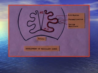
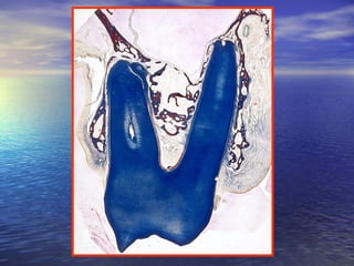
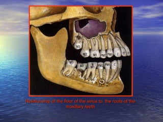
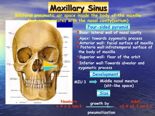
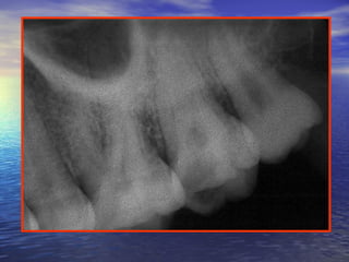
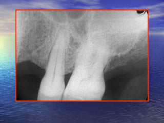
 
32)
‫هههه‬ ‫ههه‬
‫هههههه‬
‫هههههه‬
22
BYBY
DR. Fawzy DarweeshDR. Fawzy Darweesh
Is the pneumatic space that is lodgedIs the pneumatic space that is lodged
inside the body of the maxilla and thatinside the body of the maxilla and that
communicates with the environment bycommunicates with the environment by
the way of the middle nasal meatusthe way of the middle nasal meatus
and the nasal vestibules.and the nasal vestibules.
1.1. Palate formation.Palate formation.
2.2. Expansion and folding of the lateral nasalExpansion and folding of the lateral nasal
wall and 3 nasal conchae and 3 subjacentwall and 3 nasal conchae and 3 subjacent
meatuses arise.meatuses arise.
3.3. The inferior and superior meatuses remainThe inferior and superior meatuses remain
as shallow depressions.as shallow depressions.
4.4. The middle meatus expands into the lateralThe middle meatus expands into the lateral
nasal wall in an inferior direction into thenasal wall in an inferior direction into the
maxillary body.maxillary body.
5.5. The sinus then expands into the maxillryThe sinus then expands into the maxillry
body as the CRL increases.body as the CRL increases.
6.6. The sinus expands and modify in formThe sinus expands and modify in form
until the time of eruption of alluntil the time of eruption of all
permanent teethpermanent teeth..
7.7. In the adult the sinus measures aboutIn the adult the sinus measures about 3434
mmmm in thein the anteropoterioranteropoterior direction ,direction , 3333
mmmm inin superoinferiorsuperoinferior direction , anddirection , and 2323
mmmm in thein the mediolateralmediolateral direction.direction.
NoteNote ::
•The hard palate dose not enter into theThe hard palate dose not enter into the
formation of the floor of the sinus. It formsformation of the floor of the sinus. It forms
the floor of the nasal fossa. The sinusthe floor of the nasal fossa. The sinus
floor is lateral to the hard palate.floor is lateral to the hard palate.
The sinus is described as a four-sidedThe sinus is described as a four-sided
pyramid ;pyramid ;
- The base: facing medially toward the- The base: facing medially toward the
nasal cavity.nasal cavity.
- The apex : pointed laterally toward the- The apex : pointed laterally toward the
body of thebody of the
zygomatic bone. The 4 sides are:zygomatic bone. The 4 sides are:
1. Anterior, to the facial surface of the1. Anterior, to the facial surface of the
body.body.
2. Inferior, to the alveolar and zygomatic2. Inferior, to the alveolar and zygomatic
processes.processes.
3. Superior , to the orbital surface.3. Superior , to the orbital surface.
The base of the sinus presents a perforationThe base of the sinus presents a perforation
which named the ostium maxillares at thewhich named the ostium maxillares at the
level of the middle nasal meatus.level of the middle nasal meatus.
Osteum maxillares present in a recess namedOsteum maxillares present in a recess named
hiatus semilunareshiatus semilunares
In some individuals 2 or many moreIn some individuals 2 or many more
accessory ostia connect the middle nasalaccessory ostia connect the middle nasal
meatus (in 23% of cases). The accessorymeatus (in 23% of cases). The accessory
ostia occur rarely in the inferior meatus.ostia occur rarely in the inferior meatus.
During development the sinus pneumatizesDuring development the sinus pneumatizes
the maxilla beyond the boundry of thethe maxilla beyond the boundry of the
maxillary body. These expansions are foundmaxillary body. These expansions are found
in the :in the :
1. Alveolar process .1. Alveolar process .
2. Zygomatic process .2. Zygomatic process .
3. Frontal process .3. Frontal process .
4. Maxillary tuberosity .4. Maxillary tuberosity .
5. Palatine process of the maxilla .5. Palatine process of the maxilla .
The extension of sinus floor into theThe extension of sinus floor into the
alveolar process may make this floor to bealveolar process may make this floor to be
found between the root of adjacent teeth asfound between the root of adjacent teeth as
well as the roots of all individual tooth.well as the roots of all individual tooth.
The apices may become protruded into theThe apices may become protruded into the
sinus cavity and are covered by asinus cavity and are covered by a
combination of the PDL and mucouscombination of the PDL and mucous
membrane that lines the sinus.membrane that lines the sinus.
Zygomatic expantion brings theZygomatic expantion brings the superiorsuperior
alveolar neurovascular bundlesalveolar neurovascular bundles intointo
proximity with the space of the sinus .proximity with the space of the sinus .
The teeth which are commonly related toThe teeth which are commonly related to
the sinus arethe sinus are most oftenmost often thethe max. molarmax. molar
rootsroots,, less frequentlyless frequently thethe premolarspremolars andand
rarelyrarely thethe canine rootscanine roots ..
Note:
In the deciduous teeth the related teeth are the max.
first and second molars – rarely the canine.
Relationship of the floor of the sinus to the roots of theRelationship of the floor of the sinus to the roots of the
maxillary teethmaxillary teeth
Enlargement of the sinus toward theEnlargement of the sinus toward the
alveolar process may place the floor of thealveolar process may place the floor of the
sinus in a position :sinus in a position :
lower than that of the floor of the nasallower than that of the floor of the nasal
cavity .cavity .
Maxillary SinusMaxillary Sinus
Bilateral pneumatic air space inside the body of the maxilla
that communicates with the nasal cavity(ostium)
Four-sided pyramid
Base: lateral wall of nasal cavity
Apex: towards zygomatic process
Anterior wall: facial surface of maxilla
 Posterio wall:infratemporal surface of
the body of maxilla
Superior wall: floor of the orbit
Inferior wall:Towards alveolar and
zygomatic process
Development
3MIU Middle nasal meatus
(slit-like space)
Size
Newborn
8x 4 x 6 mm
growth by
pneumatization
Adult
2.3x3.4 x3.3 cm
1.1. AgenesisAgenesis : complete absence.: complete absence.
2.2. AplasiaAplasia andand hypoplasiahypoplasia : altered: altered
development or underdevelopment of thedevelopment or underdevelopment of the
sinus which occurs either alone or insinus which occurs either alone or in
association with other anomalies such as:association with other anomalies such as:
- Cleft palate.- Cleft palate.
- High palate.- High palate.
- Septal deformity.- Septal deformity.
- Absence of a concha.- Absence of a concha.
- Mandibulofacial dysostosis.- Mandibulofacial dysostosis.
-Malformation of the exrernal nose.-Malformation of the exrernal nose.
3.3. Supernumerary sinusSupernumerary sinus may occur on themay occur on the
same side by outpocketing of the nasalsame side by outpocketing of the nasal
mucosa into the primordium of themucosa into the primordium of the
maxillary body from two points either inmaxillary body from two points either in
the middle nasal meatus or in the middlethe middle nasal meatus or in the middle
and superior or middle and inferiorand superior or middle and inferior
meatuses, respectively. The result is 2meatuses, respectively. The result is 2
permanently separated ostia of the sinus.permanently separated ostia of the sinus.
The maxillary sinus is lined by a thinThe maxillary sinus is lined by a thin
delicate mucous membrane .delicate mucous membrane .
Pseudostratified ,columnar ,ciliated typePseudostratified ,columnar ,ciliated type
with goblet cells (secrets mucuswith goblet cells (secrets mucus
substances).substances).
Derived from the olfactory epithelium ofDerived from the olfactory epithelium of
the middle nasal meatus.the middle nasal meatus.
The epithelial layer
The basal lamina
Lamina lucidaLamina lucida
Lamina densaLamina densa
Containing subepithelial mixed glands or aContaining subepithelial mixed glands or a
pure population of cells of either secretorypure population of cells of either secretory
type.type.
Secretion reaches the epithelial surface andSecretion reaches the epithelial surface and
sinus lumen by way of excretory duct aftersinus lumen by way of excretory duct after
piercing the basal lamina. The
Consists of loose bundles of collagenousConsists of loose bundles of collagenous
fibers withfibers with
few elastic fibers (moderatly vascular).few elastic fibers (moderatly vascular).
The subepithelial layer (The lamina
propria)
including the periostium
Goblet cells
Lamina propria
Epithelium
Mixed glands
Lining of maxillary
Epithelium
Lamina Propria
1.1. Lightening of the skull weight.Lightening of the skull weight.
2.2. Resonance of voice.Resonance of voice.
3.3. Enhancement of faciocranial resistanceEnhancement of faciocranial resistance
to mechanical shock.to mechanical shock.
4.4. Humidification and warming ofHumidification and warming of
inspired air.inspired air.
5.5. Protects the internal structures (brain)Protects the internal structures (brain)
against exposure to cold air.against exposure to cold air.
6.6. Contribution to the olfaction.Contribution to the olfaction.
7.7. Production of bactericidal lysozyme toProduction of bactericidal lysozyme to
the nasal cavity.the nasal cavity.
8.8. Cillia, withdraw out any foreignCillia, withdraw out any foreign
1.1. In pitutary gaintism all sinuses assum aIn pitutary gaintism all sinuses assum a
much large volume than in healthymuch large volume than in healthy
individuals .individuals .
2.2. Congenital infections (by spirochetes inCongenital infections (by spirochetes in
congenital syphilis) the pneumaticcongenital syphilis) the pneumatic
processes are greatly suppressed, resultingprocesses are greatly suppressed, resulting
in small sinuses .in small sinuses .
3.3. Transfer of a pathologic condition from theTransfer of a pathologic condition from the
sinus to thesinus to the
orodental apparatus , or vice versa by :orodental apparatus , or vice versa by :
- Mechanical connection .- Mechanical connection .
- Way of the blood or lymphatic pathways .- Way of the blood or lymphatic pathways .
4.4. Surgical manipulation on upper 1Surgical manipulation on upper 1stst
molarmolar
may damage the sinusmay damage the sinus
(oroantral fistula) .(oroantral fistula) .
5.5. Extraction of hypercemetosed tooth mayExtraction of hypercemetosed tooth may
also lead to a perforation (radiographalso lead to a perforation (radiograph
should be considered before extraction) .should be considered before extraction) .
6.6. Chronic infections of mucoperiostealChronic infections of mucoperiosteal
layer of the sinus might involve superiorlayer of the sinus might involve superior
alveolar nerves, and may neuralgia thatalveolar nerves, and may neuralgia that
mimics possible dental origin .mimics possible dental origin .
7.7. The neuralgia of the maxillary nerve couldThe neuralgia of the maxillary nerve could
have an etiologic origin in the superiorhave an etiologic origin in the superior
dental apparatus or the mucopriostealdental apparatus or the mucopriosteal
layer of the sinus or both . It is importantlayer of the sinus or both . It is important
to determine percisely the cause.to determine percisely the cause.
8.8. Due to vascular connection between theDue to vascular connection between the
sinus and teeth by the superior alveolarsinus and teeth by the superior alveolar
vessels, bacterial sinusitis may bevessels, bacterial sinusitis may be
followed by some oral manifestation .followed by some oral manifestation .
9.9. A fractured root may be forcedA fractured root may be forced
accidentally during surgical manipulationaccidentally during surgical manipulation
into the sinus if the bone is thin .into the sinus if the bone is thin .
10.10. Mlignant lesions of the sinus mayMlignant lesions of the sinus may
produce their primary manifestation inproduce their primary manifestation in
the maxillary teeth (pain, loosening,the maxillary teeth (pain, loosening,
supraeruption, or gingival bleeding) .supraeruption, or gingival bleeding) .
THANK YOU

Maxillary sinus

  • 1.
  • 2.
  • 3.
    Is the pneumaticspace that is lodgedIs the pneumatic space that is lodged inside the body of the maxilla and thatinside the body of the maxilla and that communicates with the environment bycommunicates with the environment by the way of the middle nasal meatusthe way of the middle nasal meatus and the nasal vestibules.and the nasal vestibules.
  • 6.
    1.1. Palate formation.Palateformation. 2.2. Expansion and folding of the lateral nasalExpansion and folding of the lateral nasal wall and 3 nasal conchae and 3 subjacentwall and 3 nasal conchae and 3 subjacent meatuses arise.meatuses arise. 3.3. The inferior and superior meatuses remainThe inferior and superior meatuses remain as shallow depressions.as shallow depressions. 4.4. The middle meatus expands into the lateralThe middle meatus expands into the lateral nasal wall in an inferior direction into thenasal wall in an inferior direction into the maxillary body.maxillary body. 5.5. The sinus then expands into the maxillryThe sinus then expands into the maxillry body as the CRL increases.body as the CRL increases.
  • 7.
    6.6. The sinusexpands and modify in formThe sinus expands and modify in form until the time of eruption of alluntil the time of eruption of all permanent teethpermanent teeth.. 7.7. In the adult the sinus measures aboutIn the adult the sinus measures about 3434 mmmm in thein the anteropoterioranteropoterior direction ,direction , 3333 mmmm inin superoinferiorsuperoinferior direction , anddirection , and 2323 mmmm in thein the mediolateralmediolateral direction.direction.
  • 8.
    NoteNote :: •The hardpalate dose not enter into theThe hard palate dose not enter into the formation of the floor of the sinus. It formsformation of the floor of the sinus. It forms the floor of the nasal fossa. The sinusthe floor of the nasal fossa. The sinus floor is lateral to the hard palate.floor is lateral to the hard palate.
  • 11.
    The sinus isdescribed as a four-sidedThe sinus is described as a four-sided pyramid ;pyramid ; - The base: facing medially toward the- The base: facing medially toward the nasal cavity.nasal cavity. - The apex : pointed laterally toward the- The apex : pointed laterally toward the body of thebody of the zygomatic bone. The 4 sides are:zygomatic bone. The 4 sides are: 1. Anterior, to the facial surface of the1. Anterior, to the facial surface of the body.body. 2. Inferior, to the alveolar and zygomatic2. Inferior, to the alveolar and zygomatic processes.processes. 3. Superior , to the orbital surface.3. Superior , to the orbital surface.
  • 13.
    The base ofthe sinus presents a perforationThe base of the sinus presents a perforation which named the ostium maxillares at thewhich named the ostium maxillares at the level of the middle nasal meatus.level of the middle nasal meatus. Osteum maxillares present in a recess namedOsteum maxillares present in a recess named hiatus semilunareshiatus semilunares In some individuals 2 or many moreIn some individuals 2 or many more accessory ostia connect the middle nasalaccessory ostia connect the middle nasal meatus (in 23% of cases). The accessorymeatus (in 23% of cases). The accessory ostia occur rarely in the inferior meatus.ostia occur rarely in the inferior meatus.
  • 14.
    During development thesinus pneumatizesDuring development the sinus pneumatizes the maxilla beyond the boundry of thethe maxilla beyond the boundry of the maxillary body. These expansions are foundmaxillary body. These expansions are found in the :in the : 1. Alveolar process .1. Alveolar process . 2. Zygomatic process .2. Zygomatic process . 3. Frontal process .3. Frontal process . 4. Maxillary tuberosity .4. Maxillary tuberosity . 5. Palatine process of the maxilla .5. Palatine process of the maxilla .
  • 15.
    The extension ofsinus floor into theThe extension of sinus floor into the alveolar process may make this floor to bealveolar process may make this floor to be found between the root of adjacent teeth asfound between the root of adjacent teeth as well as the roots of all individual tooth.well as the roots of all individual tooth. The apices may become protruded into theThe apices may become protruded into the sinus cavity and are covered by asinus cavity and are covered by a combination of the PDL and mucouscombination of the PDL and mucous membrane that lines the sinus.membrane that lines the sinus.
  • 17.
    Zygomatic expantion bringstheZygomatic expantion brings the superiorsuperior alveolar neurovascular bundlesalveolar neurovascular bundles intointo proximity with the space of the sinus .proximity with the space of the sinus . The teeth which are commonly related toThe teeth which are commonly related to the sinus arethe sinus are most oftenmost often thethe max. molarmax. molar rootsroots,, less frequentlyless frequently thethe premolarspremolars andand rarelyrarely thethe canine rootscanine roots .. Note: In the deciduous teeth the related teeth are the max. first and second molars – rarely the canine.
  • 18.
    Relationship of thefloor of the sinus to the roots of theRelationship of the floor of the sinus to the roots of the maxillary teethmaxillary teeth
  • 19.
    Enlargement of thesinus toward theEnlargement of the sinus toward the alveolar process may place the floor of thealveolar process may place the floor of the sinus in a position :sinus in a position : lower than that of the floor of the nasallower than that of the floor of the nasal cavity .cavity .
  • 20.
    Maxillary SinusMaxillary Sinus Bilateralpneumatic air space inside the body of the maxilla that communicates with the nasal cavity(ostium) Four-sided pyramid Base: lateral wall of nasal cavity Apex: towards zygomatic process Anterior wall: facial surface of maxilla  Posterio wall:infratemporal surface of the body of maxilla Superior wall: floor of the orbit Inferior wall:Towards alveolar and zygomatic process Development 3MIU Middle nasal meatus (slit-like space) Size Newborn 8x 4 x 6 mm growth by pneumatization Adult 2.3x3.4 x3.3 cm
  • 22.
    1.1. AgenesisAgenesis :complete absence.: complete absence. 2.2. AplasiaAplasia andand hypoplasiahypoplasia : altered: altered development or underdevelopment of thedevelopment or underdevelopment of the sinus which occurs either alone or insinus which occurs either alone or in association with other anomalies such as:association with other anomalies such as: - Cleft palate.- Cleft palate. - High palate.- High palate. - Septal deformity.- Septal deformity. - Absence of a concha.- Absence of a concha. - Mandibulofacial dysostosis.- Mandibulofacial dysostosis. -Malformation of the exrernal nose.-Malformation of the exrernal nose.
  • 23.
    3.3. Supernumerary sinusSupernumerarysinus may occur on themay occur on the same side by outpocketing of the nasalsame side by outpocketing of the nasal mucosa into the primordium of themucosa into the primordium of the maxillary body from two points either inmaxillary body from two points either in the middle nasal meatus or in the middlethe middle nasal meatus or in the middle and superior or middle and inferiorand superior or middle and inferior meatuses, respectively. The result is 2meatuses, respectively. The result is 2 permanently separated ostia of the sinus.permanently separated ostia of the sinus.
  • 25.
    The maxillary sinusis lined by a thinThe maxillary sinus is lined by a thin delicate mucous membrane .delicate mucous membrane .
  • 26.
    Pseudostratified ,columnar ,ciliatedtypePseudostratified ,columnar ,ciliated type with goblet cells (secrets mucuswith goblet cells (secrets mucus substances).substances). Derived from the olfactory epithelium ofDerived from the olfactory epithelium of the middle nasal meatus.the middle nasal meatus. The epithelial layer
  • 27.
    The basal lamina LaminalucidaLamina lucida Lamina densaLamina densa
  • 28.
    Containing subepithelial mixedglands or aContaining subepithelial mixed glands or a pure population of cells of either secretorypure population of cells of either secretory type.type. Secretion reaches the epithelial surface andSecretion reaches the epithelial surface and sinus lumen by way of excretory duct aftersinus lumen by way of excretory duct after piercing the basal lamina. The Consists of loose bundles of collagenousConsists of loose bundles of collagenous fibers withfibers with few elastic fibers (moderatly vascular).few elastic fibers (moderatly vascular). The subepithelial layer (The lamina propria) including the periostium
  • 29.
  • 30.
  • 31.
    1.1. Lightening ofthe skull weight.Lightening of the skull weight. 2.2. Resonance of voice.Resonance of voice. 3.3. Enhancement of faciocranial resistanceEnhancement of faciocranial resistance to mechanical shock.to mechanical shock. 4.4. Humidification and warming ofHumidification and warming of inspired air.inspired air. 5.5. Protects the internal structures (brain)Protects the internal structures (brain) against exposure to cold air.against exposure to cold air. 6.6. Contribution to the olfaction.Contribution to the olfaction. 7.7. Production of bactericidal lysozyme toProduction of bactericidal lysozyme to the nasal cavity.the nasal cavity. 8.8. Cillia, withdraw out any foreignCillia, withdraw out any foreign
  • 33.
    1.1. In pitutarygaintism all sinuses assum aIn pitutary gaintism all sinuses assum a much large volume than in healthymuch large volume than in healthy individuals .individuals . 2.2. Congenital infections (by spirochetes inCongenital infections (by spirochetes in congenital syphilis) the pneumaticcongenital syphilis) the pneumatic processes are greatly suppressed, resultingprocesses are greatly suppressed, resulting in small sinuses .in small sinuses .
  • 34.
    3.3. Transfer ofa pathologic condition from theTransfer of a pathologic condition from the sinus to thesinus to the orodental apparatus , or vice versa by :orodental apparatus , or vice versa by : - Mechanical connection .- Mechanical connection . - Way of the blood or lymphatic pathways .- Way of the blood or lymphatic pathways .
  • 35.
    4.4. Surgical manipulationon upper 1Surgical manipulation on upper 1stst molarmolar may damage the sinusmay damage the sinus (oroantral fistula) .(oroantral fistula) . 5.5. Extraction of hypercemetosed tooth mayExtraction of hypercemetosed tooth may also lead to a perforation (radiographalso lead to a perforation (radiograph should be considered before extraction) .should be considered before extraction) .
  • 38.
    6.6. Chronic infectionsof mucoperiostealChronic infections of mucoperiosteal layer of the sinus might involve superiorlayer of the sinus might involve superior alveolar nerves, and may neuralgia thatalveolar nerves, and may neuralgia that mimics possible dental origin .mimics possible dental origin .
  • 39.
    7.7. The neuralgiaof the maxillary nerve couldThe neuralgia of the maxillary nerve could have an etiologic origin in the superiorhave an etiologic origin in the superior dental apparatus or the mucopriostealdental apparatus or the mucopriosteal layer of the sinus or both . It is importantlayer of the sinus or both . It is important to determine percisely the cause.to determine percisely the cause.
  • 40.
    8.8. Due tovascular connection between theDue to vascular connection between the sinus and teeth by the superior alveolarsinus and teeth by the superior alveolar vessels, bacterial sinusitis may bevessels, bacterial sinusitis may be followed by some oral manifestation .followed by some oral manifestation . 9.9. A fractured root may be forcedA fractured root may be forced accidentally during surgical manipulationaccidentally during surgical manipulation into the sinus if the bone is thin .into the sinus if the bone is thin .
  • 41.
    10.10. Mlignant lesionsof the sinus mayMlignant lesions of the sinus may produce their primary manifestation inproduce their primary manifestation in the maxillary teeth (pain, loosening,the maxillary teeth (pain, loosening, supraeruption, or gingival bleeding) .supraeruption, or gingival bleeding) .
  • 42.