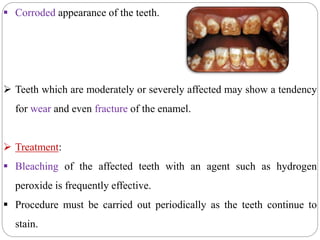
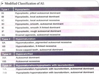
ENAMEL
Made By:
Dr. Akshat Sachdeva
MDS Ist Year
Dept. of Conservative Dentistry and Endodontics
Sudha Rustagi College of Dental Sciences & Research
INTRODUCTION
 Hardest calcified tissue in the human body that covers the anatomic
crown of the tooth.
 Only ectodermal derivative of the tooth.
 Varies in thickness in different areas.
 Thicker at incisal and occlusal areas and becomes progressively
thinner until it terminates at CEJ.
Enamel
Dentin
Pulp Chamber
Pulp Canal
Periodontal Fibers
Cementum
Fig. 1: Showing supporting structures of tooth
Source: Sturdevant’s Art and Science of Operative Dentistry
DEVELOPMENT OF ENAMEL
 The enamel organ consists of 4 distinct layers:
 Inner and outer enamel epithelium are separated by a large mass of
cells differentiated into two distinct layers.
 Layer that is close to the inner enamel epithelium consists of two or
three rows of flat polyhedral cells called stratum intermedium.
 The other layer, which is more loosely arranged, constitutes stellate
reticulum.
OUTER ENAMEL EPITHELIUM
 Consists of a single layer of cuboid cells, separated from the
surrounding connective tissue by a delicate basement membrane.
 Cells of the outer enamel epithelium become irregular in shape at the
highest convexity of the enamel organ.
 During enamel formation, cells of the outer enamel epithelium
develop villi and cytoplasmic vesicles and large numbers of
mitochondria.
STELLATE RETICULUM
 Cells in this layer are star shaped with long processes reaching in all
directions from a central body.
 Connected with each other and with the cells of the outer enamel
epithelium and the stratum intermedium by desmosomes.
 Stellate reticulum is noticeably reduced in thickness when the first
layers of dentin are laid down.
STRATUM INTERMEDIUM
 Cells are flat to cuboidal in shape and are arranged in one to three
layers.
 Connected with each other and with the neighboring cells of stellate
reticulum and inner enamel epithelium by desmosomes.
 Tonofibrils are found in the cytoplasm.
 Cells in this layer show mitotic division even after the cells of inner
enamel epithelium cease to divide.
INNER ENAMEL EPITHELIUM
 Cells of inner enamel epithelium are derived from the basal cell layer
of oral epithelium.
 These cells assume a columnar form and differentiate into ameloblasts
that produce the enamel matrix.
 Cell differentiation occurs earlier in the region of incisal edge or cusps
than in the area of cervical loop.
CERVICAL LOOP
 At the free border of the enamel organ, the outer and inner enamel
epithelial layers are continuous and reflected into one another as the
cervical loop.
 When the crown has been formed, the cells of this portion give rise to
Hertwig’s epithelial root sheath.
LIFE CYCLE OFAMELOBLAST
 According to function, the life span of cells can be divided into six
stages:
 Morphogenic.
 Organizing.
 Formative.
 Maturative.
 Protective.
 Desmolytic.
MORPHOGENIC STAGE
 The cells are short and columnar, with large oval nuclei that almost
fill the cell body.
 The ameloblasts before differentiation interact with mesenchymal
cells and determine shape of the DEJ and the crown.
 Golgi apparatus and the centrioles are located in the proximal end of
the cell, whereas the mitochondria are evenly dispersed throughout
the cytoplasm.
ORGANIZING STAGE
 Inner enamel epithelium interacts with the adjacent connective tissue
cells, which differentiate into odontoblasts.
 Cells of inner enamel epithelium become longer, and nucleus-free
zones at distal ends of the cells become almost as long as the proximal
parts containing nuclei.
 Staining methods reveal presence of acidophil granules in proximal
part of the cell.
 Epithelial cells come into close contact with connective tissue cells of
the pulp, which differentiate into odontoblasts.
 Formation of dentin by odontoblasts begins during terminal phase of
the organizing stage.
 When dentin forms, it cuts off ameloblasts from their original source
of nourishment, and from then on they are supplied by capillaries that
surround the outer enamel epithelium.
Source: Essentials of Oral Biology by Maji Jose
FORMATIVE STAGE
 Ameloblasts enter this stage after the first layer of dentin has been
formed.
 Presence of dentin is necessary for the beginning of enamel matrix
formation.
 During formation of enamel matrix, ameloblasts retain approximately
the same length and arrangement.
 Earliest apparent change to appear is the development of blunt cell
processes on ameloblast surfaces, which penetrate the basal lamina
and enter predentin.
MATURATIVE STAGE
 Enamel maturation i.e. full mineralization occurs after most of the
thickness of the enamel matrix has been formed in the occlusal or
incisal area.
 During enamel maturation, the ameloblasts are slightly reduced in
length and are closely attached to enamel matrix.
 Ameloblasts display microvilli at their distal extremities, and
cytoplasmic vacuoles containing material resembling enamel matrix
are present.
PROTECTIVE STAGE
 When the enamel has completely developed and has fully calcified,
the ameloblasts cease to be arranged in a well-defined layer.
 Cell layers then form a stratified epithelial covering of the enamel, the
so-called reduced enamel epithelium, which functions to protect the
mature enamel by separating it from connective tissue until the tooth
erupts.
 Anomalies may develop if connective tissue comes in contact with the
enamel.
DESMOLYTIC STAGE
 Reduced enamel epithelium proliferates and seems to induce atrophy
of the connective tissue.
 It has been suggested that epithelial cells elaborate enzymes that are
able to destroy connective tissue fibers by desmolysis.
 Premature degeneration of the reduced enamel epithelium may
prevent the eruption of a tooth.
Source: Essentials of Oral Biology by Maji Jose
AMELOGENESIS
 Two processes are involved in the development of enamel:
 Organic matrix formation.
 Mineralization.
ENAMEL MATRIX FORMATION
 Ameloblasts begin their secretory activity when a small amount of
dentin has been laid down.
 Ameloblasts lose their projections separating them from predentin.
 Islands of enamel matrix are deposited along the predentin.
 A thin, continuous layer of enamel is formed along the dentin.
 Amelogenin is the major component of enamel matrix proteins.
 Amelogenins have been shown to form minute nanospheres between
which enamel crystals form.
 Absence of amelogenin has been found to result in the formation of
hypoplastic teeth.
 Ameloblastin and enamelin are the other important proteins of the
enamel matrix.
 A new protein, amelotin is suggested to help in enamel formation.
DEVELOPMENT OF TOMES’ PROCESS
 Projections of ameloblasts into the enamel matrix have been named
Tomes’ process.
 They contain typical secretion granules as well as rough endoplasmic
reticulum and mitochondria.
 Tomes’ process is partially separated from cell body by an incomplete
septa formed by microfilaments and tonofilaments.
 Ameloblasts over maturing enamel are considerably shorter than the
ameloblasts over incompletely formed enamel.
 Changes occurring in the ameloblasts prior to the onset of maturation
process are called transition stage.
 During this stage:
 Ameloblasts reduce in height
 Enamel secretion stops completely and
 Process of amelogenin removal starts.
 Ameloblasts attach to the basal lamina by hemidesmosomes.
MINERALIZATION OF ENAMEL MATRIX
 Mineralization of the enamel matrix takes place in two stages.
 In the first stage, an immediate partial mineralization occurs in the
matrix segments.
 First mineral is in the form of crystalline apatite.
 Studies have shown that the initial mineral is octacalcium phosphate,
which may act as a template for hydroxyapatite.
 Second stage or maturation, is characterized by the gradual
completion of mineralization.
 Maturation process starts from height of the crown and progresses
cervically.
 The rate of formation of enamel is 4 μm/day.
 Organic matrix gradually becomes thinned and more widely spaced to
make room for the growing crystals.
 Ameloblasts undergo apoptosis after formation of enamel, hence
enamel formation does not occur later on while formation of other
hard tissues continues throughout life.
PHYSICAL CHARACTERISTICS
 Enamel forms a protective covering of variable thickness over the
entire surface of the crown.
 Color of enamel – covered crown ranges from yellowish white to
grayish white.
 Color is determined by differences in the translucency of enamel.
 Yellowish teeth have a thin, translucent enamel through which the
yellow color of the dentin is visible and grayish teeth have a more
opaque enamel.
Thickest over the cusps and incisal edges and thinnest at the cervical
margins.
Attains a maximum thickness of about 2 to 2.5 mm on the cusps of
molars and premolars.
Specific gravity of enamel is 2.8.
It has been found that enamel can act like a semipermeable membrane,
permitting complete or partial passage of certain molecules.
CHEMICAL CHARACTERISTICS
ENAMEL
Inorganic Content (96%) Organic content + water (4%)
Proteins
Amelogenins Nonamelogenins
(90%) (10%)
Amelogenins are low molecular weight proteins and are rich in
proline, histidine, glutamine and leucine.
Nonamelogenins are high molecular weight proteins and are rich in
glycine, aspartic acid and serine.
 Inorganic material of enamel is hydroxyapatite.
 Crystals of hydroxyapatite are hexagonal in cross-section.
 Average concentrations(%) of three major constituents namely
oxygen, calcium and phosphorus, are 43.4, 36.6, and 17.7
respectively.
 Minor constituents together account for 2.3%, of which sodium
carbon and magnesium are the principal constituents.
STRUCTURE
 Enamel is composed of enamel rods or prisms, rod sheath and an
interprismatic substance.
 Enamel rods normally have a clear crystalline appearance, permitting
light to pass through them.
 In cross-sections of human enamel, many rods resemble fish scales.
ULTRASTRUCTURE
 A more common pattern is a keyhole or paddle-shaped prism in
human enamel.
 Rods measure about 5 μm in breadth and 9 μm in length.
 Each enamel rod is built up of segments separated by dark lines that
give it a striated appearance.
 Striations are suggested to be due to a diurnal rhythm in the enamel
formation and that in these areas rods show variation in composition.
Source: Manual of Oral Histology and Oral Pathology by Maji Jose
HUNTER – SCHREGER BANDS
 Alternating dark and light strips of varying widths.
 Occur due to abrupt change in direction of enamel rods.
 Prisms cut longitudinally to produce dark bands are called parazones,
while those cut transversely to produce light bands are called
diazones.
 Angle between parazones and diazones is about 40 degrees.
Source: Manual of Oral Histology and Oral Pathology by Maji Jose
INCREMENTAL LINES OF RETZIUS
 Appear as brownish bands in ground sections of enamel.
 Illustrate the incremental pattern of enamel, i.e. the successive
apposition of layers of enamel during formation of the crown.
 Incremental lines of Retzius appear as concentric circles.
 Reflect variations in structure and mineralization of enamel.
Source: Manual of Oral Histology and Oral Pathology by Maji Jose
GNARLED ENAMEL
 Arrangement of enamel rods becomes more complicated in the region
of cusps and incisal edges.
 Enamel rods become more irregular and intertwine with each other
especially near the DEJ.
 This creates an optical appearance called as gnarled enamel.
Source: Manual of Oral Histology and Oral Pathology by Maji Jose
SURFACE STRUCTURES
 Structure less layer of enamel, called prismless enamel has been
described in 70% of permanent teeth and all deciduous teeth.
 Found least often over the cusp tips and most commonly toward the
cervical areas of the enamel surface.
 In this layer, the apatite crystals are parallel to one another and
perpendicular to the striae of Retzius.
 Perikymata are transverse, wave-like grooves, believed to be the
external manifestations of the striae of Retzius.
 They are continuous around a tooth with a fairly regular course and
usually lie parallel to each other and to the CEJ.
 Pits of about 1–1.5 μm in diameter and small elevations of about 10–
15 μm called enamel caps are seen on the irregular enamel surface.
 Larger enamel elevations are termed enamel brochs.
NEONATAL LINE
 Prominent incremental line separating prenatal and postnatal enamel.
 Prenatal enamel usually is better developed than the postnatal enamel.
 Fetus develops in a well-protected environment with an adequate
supply of all the essential materials.
 Found to be more frequently absent in permanent first molars of boys
than girls.
Source: Manual of Oral Histology and Oral Pathology by Maji Jose
ENAMEL CUTICLE
 Primary enamel cuticle or Nasmyth’s membrane covers the
entire crown of newly erupted tooth but is probably soon
removed by mastication.
 Erupted enamel is covered by a pellicle, a precipitate of salivary
proteins.
 Within a day or two after pellicle formation, it becomes
colonized by microorganisms to form bacterial plaque.
ENAMEL LAMELLAE
 Leaf like structures extending from outer surface of enamel towards
dentin.
 Three types of lamellae are seen:
i. Type A: Composed of poorly calcified enamel rods. Restricted to
enamel.
ii. Type B: Consists of degenerated cells and may extend into dentin.
iii. Type C: Filled with organic matter derived from saliva and maybe
extended into dentin.
ENAMEL TUFTS
 Ribbon like structures extending from DEJ into enamel.
 Resemble tufts of grass when viewed in ground sections.
 Consist of hypocalcified enamel rods and interprismatic substance.
 Extend in the direction of long axis of the crown.
Source: Manual of Oral Histology and Oral Pathology by Maji Jose
ENAMEL SPINDLE
 Odontoblastic processes crossing DEJ and extending to the enamel.
 Appear as dark spindle shaped structures.
 In ground sections of dried teeth, the organic content of the spindles
disintegrates and is replaced by air, and the spaces appear dark in
transmitted light.
 Found mainly in cusp tip region.
Source: Manual of Oral Histology and Oral Pathology by Maji Jose
DENTINO-ENAMEL JUNCTION
 Appears as a scalloped line with convexity directed towards dentin.
 Crystals of dentin and enamel mix with each other.
 More pronounced in the occlusal area, where masticatory stresses are
greater.
AGE CHANGES
 Most apparent age change in enamel is attrition or wear of the
occlusal surfaces and proximal contact points as a result of
mastication.
 This is evidenced by a loss of vertical dimension of the crown
and by flattening of the proximal contour.
 It has been seen that facial and lingual surfaces loose their
structure rapidly than do proximal surfaces, and anterior teeth
loose their structure more rapidly than do posterior teeth.
 Tooth Darkening:
 Teeth appear to darken with age.
 Darkening may be caused by deepening of dentin color seen through
the progressively thinning layer of translucent enamel.
 Permeability:
 Enamel becomes less permeable with advancing age.
 Decrease in permeability is caused due to increase in the size of the
crystals which in turn decreases the pores between them causing a
reduction in permeability.
CLINICAL CONSIDERATIONS
 Course of the enamel rods is of importance in cavity preparations.
 During cavity preparation, it is important that unsupported enamel
rods are not left at the cavity margins because they would soon break
and produce leakage.
 Bacteria would lodge in these spaces thus inducing secondary dental
caries.
Deep enamel fissures predispose teeth to caries.
Caries penetrate the floor of fissures rapidly because the enamel in
these areas is very thin.
Eventually, an area of dentin becomes carious because the entrance to
the cavity is minute.
Careful examination is necessary to discover such cavities because
most enamel fissures are minute.
TETRACYCLINE STAINING
 Discoloration of teeth due to tetracycline may occur if therapeutic
regimens are taken either during pregnancy or following childbirth.
 Forms a complex with calcium ions in the surface of hydroxyapatite
crystals.
 Usually avoided during pregnancy or till the child completes 8 years.
ECTOPIC ENAMEL
 Also called as enamel pearl or enameloma.
 Presence of enamel in unusual locations, mainly the tooth root.
 Usually develops on roots of maxillary permanent molars.
 Most incidental findings require no therapy.
 Meticulous oral hygiene should be maintained to prevent localized
loss of periodontal support.
CLINICAL CONSIDERATIONS
 The principal expressions of pathologic amelogenesis are:
 Hypoplasia occurs when matrix formation is affected and is
manifested as pitting, furrowing, or even total absence of the enamel.
 Hypocalcification results when maturation is lacking or incomplete
and can be seen in the form of opaque or chalky areas on normally
contoured enamel surfaces.
 Causes of such defective enamel formation can be generally classified
as systemic, local, or genetic.
 If drinking water contains fluoride in excess of 1.5 parts per million,
chronic endemic fluorosis may occur as a result of continuous use
throughout the period of amelogenesis.
 It is important to urge substitution of water with levels of fluoride
(about 1 part per million) well below the threshold for fluorosis.
 Teeth most frequently affected are incisors, canines and first molars.
 A small amount of fluoride (about 1 to 1.2 parts per million) reduces
susceptibility to dental caries without causing mottling.
 If an injury occurs in the formative stage of enamel development,
hypoplasia of the enamel will result.
 An injury during the maturation stage will cause a deficiency in
calcification.
AMELOGENESIS IMPERFECTA
 Structural defect of tooth enamel.
 Also called as:
 Hereditary enamel dysplasia.
 Hereditary brown enamel.
 Hereditary brown opalescent teeth.
 Prevalence of this condition has been estimated to range from 1 in 718
to 1 in 14,000, depending on the population studied.
 Classification of amelogenesis imperfecta (AI) according to Witkop
(1989):
 Type – I: Hypoplastic.
 Type – II: Hypomaturation.
 Type – III: Hypocalcified.
 Type – IV: Hypomaturation-hypoplastic with taurodontism.
 Hypoplastic AI represents 60–73% of all cases, hypomaturation AI
represents 20–40%, and hypocalcification AI represents 7%.
 Modified Classification of AI:
Etiology of AI is related to the alteration of genes involved in the
process of formation and maturation of the enamel.
Defective gene has been found to be closely linked to the locus
DXS85 at Xp22.
This also has been identified as the general location of the human gene
for amelogenin, the principal protein in developing enamel.
 Hypoplastic AI is characterized by:
 Thin enamel possessing yellowish-brown color.
 Glossy square-shaped crown.
 Lack of contact between adjacent teeth.
 Flat occlusal surfaces of posterior teeth due to attrition.
 Radiographically, there is a presence of thin radiopaque layer of
enamel with normal radiodensity.
 Histologically, defect in the enamel matrix formation is seen.
Hypoplastic AI
Hypocalcified form of AI is characterized by:
 Softer enamel which wears down rapidly.
 Pigmented dark brown colored enamel.
Radiographically, thickness of enamel is normal but radiodensity of
enamel is less than that of dentin.
Histologically, defects of matrix structure and mineralization is seen.
Hypocalcified AI
 Hypomaturation form of AI is characterized by:
 Thickness of enamel is harder than hypocalcified type and may crack
away from the crown.
 Mottled-colored cloudy white/yellow/brown/snow capped.
 Radiographically, radiodensity of enamel is similar to that of dentin.
 Histologically, alterations in enamel rod and rod sheath structures had
been noted.
Hypomaturation AI
 There is no specific treatment for the condition.
 Main objectives of treatment include preserving patient's remaining
dentition, and to treat and preserve the patient's occlusal vertical
height.
 Esthetic issues should be considered since the color of tooth crown is
yellow from exposure of dentin due to enamel loss.
 Factors to be considered in deciding treatment options include
classification and severity of AI, the patient's social history, clinical
findings etc.
Full-coverage crowns can be used to compensate for the abraded
enamel in adults.
Aesthetics may be addressed via placement of composite or porcelain
veneers.
Patient's oral hygiene and diet should be controlled as they play an
important role in the success of retaining future restorations.
Teeth may have to be extracted in worst cases and implants or dentures
can be considered for replacement.
ENAMEL HYPOPLASIA
 Incomplete or defective formation of the organic enamel matrix of
teeth.
Hereditary type Environmental factors
- Usually involves both deciduous - Involves either dentition
and permanent dentition. and sometimes even a single
tooth.
- Generally affects only enamel. - Both enamel and dentin
affected to some degree.
 In mild environmental hypoplasia, there may be only a few small
grooves, pits, or fissures on the enamel surface.
 If the condition is more severe, the enamel may exhibit rows of deep
pits arranged horizontally across the surface of the tooth.
HEREDITARY ENAMEL HYPOPLASIA
 Genetic conditions exhibiting enamel defects:
 Treacher Collins Syndrome.
 Congenital erythropoietic porphyria.
 Ectodermal dysplasia.
 Epidermolysis bullosa.
 Tuberous sclerosis.
 DiGeorge syndrome
 Kenny-Caffrey syndrome.
 Hypoplasia results only if the injury occurs during the time the teeth
are developing, or more specifically, during the formative stage of
enamel development.
 Different factors may give rise to the condition which include:
 Nutritional deficiency (vitamins A, C, and D).
 Congenital syphilis (Hutchinson’s teeth/mulberry molars).
 Local infection or trauma (Turner’s hypoplasia).
 Ingestion of chemicals (chiefly fluoride called mottled enamel).
 Idiopathic causes.
 Treatment includes veneers for teeth affected by the condition.
MOTTLED ENAMEL
 Occurs due to intake of fluoride-containing drinking water causing
disturbance of ameloblasts during the formative stage.
 Wide range of severity exists in the appearance of mottled teeth:
 Questionable changes characterized by occasional white flecking
or spotting of the enamel.
 Mild changes manifested by white opaque areas involving more of the
tooth surface.
 Moderate and severe changes show pitting and brownish staining of
the tooth surface.
 Corroded appearance of the teeth.
 Teeth which are moderately or severely affected may show a tendency
for wear and even fracture of the enamel.
 Treatment:
 Bleaching of the affected teeth with an agent such as hydrogen
peroxide is frequently effective.
 Procedure must be carried out periodically as the teeth continue to
stain.
EFFECT OF ACID ETCHING
 Acid etching of the enamel surface has become an important
technique in clinical practice.
 Enamel surface is etched with an acid to remove the smear layer on
enamel that was created during cavity preparation, making the
relatively smooth enamel surface pitted and irregular.
 When a composite resin is placed on this irregular surface, it can
achieve mechanical bonding with the enamel.
 Depending on the crystal orientation to the surface, three types of
etching patterns are produced.
 The most common is type I, characterized by preferential removal of
rods.
 In type II pattern, the interrod crystals are preferentially removed.
 Occurring less frequently is type III, which is irregular and
indiscriminate.
 Phenomenon of acid etchants producing differing surface patterns is
still debatable.
 Most commonly held view is that the etching pattern depends on
crystal orientation.
ENAMEL RENAL GINGIVAL SYNDROME
 First described by MacGibbon in 1972.
 Extremely rare disorder characterized by an autosomal recessive
pattern featuring:
 Severe enamel hypoplasia.
 Intrapulpal calcification.
 Failed tooth eruption.
 Nephrocalcinosis.
 Suggestive cause: Mutation in gene FAM20A.
CONCLUSION
 Enamel is the hardest calcified tissue in the human body that covers
anatomic crown of the tooth.
 Cells responsible for enamel formation are the ameloblasts.
 Enamel is an important structural entity and its protection is of utmost
importance.
 Aspire to inspire before you expire!
THANK YOU!

Enamel

  • 1.
    ENAMEL Made By: Dr. AkshatSachdeva MDS Ist Year Dept. of Conservative Dentistry and Endodontics Sudha Rustagi College of Dental Sciences & Research
  • 2.
    INTRODUCTION  Hardest calcifiedtissue in the human body that covers the anatomic crown of the tooth.  Only ectodermal derivative of the tooth.  Varies in thickness in different areas.  Thicker at incisal and occlusal areas and becomes progressively thinner until it terminates at CEJ.
  • 3.
    Enamel Dentin Pulp Chamber Pulp Canal PeriodontalFibers Cementum Fig. 1: Showing supporting structures of tooth Source: Sturdevant’s Art and Science of Operative Dentistry
  • 4.
    DEVELOPMENT OF ENAMEL The enamel organ consists of 4 distinct layers:  Inner and outer enamel epithelium are separated by a large mass of cells differentiated into two distinct layers.  Layer that is close to the inner enamel epithelium consists of two or three rows of flat polyhedral cells called stratum intermedium.  The other layer, which is more loosely arranged, constitutes stellate reticulum.
  • 5.
    OUTER ENAMEL EPITHELIUM Consists of a single layer of cuboid cells, separated from the surrounding connective tissue by a delicate basement membrane.  Cells of the outer enamel epithelium become irregular in shape at the highest convexity of the enamel organ.  During enamel formation, cells of the outer enamel epithelium develop villi and cytoplasmic vesicles and large numbers of mitochondria.
  • 6.
    STELLATE RETICULUM  Cellsin this layer are star shaped with long processes reaching in all directions from a central body.  Connected with each other and with the cells of the outer enamel epithelium and the stratum intermedium by desmosomes.  Stellate reticulum is noticeably reduced in thickness when the first layers of dentin are laid down.
  • 7.
    STRATUM INTERMEDIUM  Cellsare flat to cuboidal in shape and are arranged in one to three layers.  Connected with each other and with the neighboring cells of stellate reticulum and inner enamel epithelium by desmosomes.  Tonofibrils are found in the cytoplasm.  Cells in this layer show mitotic division even after the cells of inner enamel epithelium cease to divide.
  • 8.
    INNER ENAMEL EPITHELIUM Cells of inner enamel epithelium are derived from the basal cell layer of oral epithelium.  These cells assume a columnar form and differentiate into ameloblasts that produce the enamel matrix.  Cell differentiation occurs earlier in the region of incisal edge or cusps than in the area of cervical loop.
  • 9.
    CERVICAL LOOP  Atthe free border of the enamel organ, the outer and inner enamel epithelial layers are continuous and reflected into one another as the cervical loop.  When the crown has been formed, the cells of this portion give rise to Hertwig’s epithelial root sheath.
  • 10.
    LIFE CYCLE OFAMELOBLAST According to function, the life span of cells can be divided into six stages:  Morphogenic.  Organizing.  Formative.  Maturative.  Protective.  Desmolytic.
  • 11.
    MORPHOGENIC STAGE  Thecells are short and columnar, with large oval nuclei that almost fill the cell body.  The ameloblasts before differentiation interact with mesenchymal cells and determine shape of the DEJ and the crown.  Golgi apparatus and the centrioles are located in the proximal end of the cell, whereas the mitochondria are evenly dispersed throughout the cytoplasm.
  • 12.
    ORGANIZING STAGE  Innerenamel epithelium interacts with the adjacent connective tissue cells, which differentiate into odontoblasts.  Cells of inner enamel epithelium become longer, and nucleus-free zones at distal ends of the cells become almost as long as the proximal parts containing nuclei.  Staining methods reveal presence of acidophil granules in proximal part of the cell.
  • 13.
     Epithelial cellscome into close contact with connective tissue cells of the pulp, which differentiate into odontoblasts.  Formation of dentin by odontoblasts begins during terminal phase of the organizing stage.  When dentin forms, it cuts off ameloblasts from their original source of nourishment, and from then on they are supplied by capillaries that surround the outer enamel epithelium.
  • 14.
    Source: Essentials ofOral Biology by Maji Jose
  • 15.
    FORMATIVE STAGE  Ameloblastsenter this stage after the first layer of dentin has been formed.  Presence of dentin is necessary for the beginning of enamel matrix formation.  During formation of enamel matrix, ameloblasts retain approximately the same length and arrangement.  Earliest apparent change to appear is the development of blunt cell processes on ameloblast surfaces, which penetrate the basal lamina and enter predentin.
  • 16.
    MATURATIVE STAGE  Enamelmaturation i.e. full mineralization occurs after most of the thickness of the enamel matrix has been formed in the occlusal or incisal area.  During enamel maturation, the ameloblasts are slightly reduced in length and are closely attached to enamel matrix.  Ameloblasts display microvilli at their distal extremities, and cytoplasmic vacuoles containing material resembling enamel matrix are present.
  • 17.
    PROTECTIVE STAGE  Whenthe enamel has completely developed and has fully calcified, the ameloblasts cease to be arranged in a well-defined layer.  Cell layers then form a stratified epithelial covering of the enamel, the so-called reduced enamel epithelium, which functions to protect the mature enamel by separating it from connective tissue until the tooth erupts.  Anomalies may develop if connective tissue comes in contact with the enamel.
  • 18.
    DESMOLYTIC STAGE  Reducedenamel epithelium proliferates and seems to induce atrophy of the connective tissue.  It has been suggested that epithelial cells elaborate enzymes that are able to destroy connective tissue fibers by desmolysis.  Premature degeneration of the reduced enamel epithelium may prevent the eruption of a tooth.
  • 19.
    Source: Essentials ofOral Biology by Maji Jose
  • 20.
    AMELOGENESIS  Two processesare involved in the development of enamel:  Organic matrix formation.  Mineralization.
  • 21.
    ENAMEL MATRIX FORMATION Ameloblasts begin their secretory activity when a small amount of dentin has been laid down.  Ameloblasts lose their projections separating them from predentin.  Islands of enamel matrix are deposited along the predentin.  A thin, continuous layer of enamel is formed along the dentin.
  • 22.
     Amelogenin isthe major component of enamel matrix proteins.  Amelogenins have been shown to form minute nanospheres between which enamel crystals form.  Absence of amelogenin has been found to result in the formation of hypoplastic teeth.  Ameloblastin and enamelin are the other important proteins of the enamel matrix.  A new protein, amelotin is suggested to help in enamel formation.
  • 23.
    DEVELOPMENT OF TOMES’PROCESS  Projections of ameloblasts into the enamel matrix have been named Tomes’ process.  They contain typical secretion granules as well as rough endoplasmic reticulum and mitochondria.  Tomes’ process is partially separated from cell body by an incomplete septa formed by microfilaments and tonofilaments.
  • 24.
     Ameloblasts overmaturing enamel are considerably shorter than the ameloblasts over incompletely formed enamel.  Changes occurring in the ameloblasts prior to the onset of maturation process are called transition stage.  During this stage:  Ameloblasts reduce in height  Enamel secretion stops completely and  Process of amelogenin removal starts.  Ameloblasts attach to the basal lamina by hemidesmosomes.
  • 25.
    MINERALIZATION OF ENAMELMATRIX  Mineralization of the enamel matrix takes place in two stages.  In the first stage, an immediate partial mineralization occurs in the matrix segments.  First mineral is in the form of crystalline apatite.  Studies have shown that the initial mineral is octacalcium phosphate, which may act as a template for hydroxyapatite.
  • 26.
     Second stageor maturation, is characterized by the gradual completion of mineralization.  Maturation process starts from height of the crown and progresses cervically.  The rate of formation of enamel is 4 μm/day.  Organic matrix gradually becomes thinned and more widely spaced to make room for the growing crystals.  Ameloblasts undergo apoptosis after formation of enamel, hence enamel formation does not occur later on while formation of other hard tissues continues throughout life.
  • 27.
    PHYSICAL CHARACTERISTICS  Enamelforms a protective covering of variable thickness over the entire surface of the crown.  Color of enamel – covered crown ranges from yellowish white to grayish white.  Color is determined by differences in the translucency of enamel.  Yellowish teeth have a thin, translucent enamel through which the yellow color of the dentin is visible and grayish teeth have a more opaque enamel.
  • 28.
    Thickest over thecusps and incisal edges and thinnest at the cervical margins. Attains a maximum thickness of about 2 to 2.5 mm on the cusps of molars and premolars. Specific gravity of enamel is 2.8. It has been found that enamel can act like a semipermeable membrane, permitting complete or partial passage of certain molecules.
  • 29.
    CHEMICAL CHARACTERISTICS ENAMEL Inorganic Content(96%) Organic content + water (4%) Proteins Amelogenins Nonamelogenins (90%) (10%) Amelogenins are low molecular weight proteins and are rich in proline, histidine, glutamine and leucine. Nonamelogenins are high molecular weight proteins and are rich in glycine, aspartic acid and serine.
  • 30.
     Inorganic materialof enamel is hydroxyapatite.  Crystals of hydroxyapatite are hexagonal in cross-section.  Average concentrations(%) of three major constituents namely oxygen, calcium and phosphorus, are 43.4, 36.6, and 17.7 respectively.  Minor constituents together account for 2.3%, of which sodium carbon and magnesium are the principal constituents.
  • 31.
    STRUCTURE  Enamel iscomposed of enamel rods or prisms, rod sheath and an interprismatic substance.  Enamel rods normally have a clear crystalline appearance, permitting light to pass through them.  In cross-sections of human enamel, many rods resemble fish scales.
  • 32.
    ULTRASTRUCTURE  A morecommon pattern is a keyhole or paddle-shaped prism in human enamel.  Rods measure about 5 μm in breadth and 9 μm in length.  Each enamel rod is built up of segments separated by dark lines that give it a striated appearance.  Striations are suggested to be due to a diurnal rhythm in the enamel formation and that in these areas rods show variation in composition.
  • 33.
    Source: Manual ofOral Histology and Oral Pathology by Maji Jose
  • 34.
    HUNTER – SCHREGERBANDS  Alternating dark and light strips of varying widths.  Occur due to abrupt change in direction of enamel rods.  Prisms cut longitudinally to produce dark bands are called parazones, while those cut transversely to produce light bands are called diazones.  Angle between parazones and diazones is about 40 degrees.
  • 35.
    Source: Manual ofOral Histology and Oral Pathology by Maji Jose
  • 36.
    INCREMENTAL LINES OFRETZIUS  Appear as brownish bands in ground sections of enamel.  Illustrate the incremental pattern of enamel, i.e. the successive apposition of layers of enamel during formation of the crown.  Incremental lines of Retzius appear as concentric circles.  Reflect variations in structure and mineralization of enamel.
  • 37.
    Source: Manual ofOral Histology and Oral Pathology by Maji Jose
  • 38.
    GNARLED ENAMEL  Arrangementof enamel rods becomes more complicated in the region of cusps and incisal edges.  Enamel rods become more irregular and intertwine with each other especially near the DEJ.  This creates an optical appearance called as gnarled enamel.
  • 39.
    Source: Manual ofOral Histology and Oral Pathology by Maji Jose
  • 40.
    SURFACE STRUCTURES  Structureless layer of enamel, called prismless enamel has been described in 70% of permanent teeth and all deciduous teeth.  Found least often over the cusp tips and most commonly toward the cervical areas of the enamel surface.  In this layer, the apatite crystals are parallel to one another and perpendicular to the striae of Retzius.
  • 41.
     Perikymata aretransverse, wave-like grooves, believed to be the external manifestations of the striae of Retzius.  They are continuous around a tooth with a fairly regular course and usually lie parallel to each other and to the CEJ.  Pits of about 1–1.5 μm in diameter and small elevations of about 10– 15 μm called enamel caps are seen on the irregular enamel surface.  Larger enamel elevations are termed enamel brochs.
  • 42.
    NEONATAL LINE  Prominentincremental line separating prenatal and postnatal enamel.  Prenatal enamel usually is better developed than the postnatal enamel.  Fetus develops in a well-protected environment with an adequate supply of all the essential materials.  Found to be more frequently absent in permanent first molars of boys than girls.
  • 43.
    Source: Manual ofOral Histology and Oral Pathology by Maji Jose
  • 44.
    ENAMEL CUTICLE  Primaryenamel cuticle or Nasmyth’s membrane covers the entire crown of newly erupted tooth but is probably soon removed by mastication.  Erupted enamel is covered by a pellicle, a precipitate of salivary proteins.  Within a day or two after pellicle formation, it becomes colonized by microorganisms to form bacterial plaque.
  • 45.
    ENAMEL LAMELLAE  Leaflike structures extending from outer surface of enamel towards dentin.  Three types of lamellae are seen: i. Type A: Composed of poorly calcified enamel rods. Restricted to enamel. ii. Type B: Consists of degenerated cells and may extend into dentin. iii. Type C: Filled with organic matter derived from saliva and maybe extended into dentin.
  • 46.
    ENAMEL TUFTS  Ribbonlike structures extending from DEJ into enamel.  Resemble tufts of grass when viewed in ground sections.  Consist of hypocalcified enamel rods and interprismatic substance.  Extend in the direction of long axis of the crown.
  • 47.
    Source: Manual ofOral Histology and Oral Pathology by Maji Jose
  • 48.
    ENAMEL SPINDLE  Odontoblasticprocesses crossing DEJ and extending to the enamel.  Appear as dark spindle shaped structures.  In ground sections of dried teeth, the organic content of the spindles disintegrates and is replaced by air, and the spaces appear dark in transmitted light.  Found mainly in cusp tip region.
  • 49.
    Source: Manual ofOral Histology and Oral Pathology by Maji Jose
  • 50.
    DENTINO-ENAMEL JUNCTION  Appearsas a scalloped line with convexity directed towards dentin.  Crystals of dentin and enamel mix with each other.  More pronounced in the occlusal area, where masticatory stresses are greater.
  • 51.
    AGE CHANGES  Mostapparent age change in enamel is attrition or wear of the occlusal surfaces and proximal contact points as a result of mastication.  This is evidenced by a loss of vertical dimension of the crown and by flattening of the proximal contour.  It has been seen that facial and lingual surfaces loose their structure rapidly than do proximal surfaces, and anterior teeth loose their structure more rapidly than do posterior teeth.
  • 52.
     Tooth Darkening: Teeth appear to darken with age.  Darkening may be caused by deepening of dentin color seen through the progressively thinning layer of translucent enamel.  Permeability:  Enamel becomes less permeable with advancing age.  Decrease in permeability is caused due to increase in the size of the crystals which in turn decreases the pores between them causing a reduction in permeability.
  • 53.
    CLINICAL CONSIDERATIONS  Courseof the enamel rods is of importance in cavity preparations.  During cavity preparation, it is important that unsupported enamel rods are not left at the cavity margins because they would soon break and produce leakage.  Bacteria would lodge in these spaces thus inducing secondary dental caries.
  • 54.
    Deep enamel fissurespredispose teeth to caries. Caries penetrate the floor of fissures rapidly because the enamel in these areas is very thin. Eventually, an area of dentin becomes carious because the entrance to the cavity is minute. Careful examination is necessary to discover such cavities because most enamel fissures are minute.
  • 55.
    TETRACYCLINE STAINING  Discolorationof teeth due to tetracycline may occur if therapeutic regimens are taken either during pregnancy or following childbirth.  Forms a complex with calcium ions in the surface of hydroxyapatite crystals.  Usually avoided during pregnancy or till the child completes 8 years.
  • 56.
    ECTOPIC ENAMEL  Alsocalled as enamel pearl or enameloma.  Presence of enamel in unusual locations, mainly the tooth root.  Usually develops on roots of maxillary permanent molars.  Most incidental findings require no therapy.  Meticulous oral hygiene should be maintained to prevent localized loss of periodontal support.
  • 57.
    CLINICAL CONSIDERATIONS  Theprincipal expressions of pathologic amelogenesis are:  Hypoplasia occurs when matrix formation is affected and is manifested as pitting, furrowing, or even total absence of the enamel.  Hypocalcification results when maturation is lacking or incomplete and can be seen in the form of opaque or chalky areas on normally contoured enamel surfaces.  Causes of such defective enamel formation can be generally classified as systemic, local, or genetic.
  • 58.
     If drinkingwater contains fluoride in excess of 1.5 parts per million, chronic endemic fluorosis may occur as a result of continuous use throughout the period of amelogenesis.  It is important to urge substitution of water with levels of fluoride (about 1 part per million) well below the threshold for fluorosis.  Teeth most frequently affected are incisors, canines and first molars.
  • 59.
     A smallamount of fluoride (about 1 to 1.2 parts per million) reduces susceptibility to dental caries without causing mottling.  If an injury occurs in the formative stage of enamel development, hypoplasia of the enamel will result.  An injury during the maturation stage will cause a deficiency in calcification.
  • 60.
    AMELOGENESIS IMPERFECTA  Structuraldefect of tooth enamel.  Also called as:  Hereditary enamel dysplasia.  Hereditary brown enamel.  Hereditary brown opalescent teeth.  Prevalence of this condition has been estimated to range from 1 in 718 to 1 in 14,000, depending on the population studied.
  • 61.
     Classification ofamelogenesis imperfecta (AI) according to Witkop (1989):  Type – I: Hypoplastic.  Type – II: Hypomaturation.  Type – III: Hypocalcified.  Type – IV: Hypomaturation-hypoplastic with taurodontism.  Hypoplastic AI represents 60–73% of all cases, hypomaturation AI represents 20–40%, and hypocalcification AI represents 7%.
  • 62.
  • 63.
    Etiology of AIis related to the alteration of genes involved in the process of formation and maturation of the enamel. Defective gene has been found to be closely linked to the locus DXS85 at Xp22. This also has been identified as the general location of the human gene for amelogenin, the principal protein in developing enamel.
  • 64.
     Hypoplastic AIis characterized by:  Thin enamel possessing yellowish-brown color.  Glossy square-shaped crown.  Lack of contact between adjacent teeth.  Flat occlusal surfaces of posterior teeth due to attrition.  Radiographically, there is a presence of thin radiopaque layer of enamel with normal radiodensity.  Histologically, defect in the enamel matrix formation is seen.
  • 65.
  • 66.
    Hypocalcified form ofAI is characterized by:  Softer enamel which wears down rapidly.  Pigmented dark brown colored enamel. Radiographically, thickness of enamel is normal but radiodensity of enamel is less than that of dentin. Histologically, defects of matrix structure and mineralization is seen.
  • 67.
  • 68.
     Hypomaturation formof AI is characterized by:  Thickness of enamel is harder than hypocalcified type and may crack away from the crown.  Mottled-colored cloudy white/yellow/brown/snow capped.  Radiographically, radiodensity of enamel is similar to that of dentin.  Histologically, alterations in enamel rod and rod sheath structures had been noted.
  • 69.
  • 70.
     There isno specific treatment for the condition.  Main objectives of treatment include preserving patient's remaining dentition, and to treat and preserve the patient's occlusal vertical height.  Esthetic issues should be considered since the color of tooth crown is yellow from exposure of dentin due to enamel loss.  Factors to be considered in deciding treatment options include classification and severity of AI, the patient's social history, clinical findings etc.
  • 71.
    Full-coverage crowns canbe used to compensate for the abraded enamel in adults. Aesthetics may be addressed via placement of composite or porcelain veneers. Patient's oral hygiene and diet should be controlled as they play an important role in the success of retaining future restorations. Teeth may have to be extracted in worst cases and implants or dentures can be considered for replacement.
  • 72.
    ENAMEL HYPOPLASIA  Incompleteor defective formation of the organic enamel matrix of teeth. Hereditary type Environmental factors - Usually involves both deciduous - Involves either dentition and permanent dentition. and sometimes even a single tooth. - Generally affects only enamel. - Both enamel and dentin affected to some degree.
  • 73.
     In mildenvironmental hypoplasia, there may be only a few small grooves, pits, or fissures on the enamel surface.  If the condition is more severe, the enamel may exhibit rows of deep pits arranged horizontally across the surface of the tooth.
  • 74.
    HEREDITARY ENAMEL HYPOPLASIA Genetic conditions exhibiting enamel defects:  Treacher Collins Syndrome.  Congenital erythropoietic porphyria.  Ectodermal dysplasia.  Epidermolysis bullosa.  Tuberous sclerosis.  DiGeorge syndrome  Kenny-Caffrey syndrome.
  • 75.
     Hypoplasia resultsonly if the injury occurs during the time the teeth are developing, or more specifically, during the formative stage of enamel development.  Different factors may give rise to the condition which include:  Nutritional deficiency (vitamins A, C, and D).  Congenital syphilis (Hutchinson’s teeth/mulberry molars).  Local infection or trauma (Turner’s hypoplasia).  Ingestion of chemicals (chiefly fluoride called mottled enamel).  Idiopathic causes.  Treatment includes veneers for teeth affected by the condition.
  • 76.
    MOTTLED ENAMEL  Occursdue to intake of fluoride-containing drinking water causing disturbance of ameloblasts during the formative stage.  Wide range of severity exists in the appearance of mottled teeth:  Questionable changes characterized by occasional white flecking or spotting of the enamel.
  • 77.
     Mild changesmanifested by white opaque areas involving more of the tooth surface.  Moderate and severe changes show pitting and brownish staining of the tooth surface.
  • 78.
     Corroded appearanceof the teeth.  Teeth which are moderately or severely affected may show a tendency for wear and even fracture of the enamel.  Treatment:  Bleaching of the affected teeth with an agent such as hydrogen peroxide is frequently effective.  Procedure must be carried out periodically as the teeth continue to stain.
  • 79.
    EFFECT OF ACIDETCHING  Acid etching of the enamel surface has become an important technique in clinical practice.  Enamel surface is etched with an acid to remove the smear layer on enamel that was created during cavity preparation, making the relatively smooth enamel surface pitted and irregular.  When a composite resin is placed on this irregular surface, it can achieve mechanical bonding with the enamel.
  • 80.
     Depending onthe crystal orientation to the surface, three types of etching patterns are produced.  The most common is type I, characterized by preferential removal of rods.  In type II pattern, the interrod crystals are preferentially removed.
  • 81.
     Occurring lessfrequently is type III, which is irregular and indiscriminate.  Phenomenon of acid etchants producing differing surface patterns is still debatable.  Most commonly held view is that the etching pattern depends on crystal orientation.
  • 82.
    ENAMEL RENAL GINGIVALSYNDROME  First described by MacGibbon in 1972.  Extremely rare disorder characterized by an autosomal recessive pattern featuring:  Severe enamel hypoplasia.  Intrapulpal calcification.  Failed tooth eruption.  Nephrocalcinosis.  Suggestive cause: Mutation in gene FAM20A.
  • 83.
    CONCLUSION  Enamel isthe hardest calcified tissue in the human body that covers anatomic crown of the tooth.  Cells responsible for enamel formation are the ameloblasts.  Enamel is an important structural entity and its protection is of utmost importance.  Aspire to inspire before you expire!
  • 84.