Downloaded 1,866 times
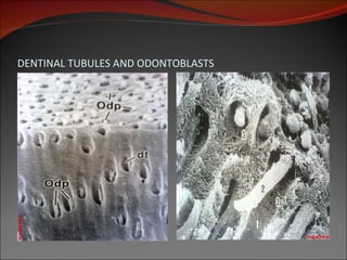
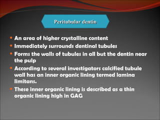


Dentin is a hard yellowish substance that forms the bulk of teeth. It is composed of 70% hydroxyapatite crystals and 30% organic materials like collagen. Dentin is formed by odontoblasts cells differentiated from dental papilla cells. It determines the shape of teeth and contains microscopic tubules that house the processes of odontoblast cells. Dentin is harder than bone but softer than enamel. It has different layers with varying properties located at different regions of the tooth.


