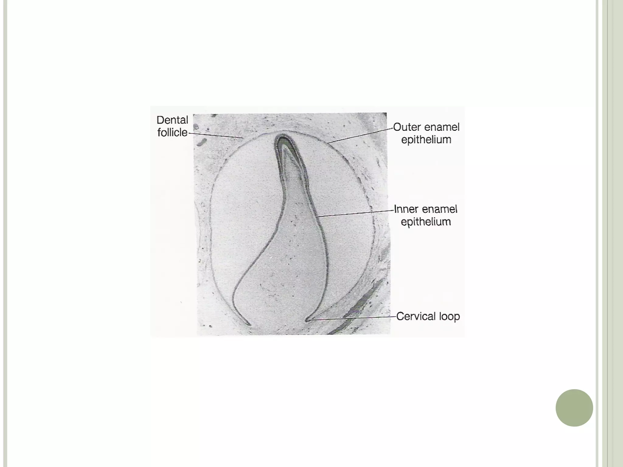
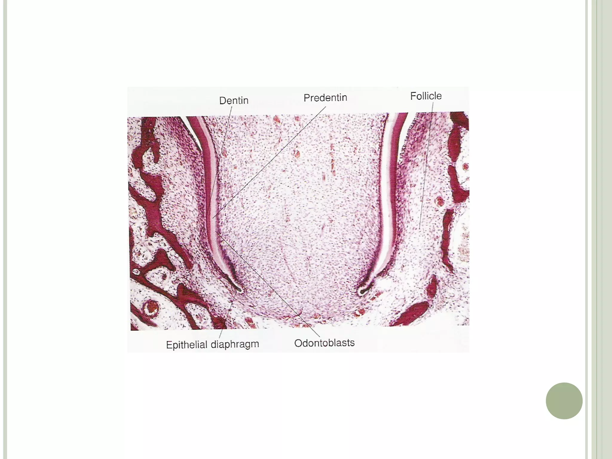
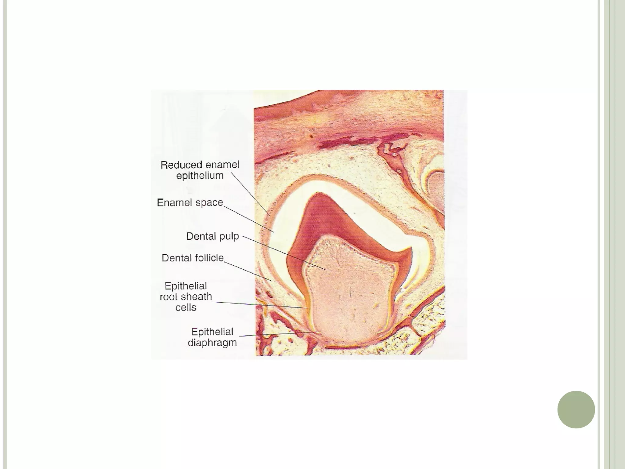
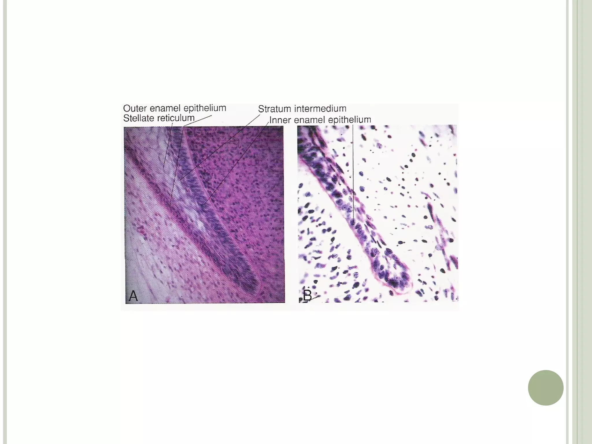
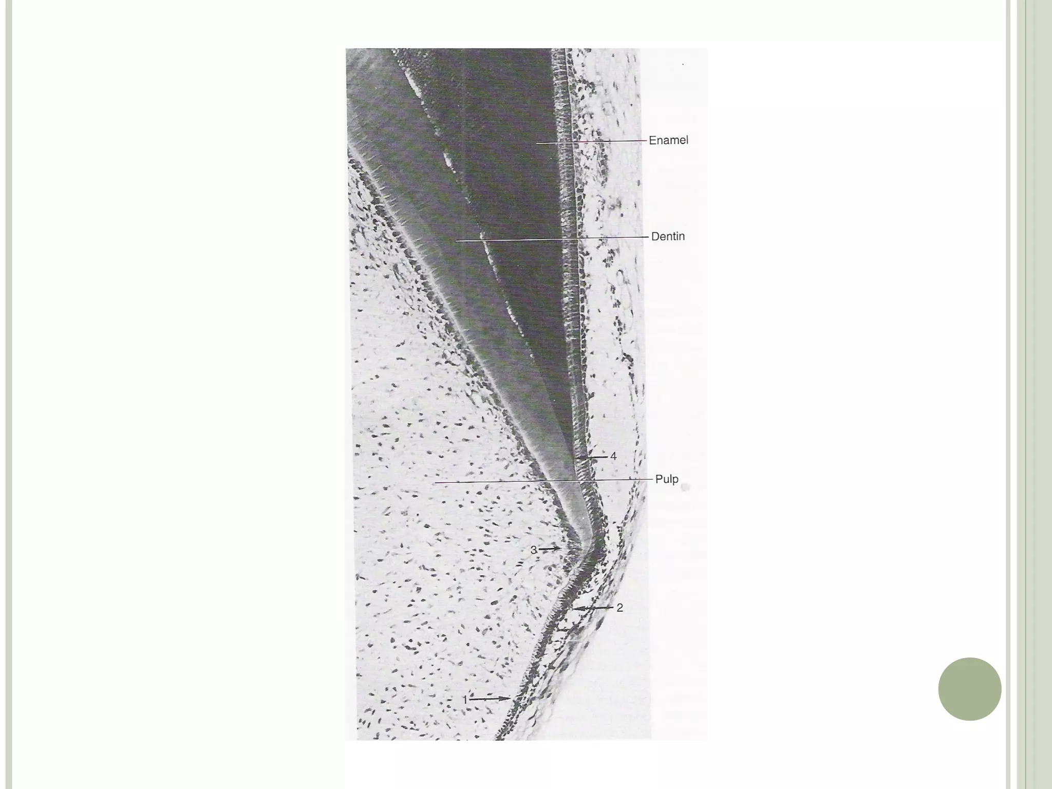
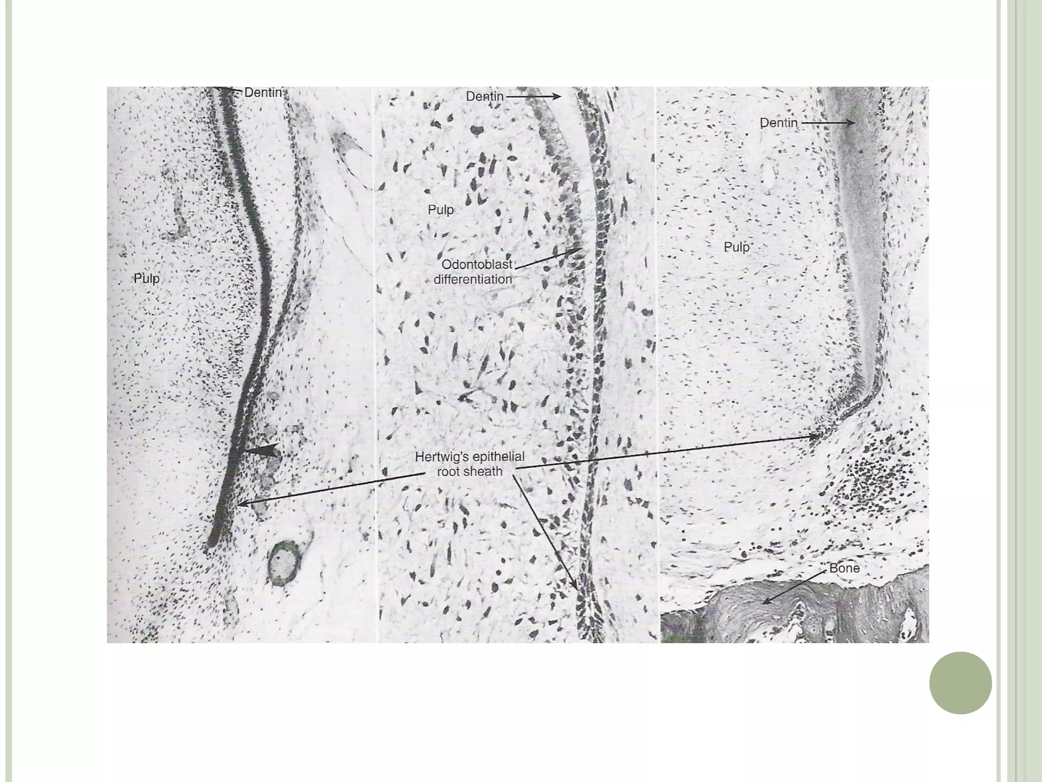
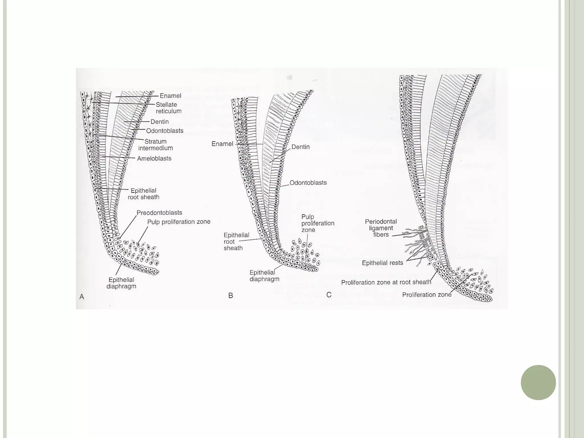
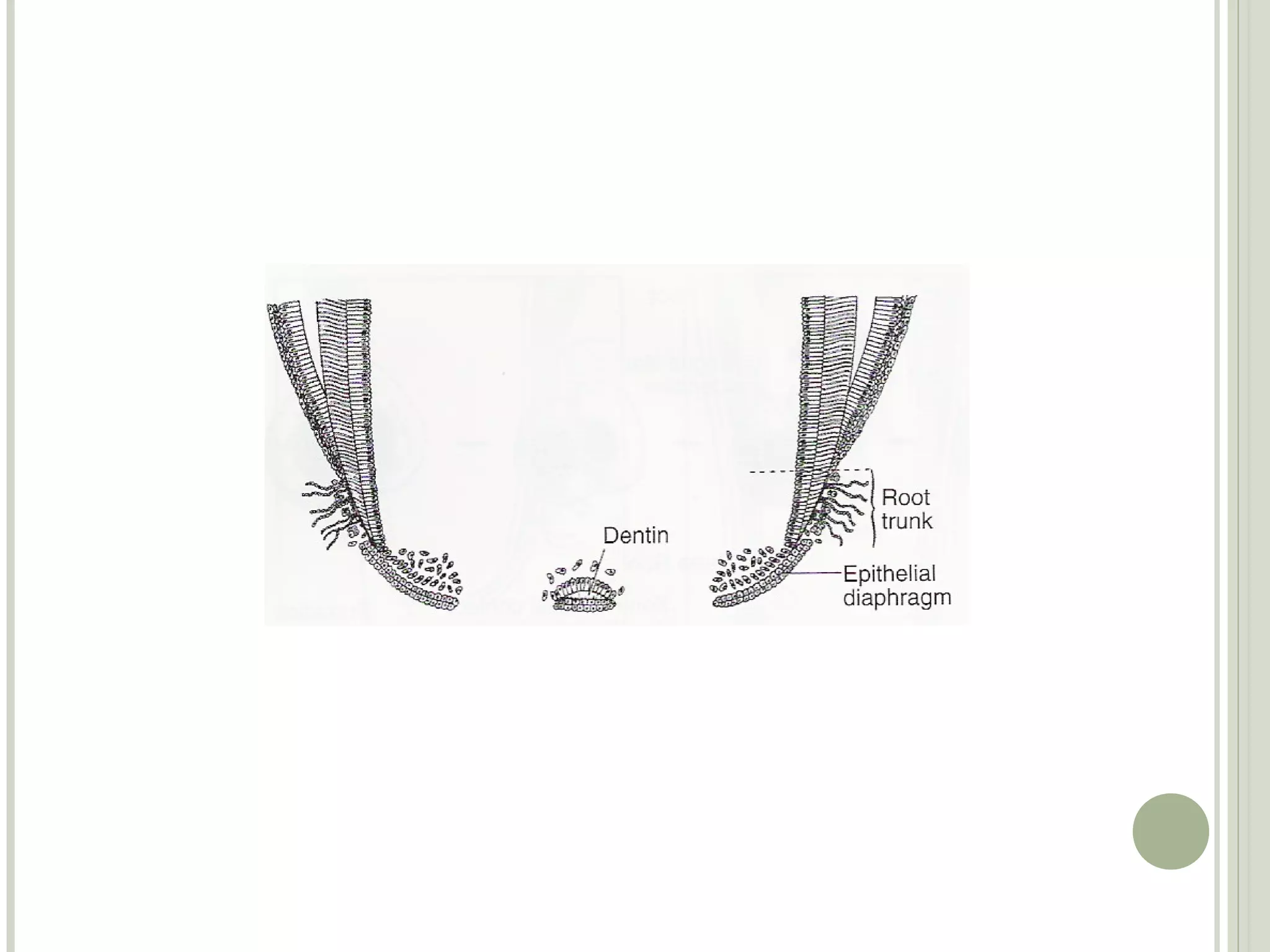
Development of Teeth: Root Formation
              Lesson - 2
Text and pictures in this presentation are taken
from Oral Histology text books: Ten Cates and
                 James Avery
Objectives
At the end of this chapter the student should be able
to understand the process of root formation.,.
including the role and function of the root sheath .


Sub topics
Topic 1. Root sheath development
Topic 2. Formation of root
Topic 3. Fate of epithelial root sheath
1 Root sheath development

The root starts to develop after the crown is completed
 Epithelial cells of the inner and outer enamel epithelium
proliferate from the cervical loop to form two layers of
epithelium called Hertwigs root sheath
 The first formed part of the root sheath bends to form a
disc like structure
The rim of this disc like structure is called the epithelial
diaphgram
The epithelial diaphgram encloses the primary apical
foramen
2 Formation of Root

After the formation of epithelial root sheath and the
epithelial diaphragm the root grows in length
The diaphragm maintains a constant size while the root
sheath grows in length at the angle of the diaphragm and
not at its tip
 The cells of the lengthening root sheath induce the
adjacent dental papilla cells to differentiate into
odontoblasts
 The newly formed odontoblasts then form the root
dentine
 As the root lengthens the crown moves occlusally
Single roots and multiple roots
For single-rooted teeth the root sheath grows like a cuff
or tube around the newly forming pulp
Development of multi-rooted teeth takes place in a same
manner until the furcation area.
 When the furcation area is reached the epithelial
diaphragm develops tongue like extensions that grow until
they contact each other
This the divides the root into two or three
Root formation anomalies
If the continuity of the root sheath is broken before the
dentine is formed it results in missing or abnormal
epithelial cells
 When the epithelial cells are missing the Odontoblasts
do not differentiate and dentine doesn’t form opposite the
defect that occurred in the root sheath
The result will be a small lateral canal. This lateral canal
is also called as supplemental canal or accessory canal
Accessory canals connect the main root canal with the
periodontal ligament
If the epithelial root sheath does not degenerate at the
proper time and remain stuck to the surface of the root
dentine, then that area becomes devoid of cementum
 Areas of root without cementum can be a cause of
sensitivity if the there is gingival recession
3 Fate of epithelial root sheath

After dentine formation in root takes place, the epithelial
root sheath breaks down and its remnants migrate away
from the dentinal surface
They lie in the periodontal ligament and are called
epithelial rests of Malassez
The epithelial rests of Malassez are found the in the
periodontal ligament through out the life
Sometimes when there is chronic inflammation the
epithelial cell rest of Malassez proliferate into cysts and
tumours
Root formation

Root formation

  • 1.
    Development of Teeth:Root Formation Lesson - 2
  • 2.
    Text and picturesin this presentation are taken from Oral Histology text books: Ten Cates and James Avery
  • 3.
    Objectives At the endof this chapter the student should be able to understand the process of root formation.,. including the role and function of the root sheath . Sub topics Topic 1. Root sheath development Topic 2. Formation of root Topic 3. Fate of epithelial root sheath
  • 4.
    1 Root sheathdevelopment The root starts to develop after the crown is completed  Epithelial cells of the inner and outer enamel epithelium proliferate from the cervical loop to form two layers of epithelium called Hertwigs root sheath  The first formed part of the root sheath bends to form a disc like structure The rim of this disc like structure is called the epithelial diaphgram The epithelial diaphgram encloses the primary apical foramen
  • 12.
    2 Formation ofRoot After the formation of epithelial root sheath and the epithelial diaphragm the root grows in length The diaphragm maintains a constant size while the root sheath grows in length at the angle of the diaphragm and not at its tip  The cells of the lengthening root sheath induce the adjacent dental papilla cells to differentiate into odontoblasts  The newly formed odontoblasts then form the root dentine  As the root lengthens the crown moves occlusally
  • 13.
    Single roots andmultiple roots For single-rooted teeth the root sheath grows like a cuff or tube around the newly forming pulp Development of multi-rooted teeth takes place in a same manner until the furcation area.  When the furcation area is reached the epithelial diaphragm develops tongue like extensions that grow until they contact each other This the divides the root into two or three
  • 16.
    Root formation anomalies Ifthe continuity of the root sheath is broken before the dentine is formed it results in missing or abnormal epithelial cells  When the epithelial cells are missing the Odontoblasts do not differentiate and dentine doesn’t form opposite the defect that occurred in the root sheath The result will be a small lateral canal. This lateral canal is also called as supplemental canal or accessory canal Accessory canals connect the main root canal with the periodontal ligament
  • 18.
    If the epithelialroot sheath does not degenerate at the proper time and remain stuck to the surface of the root dentine, then that area becomes devoid of cementum  Areas of root without cementum can be a cause of sensitivity if the there is gingival recession
  • 19.
    3 Fate ofepithelial root sheath After dentine formation in root takes place, the epithelial root sheath breaks down and its remnants migrate away from the dentinal surface They lie in the periodontal ligament and are called epithelial rests of Malassez The epithelial rests of Malassez are found the in the periodontal ligament through out the life Sometimes when there is chronic inflammation the epithelial cell rest of Malassez proliferate into cysts and tumours