
Recommended
More Related Content
What's hot
What's hot (20)
Similar to BIOLOGICAL OXIDATION L3
Similar to BIOLOGICAL OXIDATION L3 (20)
Recently uploaded
Recently uploaded (20)
BIOLOGICAL OXIDATION L3
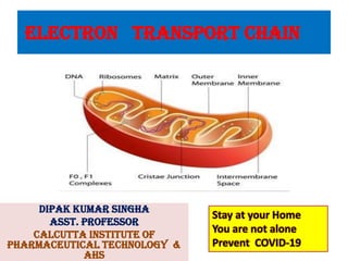
- 1. ELECTRON TRANSPORT CHAIN DIPAK KUMAR SINGHA ASST. PROFESSOR CALCUTTA INSTITUTE OF PHARMACEUTICAL TECHNOLOGY & AHS Stay at your Home You are not alone Prevent COVID-19
- 2. BIOLOGICAL OXIDATION AND ELECTRON TRANSPORT CHAIN The students must be able to answer questions on the following topics: ➢ Stages of Oxidation of Foodstuffs ➢ Redox potentials ➢ Biological oxidation ➢ Enzymes and co-enzymes involved biological oxidation ➢ High energy compounds ➢ Organization of electron transport chain ➢ Inhibitors of electron transport chain ➢ Oxidative Phosphorylation ➢ Chemiosmotic theory ➢ Proton pump ➢ ATP synthesis ➢ Inhibitors of ATP synthesis
- 3. BIOENERGETICS OR BIOCHEMICAL THERMODYNAMICS Free energy The energy actually available to do work ( utilizable)is kkown as free energy. Changes in the free energy (ΔG) are valuable in predicting the feasibility of chemical reactions. During a chemical reaction, heat may be released or absorbed. Enthalpy (ΔH) is a measure of the change in heat content of the reactants, compared to products. Entropy (ΔS) represents a change in the randomness or disorder of reactants and products. Entropy attains a maximum as the reaction approaches equilibrium. The react ions of biological systems involve a temporary decrease in entropy. The relation between the changes of free energy (ΔG), enthalpy (ΔH) and entropy (ΔS) is expressed as Δ G=ΔH-TΔS T representsth e absolutet emperaturei n Kelvin (K=273+"C). The term standard free energy represented by ΔC' (note the superscript') is often used. Lt indicates the free energy change when the reactants or products are at a concentration of 1mol/l at pH 7.0
- 4. ATP ADP Cycle • The hydrolysis of ATP is associated with the release of large amount of energy. • ATP + H2C ------+A DP + Pi + 7.3 Cal.
- 5. ATP-ADP cycle along with sources and utilization of ATP (Note that -P does not exist in free form, but is only transferred).
- 6. ORGANIZATION OF ELECTRON TRANSPORT CHAIN
- 7. Structure of Mitochondrion The electron transport chain is functioning inside the mitochondria. The mitochondrion is a subcellular organelle having the outer and inner membranes enclosing the matrix. The inner membrane is highly selective in its permeability, containing specific transport proteins. Certain enzymes are specifically localized in mitochondria. The inner membrane contains the respiratory chain and translocating systems. The knob like protrusions represent the ATP synthase system
- 9. Inner and outer mitochondrial membrane differs greatly in their composition. Inner membrane is 22% cardiolipin and contains no cholesterol, whereas outer membrane is similar to cell membrane, with less than 3% cardiolipin and 45% cholesterol. Location of enzymes in mitochondria
- 10. ORGANIZATION OF ELECTRON TRANSPORT CHAIN i. In the Electron transport chain, or respiratory chain, the electrons are transferred from NADH to a chain of electron carriers. The electrons flow from the more electronegative components to the more electropositive components. ii. All the components of electron transport chain (ETC) are located in the inner membrane of mitochondria. iii. There are four distinct multi-protein complexes; these are named as complex-I, II, III and IV. These are connected by two mobile carriers, co-enzyme Q and cytochrome c. iv. The arrangement is schematically represented.
- 11. Components and sequence of reactions of electron transport chain
- 12. NADH GENERATION The NADH is generated during intermediary metabolism. A detailed list of the reactions using NADH is given. Malate Aspartate Shuttle Mitochondrial membrane is impermeable to NADH. The NADH equivalents generated in glycolysis are therefore to be transported from cytoplasm to mitochondria for oxidation. This is achieved by malate aspartate shuttle or malate shuttle, which operates mainly in liver, kidney and heart. The cycle is operated with the help of enzymes malate dehydrogenase (MDH) and aspartate aminotransferase. From one molecule of NADH in the mitochondria, 2½ ATP molecules are generated.
- 13. Glycerol-3-phosphate Shuttle In skeletal muscle and brain, the reducing equivalents from cytoplasmic NADH are transported to mitochondria as FADH2 through glycerol-3-phosphate shuttle (Fig. 20.6). Hence only 1½ ATPs are generated when this system is operating.
- 14. ETC Complex-I i. It is also called NADH-CoQ reductase or NADH dehydrogenase complex. It is tightly bound to the inner membrane of mitochondria. ii. It contains a flavoprotein (Fp), consisting of FMN as prosthetic group and an iron-sulfur protein (Fe-S). NADH is the donor of electrons, FMN accepts them and gets reduced to FMNH2 (Fig. 20.8). Two electrons and one hydrogen ion are transferred from NADH to the flavin prosthetic group of the enzyme. NADH + H+ + FMN → FMNH2 + NAD+ iii. The electrons from FMNH2 are then transferred to Fe-S. The electrons are then transferred to co-enzyme Q (ubiquinone) (CoQ). iv. Overall function of this complex is to collect the pair of electrons from NADH and pass them to CoQ. The reactions are shown in Figure 20.8. v. There is a large negative free energy change; the energy released is 12 kcal/mol. This is utilized to drive 4 protons out of the mitochondria.
- 15. Complex II or Succinate-Q-Reductase The reaction in Complex-II is represented in Fig. 2.9. The electrons from FADH2 enter the ETC at the level of coenzyme Q. This step does not liberate enough energy to act as a proton pump. In other words, substrates oxidized by FAD-linked enzymes bypass complex-I. The three major enzyme systems that transfer their electrons directly to ubiquinone from the FAD prosthetic group are: i. Succinate dehydrogenase, ii. Fatty acyl CoA dehydrogenase iii. Mitochondrial glycerol phosphate dehydrogenase(Fig. 2.6).
- 16. Co-enzyme Q i. The ubiquinone (Q) is reduced successively to semiquinone (QH) and finally to quinol (QH2). ii. It accepts a pair of electrons from NADH or FADH2 through complex-I or complex-II respectively (Figs2.7 and 2.13).
- 17. Components and sequence of reactions of electron transport chain
- 18. iii. Co-enzyme Q is a quinone derivative having a long isoprenoid tail. The chain length of the tail is different in various species, mammals have 10 isoprene units (Fig. 2.10). Two molecules of cytochrome c are reduced. iv. The Q cycle thus facilitates the switching from the two electron carrier ubiquinol to the single electron carrier cytochrome c.
- 19. Complex III or Cytochrome Reductase i. This is a cluster of iron-sulfur proteins, cytochrome b and cytochrome c1, both contain heme prosthetic group. The sequence of reaction inside the Complex III is shown in Figure 20.11. ii. During this process of transfer of electron, the iron in heme group shuttles between Fe3+ and Fe2+ forms. iii. The free energy change is—10 kcal/mol; and 4 protons are pumped out. Cytochrome c • It is a peripheral membrane protein containing one heme prosthetic group. The term cytochrome is derived from Greek, meaning cellular colors. It is one of the highly conserved proteins among different species. Axel Theorell (Nobel • prize, 1955) isolated it. Cytochrome c collects electrons from Complex III and delivers them to Complex IV.
- 20. Complex IV or Cytochrome Oxidase i. It contains different proteins, including cytochrome a and cytochrome a3. The Complex IV is tightly bound to the mitochondrial membrane. ii. The reaction is depicted in Figure 20.12. Four electrons are accepted from cytochrome c, and passed on to molecular oxygen. 4 H+ + O2 + 4 Cyt. c-Fe++ → 2 H2O + 4 Cyt. c+++ iii. 2 protons are pumped out to the inter-membrane space. iv. Cytochrome oxidase has 4 redox centers, namely, a, a3, CuA and CuB. The electron transfer in this complex is as shown Cyto c →CuA →Cyto a →Cyto a3 →CuB Cytochrome oxidase contains two heme groups and two copper ions. The two heme groups are denoted as cytochrome-a and cytochrome a-3. The functional unit of the enzyme is a single protein, and is referred to as cytochrome a--a3. The sequential arrangement of members of electron transport chain is shown in Box 20.2 and Fig. 20.13.
- 21. INHIBITORS OF ELECTRON TRASPORT CHAIN
- 22. ANY QUESTION?