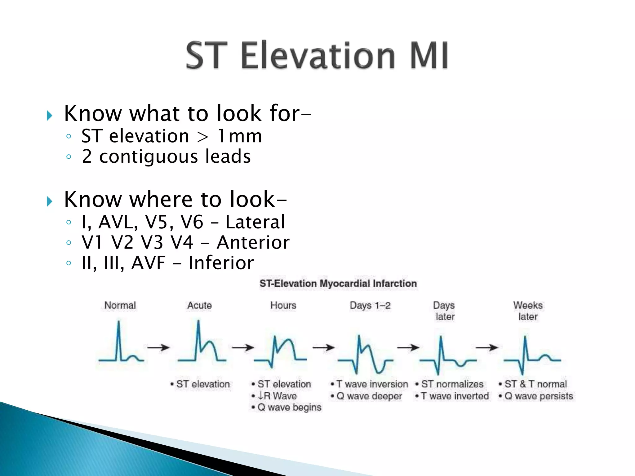
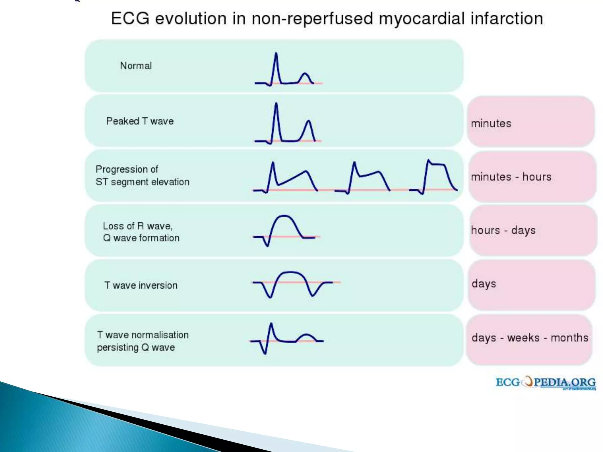
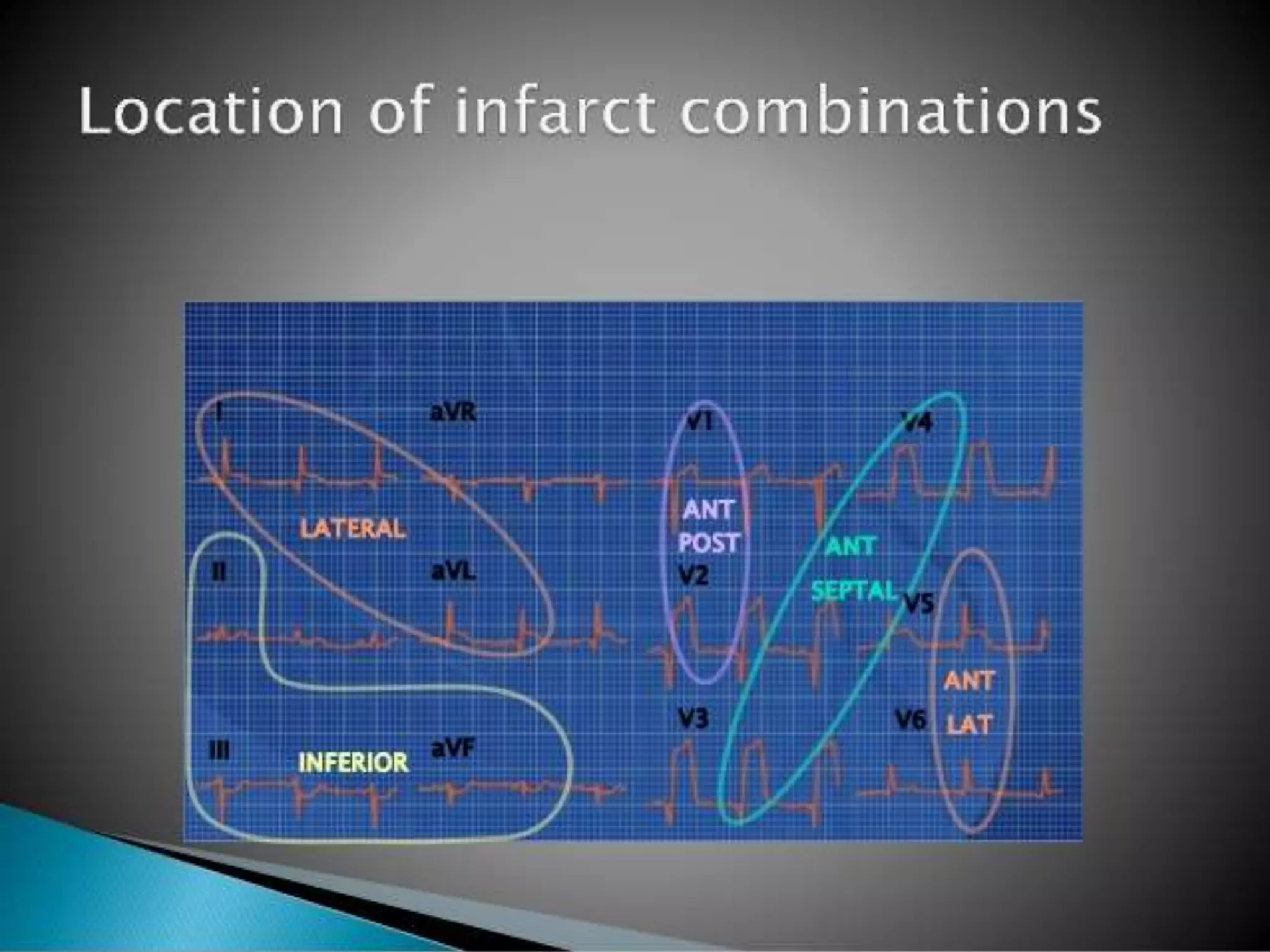
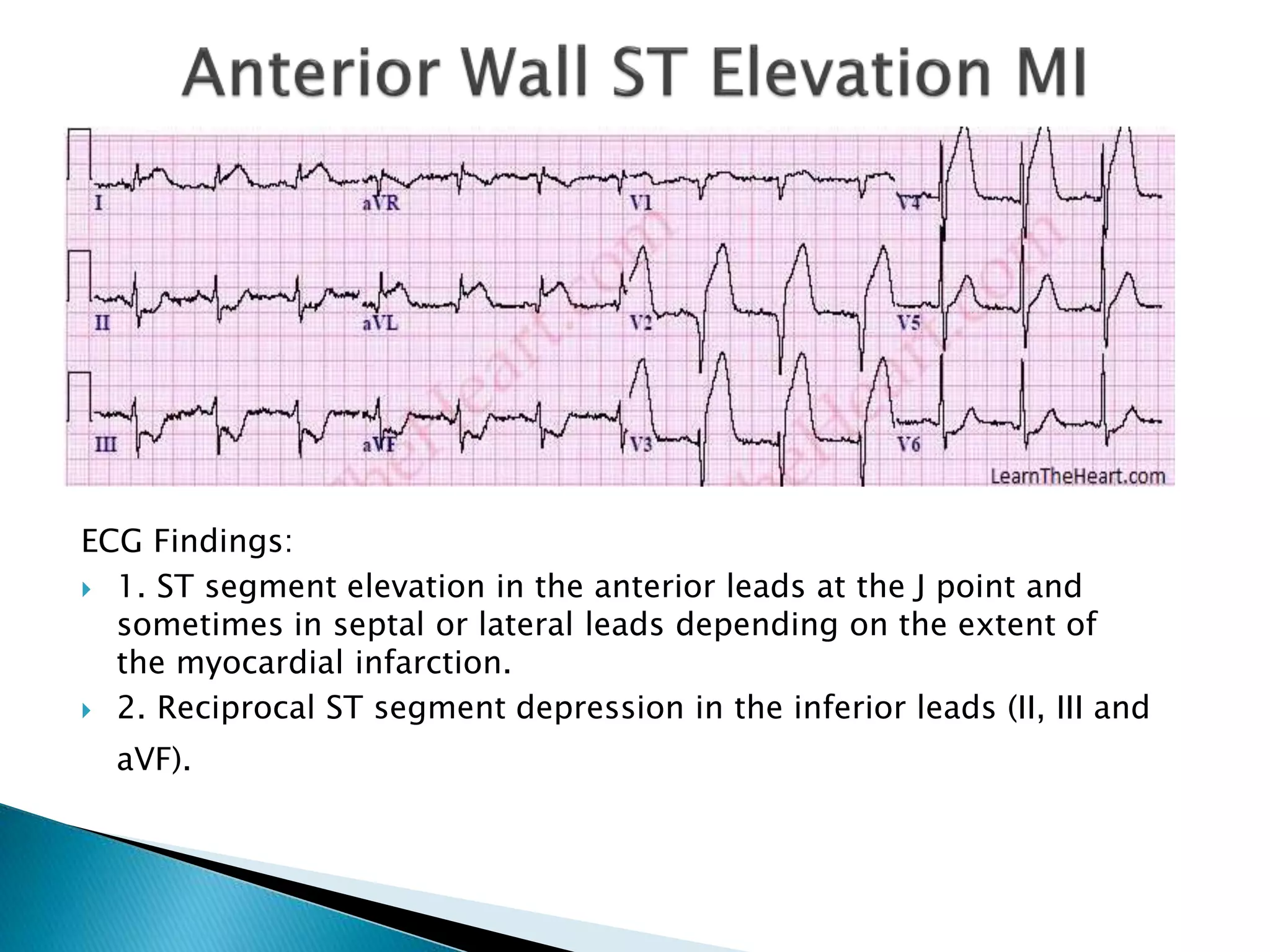
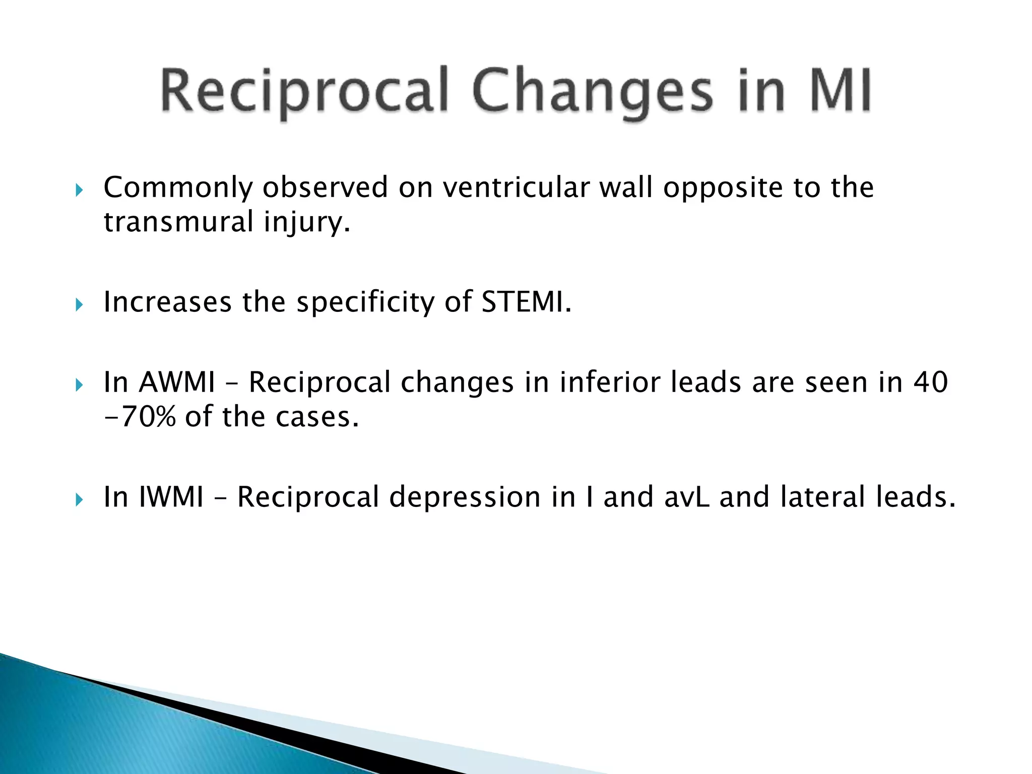
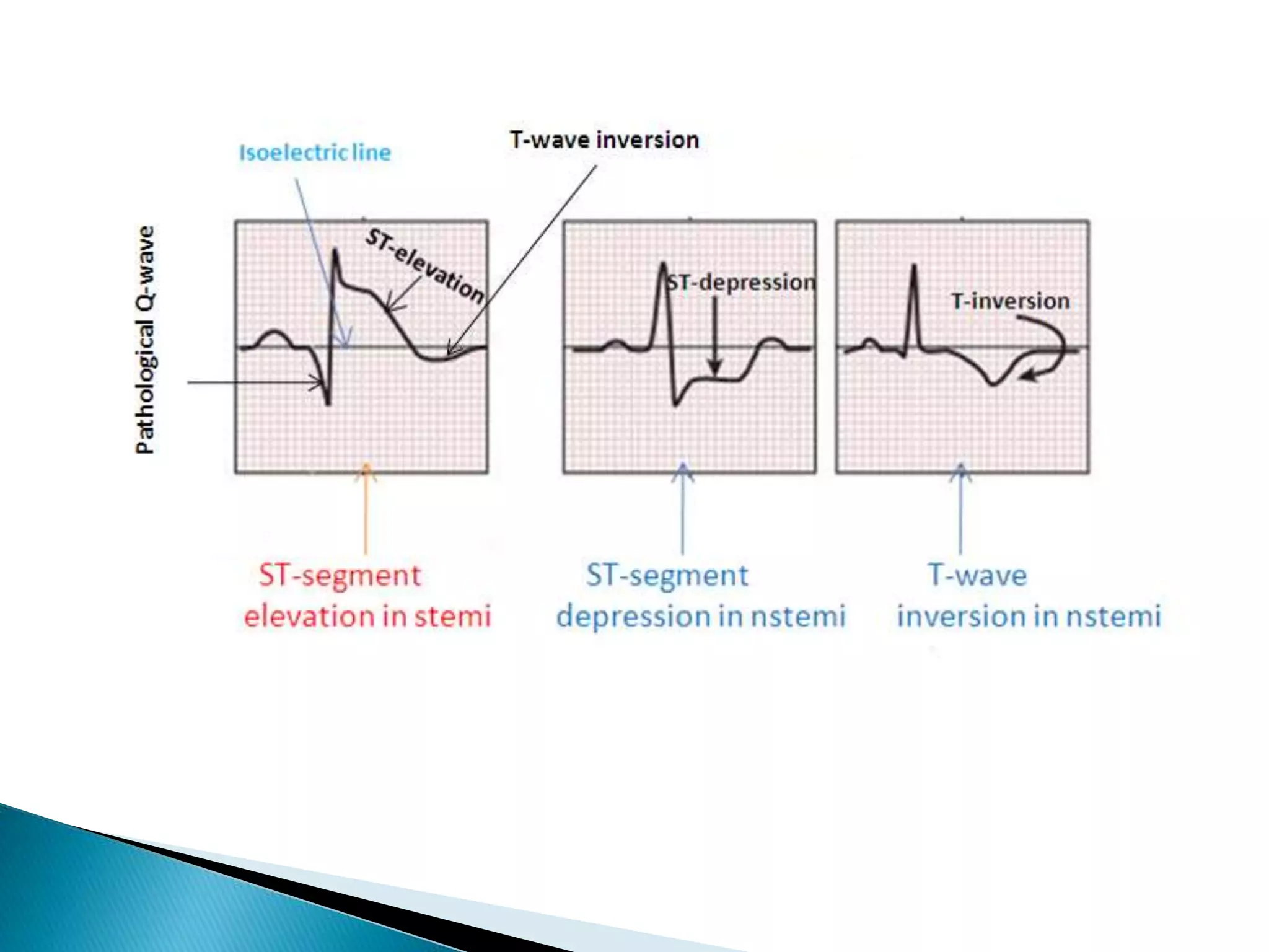
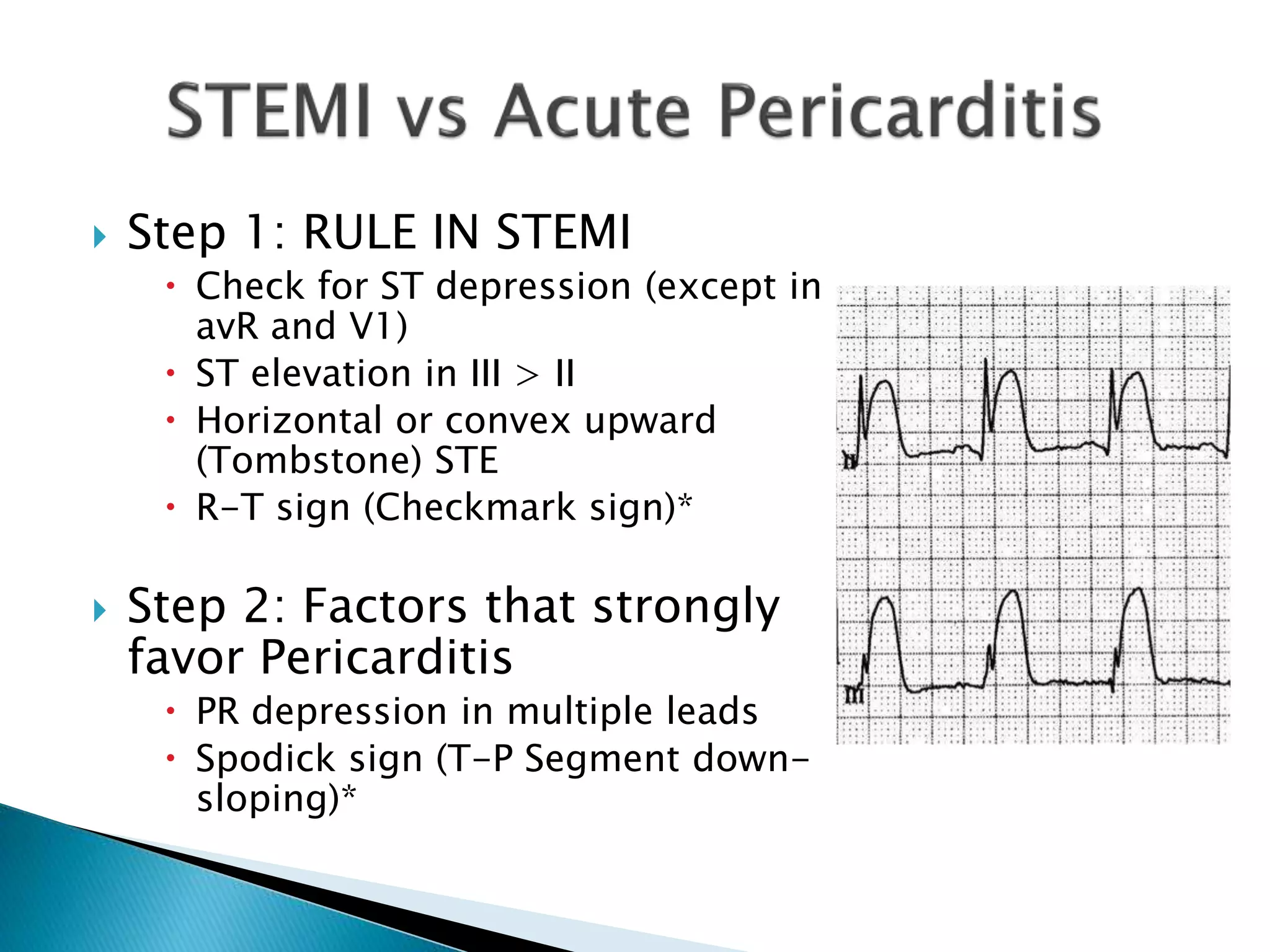
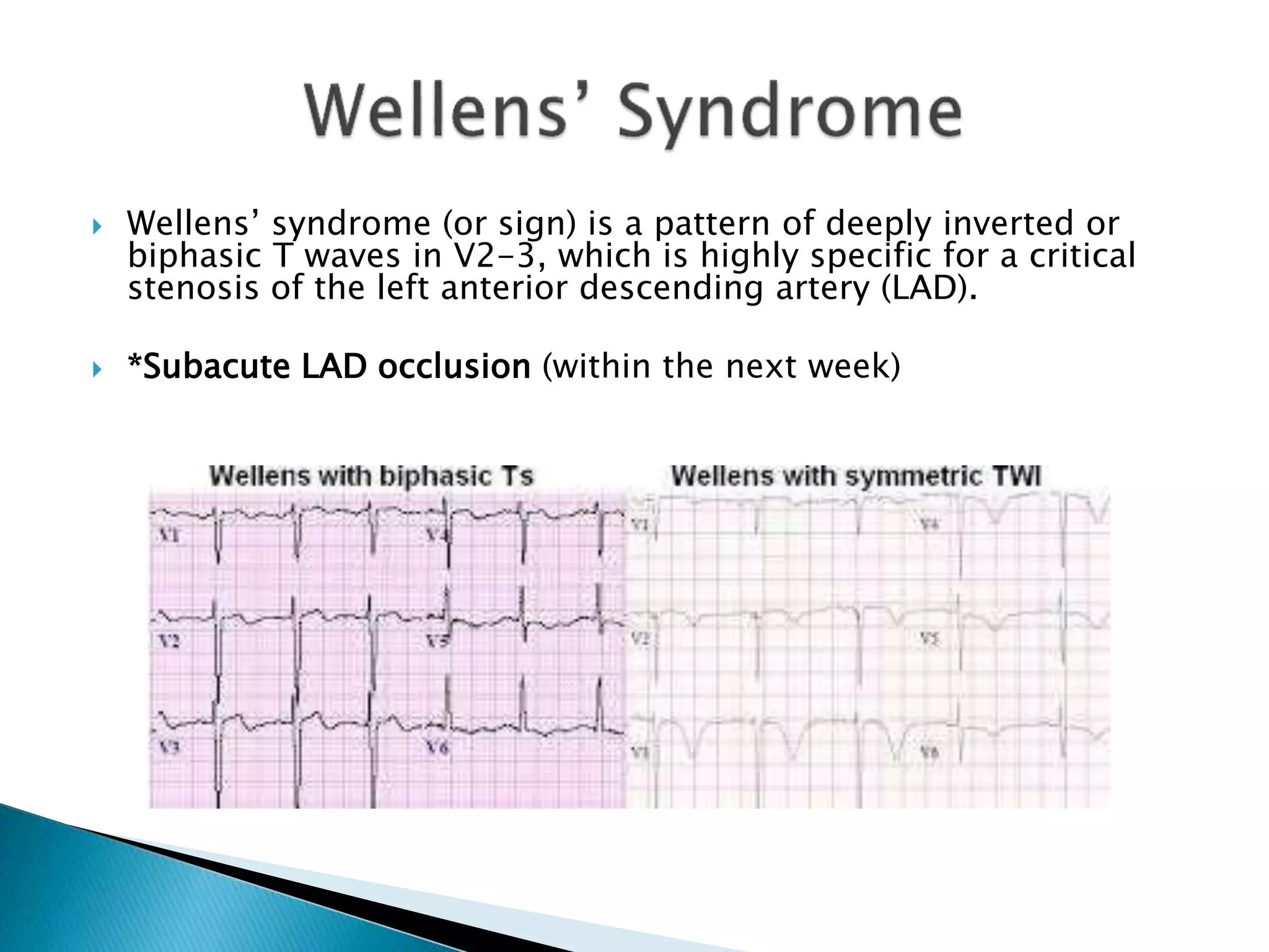
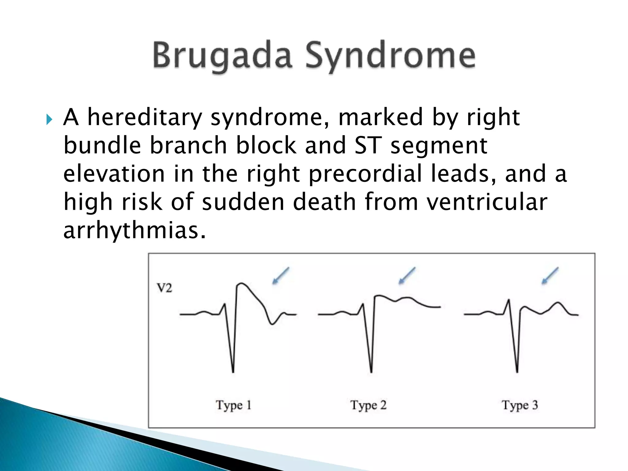
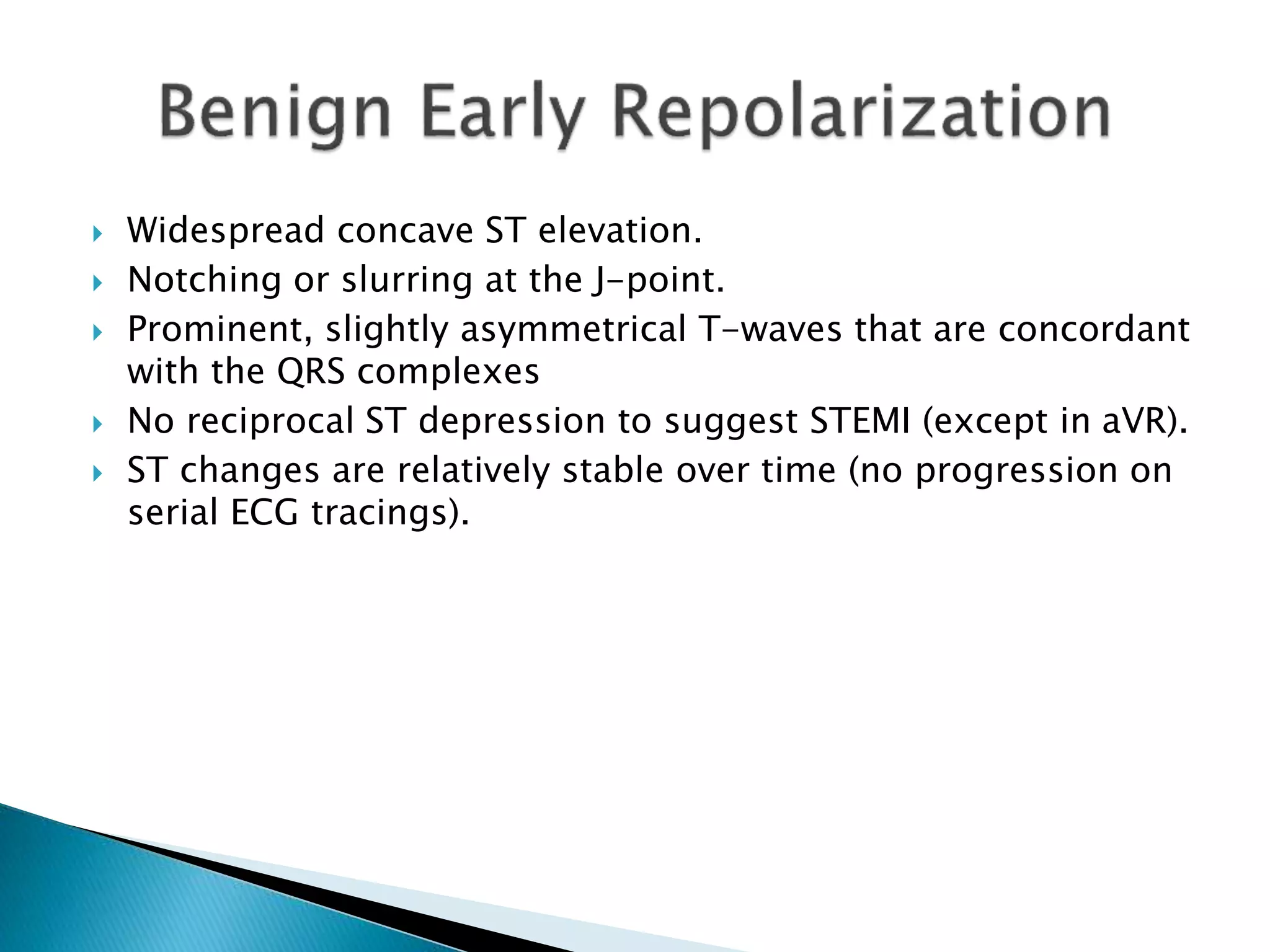
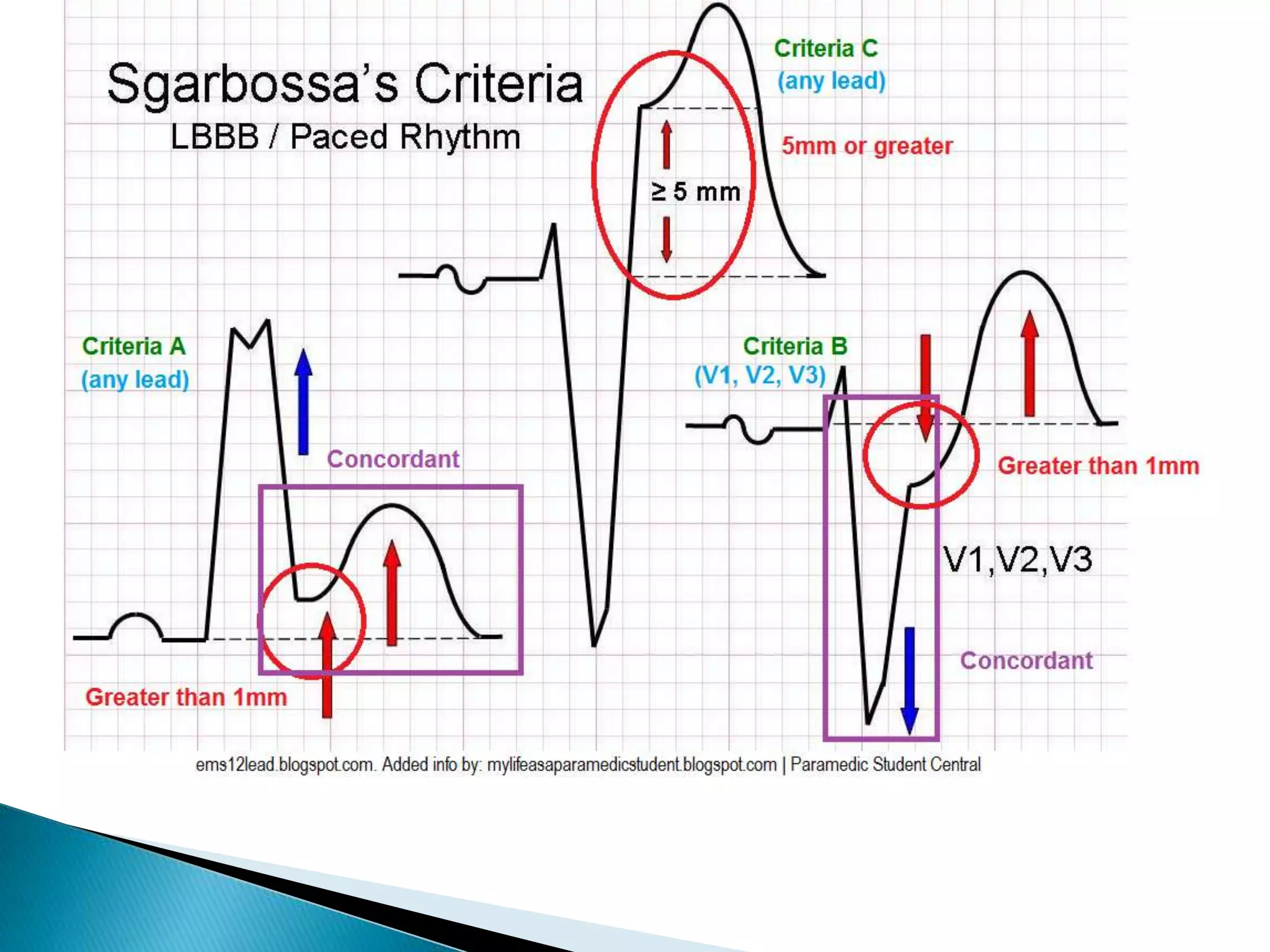
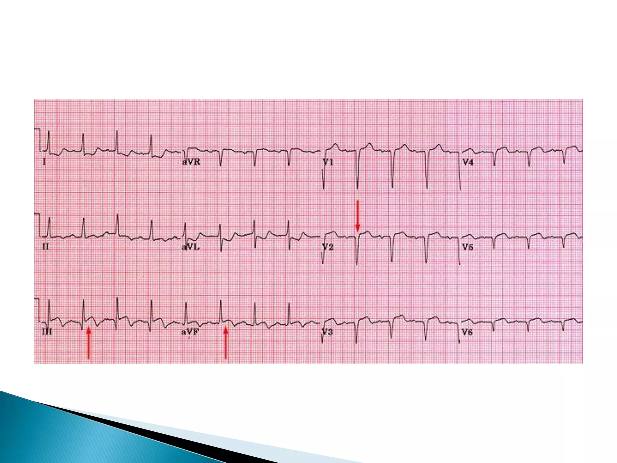
Adeboye Oluwajuyitan
 ST segment elevation over area of damage
 ST depression in leads opposite infarction
 Inverted T waves
 Pathological Q waves
 Know what to look for-
◦ ST elevation > 1mm
◦ 2 contiguous leads
 Know where to look-
◦ I, AVL, V5, V6 – Lateral
◦ V1 V2 V3 V4 - Anterior
◦ II, III, AVF - Inferior
 According to the ACC/AHA guidelines for STEMI, there must
be “New ST elevation at the J point in at least 2 contiguous
leads of ≥ 2 mm (0.2 mV) in men or 1.5 mm (0.15 mV) in
women in leads V2–V3
and/or
 of ≥1 mm (0.1 mV) in other contiguous chest leads or the
limb leads.”
 Thus, 1 mm in any 2 contiguous leads EXCEPT leads V2 or V3
where the elevation must be 2 mm in men or 1.5 mm in
women.
ECG Findings:
 1. ST segment elevation in the anterior leads at the J point and
sometimes in septal or lateral leads depending on the extent of
the myocardial infarction.
 2. Reciprocal ST segment depression in the inferior leads (II, III and
aVF).
ECG findings:
 ST segment elevation in the inferior leads (II, III, and aVF)
 Reciprocal ST segment depression in the lateral and/or high
lateral leads (I, aVL, V5 and V6)
ECG findings:
 ST segment depression in the septal and anterior precordial
leads (V1 to V4).
 The ratio of the R wave to the S wave in leads V1 or V2 is > 1.
 ST elevation in the posterior leads of a posterior ECG (leads
V7 to V9).
 ST elevation in the inferior leads (II, III, and aVF) may be seen
if an inferior MI is also present.
 Commonly observed on ventricular wall opposite to the
transmural injury.
 Increases the specificity of STEMI.
 In AWMI – Reciprocal changes in inferior leads are seen in 40
-70% of the cases.
 In IWMI – Reciprocal depression in I and avL and lateral leads.
 ECG features can be any of the following:
 1. ST depression (70-80% sensitivity)
 2. T wave inversion (10-20% sensitivity)
 3. Both ST depression and T wave inversion
 4. Normal ECG
 Step 1: RULE IN STEMI
 Check for ST depression (except in
avR and V1)
 ST elevation in III > II
 Horizontal or convex upward
(Tombstone) STE
 R-T sign (Checkmark sign)*
 Step 2: Factors that strongly
favor Pericarditis
 PR depression in multiple leads
 Spodick sign (T-P Segment down-
sloping)*
 Wellens’ syndrome (or sign) is a pattern of deeply inverted or
biphasic T waves in V2-3, which is highly specific for a critical
stenosis of the left anterior descending artery (LAD).
 *Subacute LAD occlusion (within the next week)
 ECG abnormality
described by de
Winter et al. in 1998
 Suspicious for Acute
LAD occlusion
 Characterized by 1-3
mm of ST-depression
with upright,
symmetrical T-waves
 A hereditary syndrome, marked by right
bundle branch block and ST segment
elevation in the right precordial leads, and a
high risk of sudden death from ventricular
arrhythmias.
 Widespread concave ST elevation.
 Notching or slurring at the J-point.
 Prominent, slightly asymmetrical T-waves that are concordant
with the QRS complexes
 No reciprocal ST depression to suggest STEMI (except in aVR).
 ST changes are relatively stable over time (no progression on
serial ECG tracings).
 Osborne Waves (= J waves)
 Prolonged PR, QRS and QT intervals
 Shivering artefact
 Ventricular ectopy
 Cardiac arrest due to VT, VF or asystole
 STelevation ≥1 mm in a lead with a positive QRS complex (ie:
concordance) - 5 points
 ST depression ≥1 mm in lead V1, V2, or V3 - 3 points
 ST elevation ≥5 mm in a lead with a negative (discordant) QRS
complex - 2 points
 ≥3 points = 90% specificity and 20% sensitivity
 Pathologic Q waves tend to be deeper and wider, and as
with ST/T wave changes should be seen in at least 2
contiguous (neighbouring) leads.
 ST elevation seen > 2 weeks following an acute myocardial
infarction.
 Most commonly seen in the precordial leads.
 May exhibit concave or convex morphology.
 Usually associated with well-formed Q- or QS waves.
 T-waves have a relatively small amplitude in comparison to
the QRS complex
 Factors favouring Left Ventricular Aneurysm
 ECG identical to previous ECGs (if available).
 Absence of dynamic ST segment changes.
 Absence of reciprocal ST depression.
 Factors favouring Acute STEMI
 New ST changes compared with previous ECGs.
 Dynamic / progressive ECG changes — the degree of ST elevation increases
on serial ECGs.
 Reciprocal ST depression.
 High clinical suspicion of STEMI — ongoing ischaemic chest pain, sick-
looking patient (e.g. pale, sweaty), haemodynamic instability.
Case
(Dr. Baruch Fertel, Cleaveland Clinic)
 36 yo smoker with chest pain *4 hours
◦ Sharp pain
◦ Worse sitting back
◦ Better sitting forward
◦ No radiation
◦ No relief with NTG
ECG Changes in Myocardial Infarction

ECG Changes in Myocardial Infarction

  • 1.
  • 2.
     ST segmentelevation over area of damage  ST depression in leads opposite infarction  Inverted T waves  Pathological Q waves
  • 3.
     Know whatto look for- ◦ ST elevation > 1mm ◦ 2 contiguous leads  Know where to look- ◦ I, AVL, V5, V6 – Lateral ◦ V1 V2 V3 V4 - Anterior ◦ II, III, AVF - Inferior
  • 4.
     According tothe ACC/AHA guidelines for STEMI, there must be “New ST elevation at the J point in at least 2 contiguous leads of ≥ 2 mm (0.2 mV) in men or 1.5 mm (0.15 mV) in women in leads V2–V3 and/or  of ≥1 mm (0.1 mV) in other contiguous chest leads or the limb leads.”  Thus, 1 mm in any 2 contiguous leads EXCEPT leads V2 or V3 where the elevation must be 2 mm in men or 1.5 mm in women.
  • 7.
    ECG Findings:  1.ST segment elevation in the anterior leads at the J point and sometimes in septal or lateral leads depending on the extent of the myocardial infarction.  2. Reciprocal ST segment depression in the inferior leads (II, III and aVF).
  • 8.
    ECG findings:  STsegment elevation in the inferior leads (II, III, and aVF)  Reciprocal ST segment depression in the lateral and/or high lateral leads (I, aVL, V5 and V6)
  • 10.
    ECG findings:  STsegment depression in the septal and anterior precordial leads (V1 to V4).  The ratio of the R wave to the S wave in leads V1 or V2 is > 1.  ST elevation in the posterior leads of a posterior ECG (leads V7 to V9).  ST elevation in the inferior leads (II, III, and aVF) may be seen if an inferior MI is also present.
  • 14.
     Commonly observedon ventricular wall opposite to the transmural injury.  Increases the specificity of STEMI.  In AWMI – Reciprocal changes in inferior leads are seen in 40 -70% of the cases.  In IWMI – Reciprocal depression in I and avL and lateral leads.
  • 16.
     ECG featurescan be any of the following:  1. ST depression (70-80% sensitivity)  2. T wave inversion (10-20% sensitivity)  3. Both ST depression and T wave inversion  4. Normal ECG
  • 18.
     Step 1:RULE IN STEMI  Check for ST depression (except in avR and V1)  ST elevation in III > II  Horizontal or convex upward (Tombstone) STE  R-T sign (Checkmark sign)*  Step 2: Factors that strongly favor Pericarditis  PR depression in multiple leads  Spodick sign (T-P Segment down- sloping)*
  • 21.
     Wellens’ syndrome(or sign) is a pattern of deeply inverted or biphasic T waves in V2-3, which is highly specific for a critical stenosis of the left anterior descending artery (LAD).  *Subacute LAD occlusion (within the next week)
  • 23.
     ECG abnormality describedby de Winter et al. in 1998  Suspicious for Acute LAD occlusion  Characterized by 1-3 mm of ST-depression with upright, symmetrical T-waves
  • 24.
     A hereditarysyndrome, marked by right bundle branch block and ST segment elevation in the right precordial leads, and a high risk of sudden death from ventricular arrhythmias.
  • 26.
     Widespread concaveST elevation.  Notching or slurring at the J-point.  Prominent, slightly asymmetrical T-waves that are concordant with the QRS complexes  No reciprocal ST depression to suggest STEMI (except in aVR).  ST changes are relatively stable over time (no progression on serial ECG tracings).
  • 28.
     Osborne Waves(= J waves)  Prolonged PR, QRS and QT intervals  Shivering artefact  Ventricular ectopy  Cardiac arrest due to VT, VF or asystole
  • 29.
     STelevation ≥1mm in a lead with a positive QRS complex (ie: concordance) - 5 points  ST depression ≥1 mm in lead V1, V2, or V3 - 3 points  ST elevation ≥5 mm in a lead with a negative (discordant) QRS complex - 2 points  ≥3 points = 90% specificity and 20% sensitivity
  • 31.
     Pathologic Qwaves tend to be deeper and wider, and as with ST/T wave changes should be seen in at least 2 contiguous (neighbouring) leads.
  • 33.
     ST elevationseen > 2 weeks following an acute myocardial infarction.  Most commonly seen in the precordial leads.  May exhibit concave or convex morphology.  Usually associated with well-formed Q- or QS waves.  T-waves have a relatively small amplitude in comparison to the QRS complex
  • 35.
     Factors favouringLeft Ventricular Aneurysm  ECG identical to previous ECGs (if available).  Absence of dynamic ST segment changes.  Absence of reciprocal ST depression.  Factors favouring Acute STEMI  New ST changes compared with previous ECGs.  Dynamic / progressive ECG changes — the degree of ST elevation increases on serial ECGs.  Reciprocal ST depression.  High clinical suspicion of STEMI — ongoing ischaemic chest pain, sick- looking patient (e.g. pale, sweaty), haemodynamic instability.
  • 36.
    Case (Dr. Baruch Fertel,Cleaveland Clinic)  36 yo smoker with chest pain *4 hours ◦ Sharp pain ◦ Worse sitting back ◦ Better sitting forward ◦ No radiation ◦ No relief with NTG