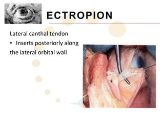
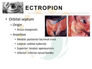
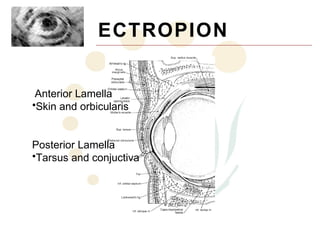
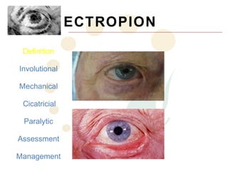
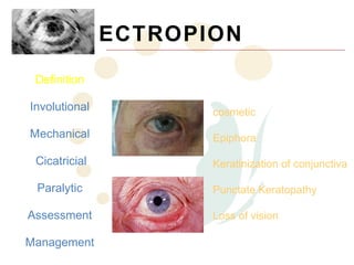
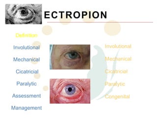
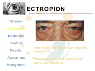
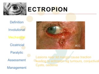
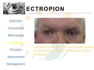
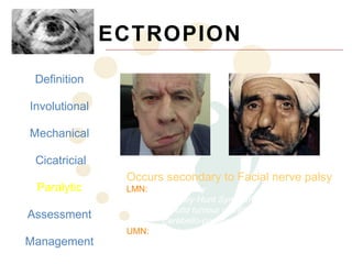
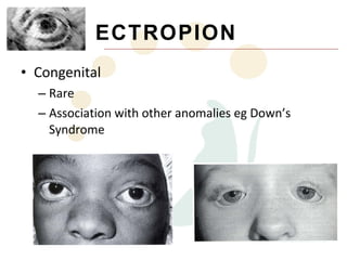
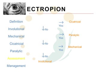
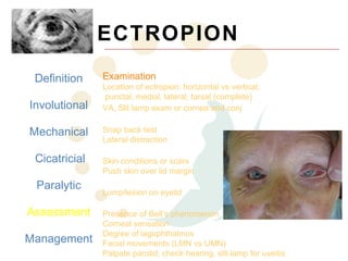
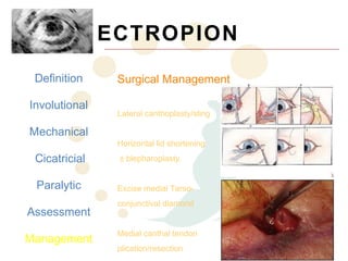
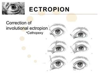
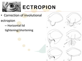
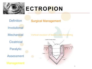
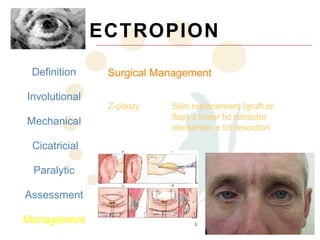
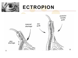
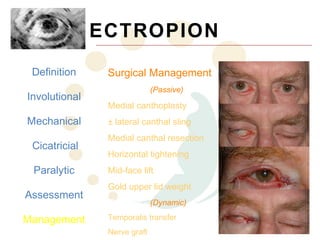
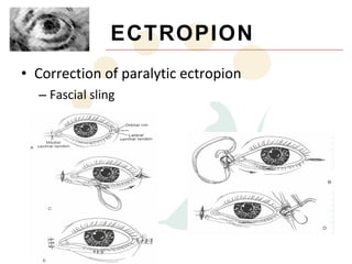
Ectropion is the abnormal eversion of the eyelid away from the globe. It can be involutional, mechanical, cicatricial, or paralytic in nature. Examination involves assessing for corneal exposure, lid laxity, lesions, and facial nerve function. Management includes lubricants, taping, scar massage, and various surgical techniques depending on the underlying cause such as lateral tarsal strip surgery for involutional ectropion or fascial sling for paralytic ectropion.
Indication, contraindication, advantage, disadvantage, types of keratoplasty, complication of keratoplasty and management, corneal graft rejection and failure
M.S ophthalmology, sarojini devi eye hospital, regional institute of ophthalmology, osmania medical college, hyderabad, telangana
Indication, contraindication, advantage, disadvantage, types of keratoplasty, complication of keratoplasty and management, corneal graft rejection and failure
M.S ophthalmology, sarojini devi eye hospital, regional institute of ophthalmology, osmania medical college, hyderabad, telangana
Eyelid laceration repair with defects.pptxSHAYRI PILLAI
PRINCIPLES OF EYELID REPAIR
Wounds should be copiously irrigated and explored, with the removal of any foreign material after local anesthesia
Reconstruction should be done in layers as per correct anatomical orientation
Wounds should not be extended to explore structures unless the exploration is for suspected foreign body
The orbital septum if damaged should never be repaired-result incompromised eyelid excursion and even lagophthalmos
LASIK or Lasik (laser-assisted in situ keratomileusis), commonly referred to as laser eye surgery or laser vision correction, is a type of refractive surgery for the correction of myopia, hyperopia, and an actual cure for astigmatism, since it is in the cornea. LASIK surgery is performed by an ophthalmologist who uses a laser or microkeratome to reshape the eye's cornea in order to improve visual acuity. For most people, LASIK provides a long-lasting alternative to eyeglasses or contact lenses.
The planning and analysis of corneal reshaping techniques such as LASIK have been standardized by the American National Standards Institute, an approach based on the Alpins method of astigmatism analysis. The FDA website on LASIK states,
"Before undergoing a refractive procedure, you should carefully weigh the risks and benefits based on your own personal value system, and try to avoid being influenced by friends that have had the procedure or doctors encouraging you to do so."
The procedure involves creating a thin flap on the eye, folding it to enable remodeling of the tissue beneath with a laser and repositioning the flap.
Ectropion
It is an outward turning of the eyelid margin . This more frequently affects the lower eyelid.Upper eyelid ectropion is uncommon.Classified in 5 types
1)Congenital 2) Involutional 3) Paralytic 4) Cicatricial 5) Mechanical
Involutional ectropion is more common.Congenital ectropion is very rare.
Symptoms Epiphora :- excessive tearing.Excessive dryness.
Foreign body sensation Irritation.Burning.Redness.Chronic conjunctivitis KeratinizationCorneal exposure
Grading
Lid margin is out rolled and depending on out rolling ectropion can be classified as under:
Grade I –only punctum is everted
Grade II –lid margin is everted and palpebral conjunctiva is visible
Grade III –fornix is also visible
Etiological factors
Horizontal lid laxity:-can be demonstrated by pulling the central part of the lid 8 mm or more from the globe, with a failure to snap back to its normal position on release without the patient first blinking.
Medial canthal tendon laxity
demonstrated by pulling the lower lid laterally and observing the position of the inferior punctum If the lid is normal the punctum should not be displaced more than 1–2 mm
Lateral canthal tendon laxity
characterized by a rounded appearance of the lateral canthus and the ability to pull the lower lid medially more than 2 mm.
>Normally, the displacement should only be 0-2 mm.
Treatment
1 medical therapy
2 surgical therapy
10. ECTROPIO
N
• Definition
– Abnormal eversion of the eyelid away from the
globe
• Key Features:
– Eyelid margin and lash drive are turned away from
the cornea
– Conjuctival surface is exposed, which can lead to
keratinisation of the epithelium
– Corneal exposure results in foreign body
sensation, corneal dryness and ulceration
11. ECTROPION
Definition
Involutional cosmetic
Mechanical Epiphora
Cicatricial Keratinization of conjunctiva
Paralytic Punctate Keratopathy
Assessment Loss of vision
Management
19. ECTROPION
Definition Full Ocular History
Involutional Onset and duration of signs/symptoms
Mechanical Trauma/burns/surgery
Cicatricial Patient’s concerns
Paralytic
Assessment
Management
20. ECTROPION
Definition Examination
Location of ectropion: horizontal vs vertical,
punctal, medial, lateral, tarsal (complete)
Involutional VA, Slit lamp exam or cornea and conj
Involutional
Mechanical Snap back test
Lateral distraction
Cicatricial
Cicatricial Skin conditions or scars
Push skin over lid margin
Paralytic Mechanical
Lump/lesion on eyelid
Paralytic
Assessment Presence of Bell’s phenomenon
Corneal sensation
Degree of lagophthalmos
Management Facial movements (LMN vs UMN)
Palpate parotid, check hearing, slit-lamp for uveitis
21. ECTROPION
Definition
Involutional Non-operative Management
Lumbricants for corneal exposure/
Mechanical conjunctival keratinization
Taping lateral canthal skin superotemporally
Cicatricial
Scar (cicatricial) massage, ? Steroid injection
Paralytic Taped lid weights for Facial nerve palsy
Advice on wiping tears (up and in toward
Assessment nose)
Management
Dense fibrous tissue that imparts structural integrity to the eyelids. Medially and laterally they taper to 2mm in height as they pass into the canthal tendons. Contain meibomian glands (25 upper, 20 lower) that as not associated with lash follicles..produce lipid layer of the precornal tear film
Striated muscles divided into 3 parts. Orbital overlies the bony orbital rim. Arise from insertions on the frontal process of maxillary bone, orbital process of frontal, medial canthal tendon. Palpebral overlies the mobile eyelid from the orbital rim to the eyelid margins. Fixed medially and laterally to canthal tendons. Preseptal over septum, arise from upper and lower borders of medial canthal tendon and insert along the lateral horizontal raphe. Pretarsal overlies tarsal plates from MCT via superficial and deep heads arc around the lids and insert into LCT..contraction aids lacrimal pump
Tarsal plates pass in to fibrous bands and form the crura of the MCT lies between orb oculi (ant) and conjuctiva (post).. The superior and inferior crura fuse to form stout common tendon that inserts via 3 limbs. Anterior onto orbital process of the maxillary bone infront of and above anteror lacrimal crest. Posterior limb passes between the canaliculi and inserts onto the posterior lacrimal crest. Superior limb arises from both ant and post limbs, inserts onto orbital process of frontal bone..posterior head of preseptal orb oculi inserts onto this limb….forms roof of lacrimal sac fossa
Laterally the tarsal pates pass into not very well developed fibrous strands that becomes crura of LCT..distinct entity separate from orb oculi. Inserts posteriorly along the lateral orbital wall…where t blends with strands of the lateral check ligament from the sheath of the lateral rectuc mm
Thin fibrous multilayered membrane begins at arcus marginalis along the orboital rim and represents a coninuation of the orbital fascial system.
Ectropion may be defined as eversion of the eyelid or when the lid rolls out away from the globe. It ranges in severity from mild punctal malposition (such as this case - note the puncta has moved out away from the globe - in this case due to medial canthal tendon laxity) to involvement of the whole lid (referred to as a tarsal ectropion.
Individuals will seek medical assistance for ectropion because it is unsightly, because they get epiphora, keratinization of conj may occur with chronic exposure, they may develop punctate keratopathy and at its extreme loss of vision
It is important to work out the cause of the ectropion as this will help direct your management. Causes of ectropion may classified as involutional, mechanical, cicatrical, paralytic or congenital
Involutional ectropion results from age related degenerative changes to the eyelid. Either medial or lateral canthal tendons become weak as does pre-tarsal orbicularis. This gentleman also has other evidence of tissue weakness (orbital fat pad herniation).
Mechanical ectropion occurs when a tractional force is applied to the lid by a discrete lesion. In this case, a BCC is pulling the lower lid down and out.
Cicatrical ectropion occurs when there is deficiency of anterior lamella tissue (including skin) on the eyelid. It may result from trauma or burns, skin conditions or may be iatrogenic (eg. Blephroplasty). In this case, the patient suffers from eczma affecting the face - you can appreciate how tight the eyelid tissue is.
Paralytic ectropion occurs from a facial nerve palsy. Loss of muscle tone and weakening contraction of orb oculi
This is a modified algorithm from Richard Collin’s book on eyelid surgery to assist in sorting out the cause of ectropion. Essentially you need only ask 3 questions to get the answer. 1. Is there shortage of skin? - if yes then they have cicatrical. If not, then 2. Do they have abnormal eyelid closure? - if yes then they have paralytic. If no, then 3. Is there a lump on the lid? - if yes then it is mechanical and if not then it must be involutional.
As with everything, history is an important part of the assessment.
MCT laxitywatch the displacement of the punctum on lateral excursion…shouldn’t be displaced greater than 1-2mm. Pulling lid>10mm from globe suggests horizontal laxity and if slow to return to position then positive snap test. Punctum usually just lateral to caruncle and below superior punctum. Also shouldn’t be visible as usually faces posteriorly towards the globe
Definitive management of ectropion is with surgery but there are some temporizing measures that may be of use: lubricants if cornea exposed, taping lateral canthus, massaging scar or steroid injection, taping wieghts to upper lid in paralytic ectropion may improve lagophthalmos, wiping tears in an up and in inward direction may reduce exacerbation of ectropion.
Surgical management differs depending on the cause. In involutional ectropion, the aim is to correct horizontal lid laxity. This may be achieved by lateral canthoplasty or sling. Horizontal lid shortening, by removing an full thickness wedge, is sometimes used. There is also either medial tarsoconjunctival (diamond shaped segment of tarsus and conjuctiva is resected directly below the punctum) or medial canthal tendon plication…plication of anterior limb or resection for medial ectropion. In resection you resect the MCT structres combined with horizontal lid shortening, then the posterior limb is recontructed with a permanent suture and the cut inferior canaliculus is marsupialised into the conjuctival sac of the lower fornix
Mechanical ectropion is usually corrected by removing the offending lesion using a vertical excision.
The aim of surgery for cicatricial ectropion is to increase the vertical height of the lid. This may be achieved with either a Z-plasty that both shorthens the lid horizontally and lengthens vertically, or skin replacement usually with lower lid retractor reinsertion.
Surgical management of paralytic ectropion may be thought of in terms of passive and dynamic procedures. Passive procedurs include medial canthoplasty with lateral canthal sling, medial canthal tendon resection or if paralysis is severe mid face lift may be of benefit. dynamic procedures, include such things as temporalis transfer and or nerve grafting and are aimed at improving lid function.