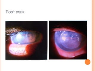
Indication, contraindication, advantage, disadvantage, types of keratoplasty, complication of keratoplasty and management, corneal graft rejection and failure
M.S ophthalmology, sarojini devi eye hospital, regional institute of ophthalmology, osmania medical college, hyderabad, telangana
3. DEFINITION
Keratoplasty refers to replacement of diseased host
corneal tissue by healthy donor cornea.
Full thickness (Penetrating keratoplasty)
Partial thickness (Anterior and Posterior Lamellar
keratoplasty
4. GENERAL INDICATIONS
Optical
To restore vision by obtaining clear visual axis
Tectonic
To restore corneal integrity and tectonic support in
severe structural changes
Therapautic
To remove the infected corneal tissue who are
unresponsive to antimicrobial therapy
Cosmetic
Rare
To improve appearance of eye with whitish corneal scar
7. PREOP EVALUATION
General systemic evaluation – HTN, DM , cardiac disease
Patient socioeconomic status and compliance for follow up
Ocular history of present & past illness and ocular
surgeries
8. Ocular examination of eye and adnexa – slit lamp, details
of corneal vascularization, tear film assessment, IOP,
presence of cataract
If anterior segment details are not visible-
Ant-seg OCT
Posterior segment evaluation – Bscan
9. PREOPERATIVE PREPARATION
Antiinfective agent – reduce the risk of endophthalmitis,
appropriate lid care
IOP control – good lid and extaocular akinesia, use of
mannitol preoperatively
Pupil –not dilated, constricting the pupil with 2% pilocarpine
intraoperatively maybe done to reduce lens damage during
trephining of phakic eye
10. PENETRATING KERATOPLASTY
Full thickness Replacement of corneal tissue with healthy
donor graft
INDICATIONS(Optical & Therapeutic)
Pseudo/aphakic corneal decompensation
Stromal corneal dystrophy
Granular, lattice, macular, Schnyder central crystallin,
central cloudy dystrophy
13. Corneal degeneration
Failed corneal graft
Non healing infectious keratitis,
Infectious keratitis with perforation,
Post chemical injury and melt
15. ADVANTAGE
Full thickness transplant
No interface related visual problem
Ability to treat all layers
Anterior segment reconstruction in single procedure
16. DISADVANTAGE
Corneal surface disease or neurotrophic cornea
leads to prolonged healing or persistent epithelial
defect.
Irregular or significant regular astigmatism
Difficulty in determining anterior corneal curvature
17. S/P OKP 2ND POD FOR RE CENTRAL CORNEAL
OPACITY
24. Intraoperative complication
Scleral perforation d/t bridle suture or sclear fixation ring
Trephination problem
Small size trephine of donor cornea leads to improper water
tight closure
Flat cornea
Hyperopia
Angle compression leading to raised IOP.
Eccentric trephination leads to increased astigmatism and
rejection
Improper trephination leads to endothelial damage d/t
repunching
25. Iridolenticular damage – avoided by globe hypotony, Ac
maintained with viscoelastics, miotics
Endothelial damage d/t improper trephination, improper
tissue handling, iris,lens,IOL touch during surgery.
Intra ocular haemorrage bleeding into AC d/t corneoiridic
scar in post infection, post traumatic case, Iol
explantation in case of PBK, iridodialysis or synechiolysis
at the the angle
Vitreous loss in case of combined cataract extraction and
keratoplasty
26. Postoperative complication
Immediate
Wound leak leads to shallow AC with low IOP.
Persistent epithelial defect and filamentary keratitis
Postoperative inflammation
Suture related infiltrates suture induced vascularization
Raised IOP
Pupillary block
Anterior synechiae formation
Choroidal detachment /haemorrhage
27.
28. Late postoperative complications
Post PK astigmatism
Graft infection
Graft rejection
Post PK glaucoma
Retrocorneal membrane formation
Late wound dehiscence
29. DEEP ANTERIOR LAMELLAR KERATOPLASTY
DALK – corneal tissue is removed almost to level of
Descemet membrane and replaced with donor
corneal tissue.
31. CONTRAINDICATIONS
Absolute:
Endothelial dysfunction – posterior dystrophies, corneal
edema(pseudophakic and aphakic), ICE syndrome
Relative:
Epithelial dysfunction- limbal stem cell deficiency, chronic
surface disease
The big-bubble technique is contraindicated if there is
a pre-existing break in the Descemet’s membrane (post
hydrops) or
there are deep scars (however small) involving the
Descemet’s membrane
32. ADVANTAGE
Non penetrating surgery –less risk of Intraocular
complications
Selective removal of pathology
More rapid visual rehabilitation
Early removal of suture, Less need of suture and wound
induced astigmatism
Minimal requirement of donor tissue
Reduced incidence of graft rejection retains normal recipient
endothelial layer
Preservation of globe integrity
33. DISADVANTAGE
Irregular or significant regular astigimatism
Irregular interface
Ocular surface disease leads to persistent epithelial
defect
Stromal opacification
Interface debris
DALK is technically demanding and time consuming.
47. Intraoperative complications
Poor microkeratome dissection of donor tissue in DSEK
Inability to strip host DM & endothelium in DMEK
Retained DM.
Excessive manipulation of donor tissue leading to cell
loss and possible primary graft failure
Tearing of donor tissue
Interface debris or blood
Choroidal hemorrhage (lower than PK)
Inversion of donor lenticule
53. DSEK DSAEK
Manual dissection- Increased donar
tissue perforation
Automated dissection – less risk of
perforation
Do not yield smooth anterior surface of
donor posterior lamella
Yields good post donor lamella of
superior optical quality
More time consuming and visual
recovery is slow
Less time consuming and visual
recovery is rapid
Adhesion is better because of greater
thickness& irregular anterior surface
Adhesion is not easy because of thin
and smooth anterior surface
Donor lenticule dislocation is less more
54. CORNEAL GRAFT FAILURE
Corneal graft is considered failed
if it fails to retain optical clarity,
if there is no impact on therapeutic attempts,
if there is severe astigmatism that could not be optically
corrected -resulting in regraft
55. CAUSES OF GRAFT FAILURE
Allograft rejection 29.2%
Post pk glaucoma 16.9%
Infection excluding endophthalmitis 15.4%
Surface problem 12.7%
Primary graft failure 6.6%
Endothelial decompensation 3.8%
Endophthalmitis 2.7%
Recurrence of host disease 2.0%
Severe astigmatism 0.6%
56. GRAFT FAILURE (2 TO 45%)
It should be differentiated from graft rejection
Significant edema of donor tissue in Non-inflamed eye
present in 1st postoperative day and does not resolve
by 2-4 weeks d/t usually not followed by period of
clear cornea
57. Prolonged death or prolonged preservation > 7days
Poor endothelial function
Elderly donor > 70 years
Iatrogenic damage to donor tissue during PKP
Defective preservation media or Intraocular fluids
RISK FACTORS
58. MECHANISM
Hyposecretion of aqueous after PK result in corneal
edema d/t decreased supply of metabolites to
endothelium
Increased positive pressure increases the chance
of contact between endothelium and other
intraocular structure– so preoperative hypotony with
digital massage or IV mannitol is mandatory.
59. Primary and secondary failure
Primary
Eye with well apposed grafts after surgery but with
persistent corneal edema
d/t inadequate endothelial function, traumatic pre or
intra operative technique
To prevent PGF, donor cornea should have atleast
200 cells/mm2 for better chance of graft survivial
60. Secondary graft failure
Donor endothelial tissue is detached from recipient
stromal cornea preventing the cornea from clearing
Causes- residual fluid or viscoelastic in the interface or
retained DM and graft rejection
Management –unresponsive to hypertonic saline or
steriods.
1.Observation for atleast 3 weeks prior to regraft
2. Regraft
61.
62. GRAFT REJECTION
Specific immunologically mediated that may be
irreversible/reversible in which corneal graft is clear
for at least 2 weeks, suddenly succumbs to graft
edema in conjunction with anterior segment
inflammation sign
63. CHARACTERISTICS
Immune mediated resulting in decompensation of
transplanted cornea
3 or more weeks
Inflammatory process limited to graft
Starts at graft margin nearest to proximal blood vessels
Movement of inflammatory reaction towards centre to
involve the entire graft
Increase in corneal thickness( edema) in previously
clear compact graft
64. PATHOLOGY
Cell mediated by CD4+ and CD8+ cells
Delayed Hypersensitivity reaction
Inflammation induce vessels, lymphatics growth into
cornea and attracts APC into central cornea
MHC antigen expression on corneal cells are
upregulated
Recognition of foreign Histocompatiblity antigens leads
to immune cascade afferent immune response
65. Donar antigen processed by host APC
(afferent rejection cascade)
Presented to host immune sytem in
conjunction with HLA II & IL 1
Host mounts immune response
against foreign antigen
(efferent rejection cascade)
Activated Thcells release IL2,
IFN macrophages
Lymphokines cytokines
migration inhibition factor
66. Anterior chamber associated immune deviation
Corneal lymph angiogenesis and ham-
angiogenesis
Lymphocytotoxic antibodies directed against HLA 1
Donor derived Langerhans cells also mediate
rejection
68. EPITHELIAL REJECTION (10 – 14%)
Quiet, asymptomatic subsides in 6to 10 days
Linear pattern from graft host junction at the site of
vascularization with gradual progression towards
center
Elevated undulating line with positive FS and rose
Bengal stain
A/w persistent epithelial defect or epithelial ring
presentation
70. CHRONIC STROMAL REJECTION (2.4 – 15%)
Low grade rejection- risk of development of other
severe Rejection
Subepithelial infiltrate - Krachmer spots for 6
weeks to 21 months
0.2 to 1 mm in diameter either in or immediately
below bowman’s layer
71.
72.
73. HYPERACUTE STROMAL REJECTION
Occurs simultaneously with / immediately following
endothelial rejection
Sudden onset of peripheral full thickness stromal haze in
previously clear graft adjacent to area of vascularization
with rapid progression to center in 24 to 48 hrs
An abscess like picture without hypopyon in early stage
Ciliary congestion a/w epithelial defect
74. CHARACTERISTICS
Migrates away from vascularized part of graft
Growth of vessels into stroma
Mimics corneal abscess in heavily vascularized cornea
Haze may invade adjacent host cornea proximity to
vascularization
Complicated by persistent epithelial defect leading to
stromal necrosis, descemetocele and perforation
75.
76.
77. CHRONIC FOCAL REJECTION/
ENDOTHELIAL REJECTION (2 – 40%)
Most common, severe form which leads to graft failure d/t
significant loss of endothelial cells
Onset within month
78. RISK FACTORS
Direct correlation with degree of vascularization
Young individual with active immune system
Large size /eccentric graft
No of quadrants of anterior synechiae
Preoperative glaucoma
Previous corneal transplant
79. Reversible – responds to corticosteroids, reduction
in edema , restoration of clarity, resolution of
inflammation
Irreversible- endothelial decompensation – graft
failure
80. SIGNS AND SYMPTOMS
Redness, photophobia, irritation, halos around of light and
dimension of vision
Conjuctival hyperemia, Ac reaction
KPs – Diffuse scattered/ lined up in a chain like fashion
forming pathognomonic endothelial rejection line of
khodadoust.
Graft edema, Stromal edema & DM folds
84. PREVENTION
Preoperative measures to minimize antigenic difference by
steroids prophylaxis
Intraoperative – meticulous surgical technique, avoiding
decentration of recipient bed trephination, optimal suturing,
good graft host apposition
Followup, timely suture management- decreases suture
related vascularization and rejection
85. TREATMENT
Corticosteroids – prednisolone acetate / prednisolone
sodium phosphate 1%
Preoperatively- 4times/ 1 week
Post operatively- hrly 3days, 2hrsly 15 days,
4 times for 3 months then tapered 1drop for every 2
months
For acute rejection hourly steroids
Systemic steroid either by oral (60 to 80 mg) or
intravenous route (500mg in 150ml of IV fluid)
87. CORNEAL GRAFT INFECTION
Predisposing factor
Suture related problem
Persistent epithelial defect
Recurrence of HSV keratitis
Ocular surface disorder
88. Graft failure
Use of soft contact lens
Lid abnormalities
Donor tissue contamination
Incomplete excision of infected tissue
89. CLINICAL FEATURES
Peripheral keratitis, ulceration in suture related
Central or paracentral keratitis, ulceration in epithelial
defect
Stromal infiltrate, Ac inflammation
Inferior part is affected – tear film insufficiency/ exposure
keratitis
Herpetic keratitis – dendritic keratitis / non healing epithelial
defect
Infectious crystalline keratopathy
90.
91. INFECTIOUS CRYSTALLINE KERATOPATHY
• Noninflammatory, intrastromal bacterial colonization of a
corneal graft
• α-hemolytic streptococci of the viridans
• Bacteria are thought to gain access to the corneal stroma via
epithelial ingrowth into a suture track or by direct access
through an epithelial defect.
• A pauci-inflammatory response is characteristic, and corneal
thinning and necrosis are not seen
92.
93. Corneal scraping –gram staining & KOH
Blood agar chocolate agar and sabauraud’s agar
Confocal microscopy
CAUSATIVE ORGANISM
INVESTIGATION
Gram positive- Coagulase negative staph aureus
Aspergillus
Strep pneumoniae, viridians
Pseudomonas aeruginosa- gram negative
99. INVESTIGATION
If corneal surface is intact- goldmann applanation
tonometer
If corneal surface is irregular- pneumatic applanation
tonometer, tonopen, dynamic contour tonometer
Gonioscopy
Anterior segment OCT- when anterior segment not visible.
Posterior segment evaluation
100. MANAGEMENT
Medical
Timolol 0.5%, betaxolol 0.5%
Brimonidine 0.1%
Lantanaprost, bimatoprost, travoprost
Acetazolamide, methazolamide
Topical use of dorzolamide and brinzolamide not
recommended as they block CA enzyme in corneal
endothelium – graft decompensation
107. KAYE DOTS
Discrete white dots in the donor corneal epithelium in a 1–
2-mm region central to the graft sutures.
Not associated with erosions or fluorescein staining,
rejection or infection, not altered or caused by drug
therapy,
onset is at 6.5 weeks postoperatively
Histologically, the dots correspond to epithelial cells in
various stages of degeneration.
After suture removal, the dots typically shift centrally and
gradually disappear over a period of 30 days