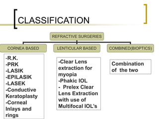
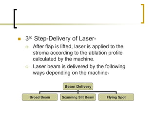
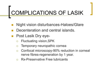
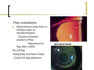
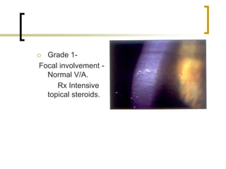
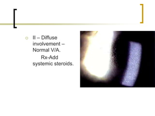
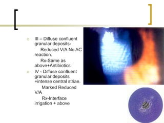
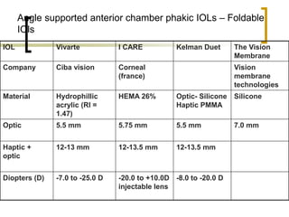
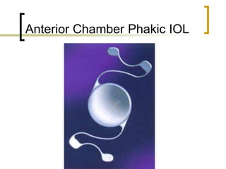
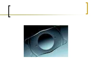
REFRACTIVE SURGERIES
Dr.Jyoti Shetty
Medical Director,
Bangalore West Lions
Superspeciality Eye Hospital
CLASSIFICATION
REFRACTIVE SURGERIES
CORNEA BASED LENTICULAR BASED COMBINED(BIOPTICS)
-R.K.
-PRK
-LASIK
-EPILASIK
-LASEK
-Conductive
Keratoplasty
-Corneal
Inlays and
rings
-Clear Lens
extraction for
myopia
-Phakic IOL
- Prelex Clear
Lens Extraction
with use of
Multifocal IOL’s
Combination
of the two
LASIK(Laser Assisted In Situ
Keratomileusis)
 Procedure using laser to ablate the
tissue from the corneal stroma to
change the refractive power of the
cornea
 Types of lasers used-
 Excimer-Excited dimer of two atoms
-an inert gas(Argon)
-Halide(Fluoride)
which releases ultraviolet energy at193nm
for corneal ablation
 Non-Excimer solid state lasers-
 210nm Q switched diode pumped laser (laser
off)
 213 nm Q switched diode pumped
laser(Pulsar)
 Advantage of Non-Excimer solid state
lasers-
 No toxic excimer gases
 Wavelength closer to absorption peak of corneal
collagen—less thermal and collateral damage
 Better pulse to pulse stability
 Not absorbed by air,water,tear fluid-so less
sensitive to humidity or room temperature
 No purging with inert gases required.
Patient selection
 Patients need to be fully informed about
potential risks,benefits and realistic
expectations
 Age should be above 18 years
 Refractive status should have been stable
for at least 1 year.
 Current FDA approval-
 Myopia-upto -12D
 Hyperopia –upto +6D
 Astigmatism-upto 7D
 CCT such that minimum safe bed
thickness left(250-270µ).Post op
Corneal thickness should not be
<410µ.
 Cornea not too flat or steep.<36D
or>49D(Poor Optics).
CONTRAINDICATIONS
 Systemic factors-
 Poorly controlled IDDM
 Pregnancy/lactation
 Autoimmune / connective tissue disorders(RA,SLE,PAN
etc)
 Clinically significant Atopy,Immunosuppressed patients
 Keloid tendency(esp PPK)
 Slow wound healing-Marfans,Ehler-Danlos
 Systemic Infection-(HIV,TB)
 Drugs-Azathioprene,Steroids(Slow wound healing)
CONTRAINDICATIONS
 Ocular Factors-
 Glaucoma,RP(Suction Pressure-ON
damage,Blebs)
 Previous h/o RD or f/h of RD.
 One eyed individual
 Pre-existing dry eye,Keratoconus.pellucid
marginal degeneration,Superficial corneal
dystrophy,RCE,Uveitis,early Lenticular changes
 h/o Herpetic Keratitis(one year prior to surgery)
PREOPERATIVE EVALUATION
PRIOR TO LASIK
 Record UCVA and BCVA Snellens V/a
 Dry and wet manifest refraction(with
1% cyclopentolate)
 Pupillometry-Infrared Pupillometer
-Aberrometer
 Large pupil-Increased HOA
perceived so increased glare
-Can change Optic Zone
 Slit Lamp Examination-
 Rule out blepharitis, miebomianitis,
pingecula, Pterygium,corneal
neovascularization
 Other contraindications for LASIK.
 IOP by applanation
 Dilated Fundus Examination to role out
holes ,tears.
 Tear film asessment-Schirmers,TBUT and
Lissamine staining
 Blink Rate-(Normal---3-7/min)
 Corneal Topography-
 Scanning slit/placido disc
 Stop RGP lenses 2 weeks prior and soft lenses I
wk prior
 To rule out early Keratoconus and other ectasias
 For mean K values
 Pachymetry -For CCT
(Ultrasound/Optical)
 Contrast Sensitivity testing for pre-
operative baseline.
BASIC STEPS AND
MACHINE SPECIFICATIONS
 Topical anasthesia-Proparacaine
0.5%, Lignocaine 4%.
 Surgical Painting and draping(Lint
Free).
 Lid speculum with aspiration.
 Corneal marking-Orientation of free
cap
Creation of flap-
 1st Step-Creation of suction by suction
pump to raise the IOP to 65 mm Hg
which is necessary for the
microkeratome to create a pass and
resect the corneal flap.
 This is crosschecked with Barraquers
tonometer.
 2nd step-Resection of corneal flap
Microkeratome Femtosecond Laser
(Intralase)
 Microkeratome-
 Uses Disposable blades
 Blade Plate can be set at
120µ,140µ,160µ and180µ.
 Nasal or superiorly hinge flaps can be
created.
 Eg.Hansatome,ACS,Carriazo Barraquer,
Moria.
 Femtosecond Laser for Flap-
 Creates photodisruption using femtosecond
solid state laser with wavelength of 1053nm.
 Needs lower vacum.
 Very short pulse with spot size of 3µ-High
precision cutting device.
 Any hinge can be made
 Can make flaps as thin as 100µ(Sub Bowmanns
Keratomileusis)
 Flap has vertical edges –so reduced
epithelial ingrowth.
 Microkeratome flap thicker in periphery
and thinner in the centre.Not so with
Intralase(Planar).
 3rd Step-Delivery of Laser-
 After flap is lifted, laser is applied to the
stroma according to the ablation profile
calculated by the machine.
 Laser beam is delivered by the following
ways depending on the machine-
Beam Delivery
Broad Beam Scanning Slit Beam Flying Spot
 Most machines employ a flying spot to
deliver laser with the help of
incorporated eye tracker.
 4th step-Reposition Of the Flap-
 After irrigating interface ,flap reposited
 Adhesion test-Striae test
ABLATION PROFILES
 Wavefront Guided or customized
ablation-to improve quality of vision by
correcting higher order aberrations.
-Wavefront analysis on entire eye
done by –Hartmann Shack
-Tracy
ABLATION PROFILES
 Aspheric Ablation-Normal LASIK converts
prolate cornea to oblate structure.(Central
flattening,steep in periphery.) which induces
higher order aberrations.
 To reduce this and preserve the prolate
structure,’Q’ value is calculated and altered
to give a more aspheric ablation.
COMPLICATIONS OF LASIK
 Under/over correction and regression (over
time).
 Post –op Keratectasia
 Presents 1-12 months
 Progressive regression
 Treatment-RGP,Corneal transplant.
 Prevention- Leave residual stromal bed
-Do surface ablation
-Don’t violatecorneal topography
diagnosis of forme-fruste keratoconus
COMPLICATIONS OF LASIK
 Night vision disturbances-Haloes/Glare
 Decenteration and central islands.
 Post Lasik Dry eye-
 Fluctuating vision,SPK
 Temporary neuropathic cornea
 Confocal microscopy-90% reduction in corneal
nerve fibres-regeneration by 1 year.
 Rx-Preservative Free lubricants
COMPLICATIONS OF LASIK
 Post op Glaucoma(Pseudo DLK)-
Steroid induced.
 Vitreoretinal Complications-
 Increased risk of RD due to alteration of
anterior vitreous by suction ring-Risk
0.08%.
 PVD(0.1% Risk)
 Macular Hemorrage(0.1% Risk)
COMPLICATIONS OF LASIK
 Flap
Complications-
 Button Hole-If
K>50D,due to
central corneal
buckling.
 .
 Irregular thin flap-
Inadequate
suction/old blade
 Short Flap-Hinge
encroaches on
visual axis-Due to
jamming of
microkeratome with
hair/FB
SHORT FLAP
 Free Cap-Due to
flat pre –op
K(<38D).
 .Flap undulations-
 Macrostriae-Linear lines in
clusters,seen on
retroillumination.
Causes-Incorrect
position of flap
-Movement of
flap after LASIK
Rx-Lift flap
-Rehydrate and float it back
-Check for flap adhesion
MACROSTRIAE
 Microstriae-Flap in
position but fine
wrinkles seen
superficially
-Due to large
myopic ablation
-Rx-
Observe.They
resolve
spontaneously
MICROSTRIAE
 Bleeding during flap cutting due to
corneal neovascularization in contact
lens users
 Interface Inflammation(Sands Of
sahara/DLK)-Non-Infective
inflammation at the interface seen in
1st week after LASIK.
 Diffuse,confluent,white granular material
at the interface 1-7 days after LASIK.
 Slight CCC
 No AC reaction
 Reduced Visual acuity
 Grade 1-
Focal involvement -
Normal V/A.
Rx Intensive
topical steroids.
 II – Diffuse
involvement –
Normal V/A.
Rx-Add
systemic steroids.
 III – Diffuse confluent
granular deposits-
Reduced V/A.No AC
reaction.
Rx-Same as
above+Antibiotics
 IV - Diffuse confluent
granular deposits
+intense central striae.
Marked Reduced
V/A
Rx-Interface
irrigation + above
 Causes-Proposed Theory
 Bacterial cell wall endotoxin
 Cleaning solution toxicity
 Talc from gloves
 Miebomian secretions
 Infection-Potential complication as any
surgical procedure
 Epithelial ingrowth-Presents 1-3
months after LASIK.
 Causes-Epithelial cells trapped under flap
 Risk factors-Peripheral epithelial defects
-Poor flap adhesion
-Buttonholed flaps
-Repeat LASIK
 Classification-
 GRADE 1-Faint white line <2mm from flap
edge
 GRADE 2-Opaque cells <2mm from flap
edge with rolled flap edge
 GRADE 3-Grey to white fine opaque line
extending >2mm from flap edge.
 GRADE 4-If ingrowth >2mm from edge with
documented progression—Lift flap and
remove the sheets of epithelium.Can use
MMC.
EPILASIK / LASEK
 Anterior stroma of cornea (ant. 1/3 rd)
has stronger interlamellar connections
than post. 2/3rd.
So surface ablation preserves the
structural integrity better than LASIK
especially in the correction of
moderate to high myopia.
 LASEK-Camellins Technique-
 20% absolute alcohol used for 20-35s. To
raise epithelial flap.
 Flap reposited after ablation
 EPILASIK- Epithelial keratome used to
lift epithelial flap of about 60-80µ thick.
 Epithelial keratomes use
- PMMA blades
-Metal Epithelial Separator
CONDUCTIVE
KERATOPLASTY
 Uses mild heat from radiofreqoency waves
to shrink collagen in the periphery of the
cornea---This steepens the paracentral
cornea.
 Used for hyperopia (1 – 2.25D) and
presbyopia.
 C.K. spots are applied with a probe in rings
with a dia. Of 6/7/8 mm.
 8 spots are given in each diameter ring.
5mm
6 7
 Drawbacks-
 Regression and retreatment in 100%
cases after 6 months.
 Induced cylinder >1D reported in many
cases.
 Usually done in one eye—Many have
intolerance to monovision.
CORNEAL INLAYS
 Increase the depth of focus by using pinhole
optics.
 Inlays have 1.6mm centre with 3.6mm
surround.
 Near vision improves by 1.5D with no loss of
distant vision.
 Used in the non –dominant eye.
 These are hydrogel based.Placed in a
tunnel 200-400 µ deep in centre of cornea.
AcuSof Corneal inlay
Phakic IOLs
 An intra-ocular lens is placed inside the
eye in front of the patient’s natural lens.
 These are available in three types
1. Anterior chamber angle fixated IOL – Nuvita
(Bausch & Lomb), Kelman duet, I care
(corneal), Vivarte (Ciba vision)
2. Iris supported phakic IOL – Verisyse/ Artisan
(AMO/Ophtec)
3. Plate lens that fits between the iris & the
crystalline lens – Starr implantable contact lens
(ICL), PRL (Ciba).
Indications
 Age above 18 years
 Stable refraction for one year
 Patients not suitable for LASIK/LASEK
due to high powers or thin corneas
 AC depth 3.0 mm
 Endothelial count >2000cells/cumm
 No other ocular pathology
Contraindications
 Myopia other than axial myopia
 Corneal dystrophy/ Endothelial cell count
<2000cells/cumm
 Anterior chamber depth less than 3.0mm
 History of uveitis
 Presence of anterior/posterior synechiae
 Glaucoma or IOP higher than 20 mmHg
 Evidence of nuclear sclerosis or developing
cataract
 Personal or family history of retinal detachment
 Diabetes mellitus
Angle supported anterior chamber phakic IOLs – Rigid lenses
IOL NuVita MA20 ZSAL-4 Phakic6
Company Bausch & Lomb Morcher M & C
Prev. model ZB5M / ZB5MF
(Baikoff)
ZSAL 1-3 ________
Material PMMA PMMA PMMA
Optic 5.0 mm 5.8 mm 6.0 mm
Eff.opt.zone 4.5 mm 5.3 mm ??
Haptic + optic 12 -13.5mm 12.0/13.5mm 12 – 14mm
Diopters (D) - 3.0 to – 23.0 D -20.0 to +10.0D
Plano concave (-20
to -3.0)
Convexo-concave
(-2.5 to +4.5)
Biconvex (+5 to
+10)
- 2.0 to -25.0D
+2.0 to +10.0D
Angle supported anterior chamber phakic IOLs – Foldable
IOls
IOL Vivarte I CARE Kelman Duet The Vision
Membrane
Company Ciba vision Corneal
(france)
Vision
membrane
technologies
Material Hydrophillic
acrylic (RI =
1.47)
HEMA 26% Optic- Silicone
Haptic PMMA
Silicone
Optic 5.5 mm 5.75 mm 5.5 mm 7.0 mm
Haptic +
optic
12-13 mm 12-13.5 mm 12-13.5 mm
Diopters (D) -7.0 to -25.0 D -20.0 to +10.0D
injectable lens
-8.0 to -20.0 D
Anterior Chamber Phakic IOL
Kelman Duet phakic IOL
 Two piece phakic IOL. The PMMA
haptic is first snaked through a 1.5mm
incision. The silicone optic is then
compressed & inserted. Once the optic
unfolds in the anterior chamber the two
tabs on either side of the optic are
snapped into projections on the haptic.
The main advantage of this lens is that
the optic can be exchanged with a new
one if the patient’s refraction changes.
Iris fixated phakic IOL – Verisyse
Phakic IOL
 Most commonly used phakic IOL
 One-piece design
Verisyse Phakic IOL
Pre-op assesment for phakic IOL
 Refraction – Objective & subjective
acceptance at 12mm vertex distance
 Anterior chamber depth – from epiuthelium
to endothelium
 Anterior & posterior segment examinations
 K-reading & Topography – Orbscan-II
 Intra-ocular pressure
 White to white measurement
 Specular microscopy
Veriflex (artiflex)
 Foldable iris claw lens. It is a
modification of Verisyse (Artisan)
phakic IOl.
Posterior chamber lenses
 These phakic IOLs are placed in the
posterior chamber between the iris &
the crystalline lens. These are
1. Starr ICL
2. Cibavision PRL
STAAR ICL
 The STAAR Collamer ICL and the TORIC
ICL are posterior chamber phakic intraocular
lenses. Made of Collamer, STARR’s
proprietary collagen copolymer
(colagen/HEMA), the lens rests behind the
iris in the ciliary sulcus.
Procedure
 The lens is gently folded and injected
into the anterior chamber through a 3.0
mm, temporal, clear corneal incision.
The ICL is then carefully positioned by
manipulating the footplates of the lens
posterior to the iris plane and and into
the sulcus. Pre-operative YAG
iridotomy is essential.
Complications
 ICL decentration
 Pupillary block
 Pigment dispersion
 Subcapsular cataract
Advantages of phakic IOLs over
laser corrective procedures
 A higher range of refractive errors can be corrected
 Reversible: Phakic IOL implantation is a potentially
reversible procedure
 Safe: No structural changes are induced. Hence it is
safe in any eye with high error & also thin corneas.
 Better quality of vision: Quality of vision (contrast
sensitivity) is better than the laser refractive
procedures in eyes with higher refractive errors and
no induced higher order aberrations. There is also a
considerable improvement in BVCA with these
lenses because of the magnification effect.
 Highly skilled procedure: Prevents misuse of the
procedure.
Bioptics
 Bioptics is a combination of phakic IOL and
LASIK. Bioptics is done for the correction of
the residual spherocylindrical power when a
spherical implant is used.
49-REFRACTIVE-SURGERIES-(2).ppt

49-REFRACTIVE-SURGERIES-(2).ppt

  • 1.
    REFRACTIVE SURGERIES Dr.Jyoti Shetty MedicalDirector, Bangalore West Lions Superspeciality Eye Hospital
  • 2.
    CLASSIFICATION REFRACTIVE SURGERIES CORNEA BASEDLENTICULAR BASED COMBINED(BIOPTICS) -R.K. -PRK -LASIK -EPILASIK -LASEK -Conductive Keratoplasty -Corneal Inlays and rings -Clear Lens extraction for myopia -Phakic IOL - Prelex Clear Lens Extraction with use of Multifocal IOL’s Combination of the two
  • 3.
    LASIK(Laser Assisted InSitu Keratomileusis)  Procedure using laser to ablate the tissue from the corneal stroma to change the refractive power of the cornea
  • 4.
     Types oflasers used-  Excimer-Excited dimer of two atoms -an inert gas(Argon) -Halide(Fluoride) which releases ultraviolet energy at193nm for corneal ablation
  • 5.
     Non-Excimer solidstate lasers-  210nm Q switched diode pumped laser (laser off)  213 nm Q switched diode pumped laser(Pulsar)
  • 6.
     Advantage ofNon-Excimer solid state lasers-  No toxic excimer gases  Wavelength closer to absorption peak of corneal collagen—less thermal and collateral damage  Better pulse to pulse stability  Not absorbed by air,water,tear fluid-so less sensitive to humidity or room temperature  No purging with inert gases required.
  • 7.
    Patient selection  Patientsneed to be fully informed about potential risks,benefits and realistic expectations  Age should be above 18 years  Refractive status should have been stable for at least 1 year.  Current FDA approval-  Myopia-upto -12D  Hyperopia –upto +6D  Astigmatism-upto 7D
  • 8.
     CCT suchthat minimum safe bed thickness left(250-270µ).Post op Corneal thickness should not be <410µ.  Cornea not too flat or steep.<36D or>49D(Poor Optics).
  • 9.
    CONTRAINDICATIONS  Systemic factors- Poorly controlled IDDM  Pregnancy/lactation  Autoimmune / connective tissue disorders(RA,SLE,PAN etc)  Clinically significant Atopy,Immunosuppressed patients  Keloid tendency(esp PPK)  Slow wound healing-Marfans,Ehler-Danlos  Systemic Infection-(HIV,TB)  Drugs-Azathioprene,Steroids(Slow wound healing)
  • 10.
    CONTRAINDICATIONS  Ocular Factors- Glaucoma,RP(Suction Pressure-ON damage,Blebs)  Previous h/o RD or f/h of RD.  One eyed individual  Pre-existing dry eye,Keratoconus.pellucid marginal degeneration,Superficial corneal dystrophy,RCE,Uveitis,early Lenticular changes  h/o Herpetic Keratitis(one year prior to surgery)
  • 11.
    PREOPERATIVE EVALUATION PRIOR TOLASIK  Record UCVA and BCVA Snellens V/a  Dry and wet manifest refraction(with 1% cyclopentolate)  Pupillometry-Infrared Pupillometer -Aberrometer  Large pupil-Increased HOA perceived so increased glare -Can change Optic Zone
  • 12.
     Slit LampExamination-  Rule out blepharitis, miebomianitis, pingecula, Pterygium,corneal neovascularization  Other contraindications for LASIK.  IOP by applanation  Dilated Fundus Examination to role out holes ,tears.
  • 13.
     Tear filmasessment-Schirmers,TBUT and Lissamine staining  Blink Rate-(Normal---3-7/min)  Corneal Topography-  Scanning slit/placido disc  Stop RGP lenses 2 weeks prior and soft lenses I wk prior  To rule out early Keratoconus and other ectasias  For mean K values
  • 14.
     Pachymetry -ForCCT (Ultrasound/Optical)  Contrast Sensitivity testing for pre- operative baseline.
  • 15.
    BASIC STEPS AND MACHINESPECIFICATIONS  Topical anasthesia-Proparacaine 0.5%, Lignocaine 4%.  Surgical Painting and draping(Lint Free).  Lid speculum with aspiration.  Corneal marking-Orientation of free cap
  • 16.
    Creation of flap- 1st Step-Creation of suction by suction pump to raise the IOP to 65 mm Hg which is necessary for the microkeratome to create a pass and resect the corneal flap.  This is crosschecked with Barraquers tonometer.
  • 17.
     2nd step-Resectionof corneal flap Microkeratome Femtosecond Laser (Intralase)
  • 18.
     Microkeratome-  UsesDisposable blades  Blade Plate can be set at 120µ,140µ,160µ and180µ.  Nasal or superiorly hinge flaps can be created.  Eg.Hansatome,ACS,Carriazo Barraquer, Moria.
  • 19.
     Femtosecond Laserfor Flap-  Creates photodisruption using femtosecond solid state laser with wavelength of 1053nm.  Needs lower vacum.  Very short pulse with spot size of 3µ-High precision cutting device.  Any hinge can be made  Can make flaps as thin as 100µ(Sub Bowmanns Keratomileusis)
  • 20.
     Flap hasvertical edges –so reduced epithelial ingrowth.  Microkeratome flap thicker in periphery and thinner in the centre.Not so with Intralase(Planar).
  • 21.
     3rd Step-Deliveryof Laser-  After flap is lifted, laser is applied to the stroma according to the ablation profile calculated by the machine.  Laser beam is delivered by the following ways depending on the machine- Beam Delivery Broad Beam Scanning Slit Beam Flying Spot
  • 22.
     Most machinesemploy a flying spot to deliver laser with the help of incorporated eye tracker.
  • 23.
     4th step-RepositionOf the Flap-  After irrigating interface ,flap reposited  Adhesion test-Striae test
  • 24.
    ABLATION PROFILES  WavefrontGuided or customized ablation-to improve quality of vision by correcting higher order aberrations. -Wavefront analysis on entire eye done by –Hartmann Shack -Tracy
  • 25.
    ABLATION PROFILES  AsphericAblation-Normal LASIK converts prolate cornea to oblate structure.(Central flattening,steep in periphery.) which induces higher order aberrations.  To reduce this and preserve the prolate structure,’Q’ value is calculated and altered to give a more aspheric ablation.
  • 26.
    COMPLICATIONS OF LASIK Under/over correction and regression (over time).  Post –op Keratectasia  Presents 1-12 months  Progressive regression  Treatment-RGP,Corneal transplant.  Prevention- Leave residual stromal bed -Do surface ablation -Don’t violatecorneal topography diagnosis of forme-fruste keratoconus
  • 27.
    COMPLICATIONS OF LASIK Night vision disturbances-Haloes/Glare  Decenteration and central islands.  Post Lasik Dry eye-  Fluctuating vision,SPK  Temporary neuropathic cornea  Confocal microscopy-90% reduction in corneal nerve fibres-regeneration by 1 year.  Rx-Preservative Free lubricants
  • 28.
    COMPLICATIONS OF LASIK Post op Glaucoma(Pseudo DLK)- Steroid induced.  Vitreoretinal Complications-  Increased risk of RD due to alteration of anterior vitreous by suction ring-Risk 0.08%.  PVD(0.1% Risk)  Macular Hemorrage(0.1% Risk)
  • 29.
    COMPLICATIONS OF LASIK Flap Complications-  Button Hole-If K>50D,due to central corneal buckling.  .
  • 30.
     Irregular thinflap- Inadequate suction/old blade  Short Flap-Hinge encroaches on visual axis-Due to jamming of microkeratome with hair/FB SHORT FLAP
  • 31.
     Free Cap-Dueto flat pre –op K(<38D).
  • 32.
     .Flap undulations- Macrostriae-Linear lines in clusters,seen on retroillumination. Causes-Incorrect position of flap -Movement of flap after LASIK Rx-Lift flap -Rehydrate and float it back -Check for flap adhesion MACROSTRIAE
  • 33.
     Microstriae-Flap in positionbut fine wrinkles seen superficially -Due to large myopic ablation -Rx- Observe.They resolve spontaneously MICROSTRIAE
  • 34.
     Bleeding duringflap cutting due to corneal neovascularization in contact lens users
  • 35.
     Interface Inflammation(SandsOf sahara/DLK)-Non-Infective inflammation at the interface seen in 1st week after LASIK.  Diffuse,confluent,white granular material at the interface 1-7 days after LASIK.  Slight CCC  No AC reaction  Reduced Visual acuity
  • 36.
     Grade 1- Focalinvolvement - Normal V/A. Rx Intensive topical steroids.
  • 37.
     II –Diffuse involvement – Normal V/A. Rx-Add systemic steroids.
  • 38.
     III –Diffuse confluent granular deposits- Reduced V/A.No AC reaction. Rx-Same as above+Antibiotics  IV - Diffuse confluent granular deposits +intense central striae. Marked Reduced V/A Rx-Interface irrigation + above
  • 39.
     Causes-Proposed Theory Bacterial cell wall endotoxin  Cleaning solution toxicity  Talc from gloves  Miebomian secretions
  • 40.
     Infection-Potential complicationas any surgical procedure
  • 41.
     Epithelial ingrowth-Presents1-3 months after LASIK.  Causes-Epithelial cells trapped under flap  Risk factors-Peripheral epithelial defects -Poor flap adhesion -Buttonholed flaps -Repeat LASIK
  • 42.
     Classification-  GRADE1-Faint white line <2mm from flap edge  GRADE 2-Opaque cells <2mm from flap edge with rolled flap edge  GRADE 3-Grey to white fine opaque line extending >2mm from flap edge.  GRADE 4-If ingrowth >2mm from edge with documented progression—Lift flap and remove the sheets of epithelium.Can use MMC.
  • 43.
    EPILASIK / LASEK Anterior stroma of cornea (ant. 1/3 rd) has stronger interlamellar connections than post. 2/3rd. So surface ablation preserves the structural integrity better than LASIK especially in the correction of moderate to high myopia.
  • 44.
     LASEK-Camellins Technique- 20% absolute alcohol used for 20-35s. To raise epithelial flap.  Flap reposited after ablation
  • 45.
     EPILASIK- Epithelialkeratome used to lift epithelial flap of about 60-80µ thick.  Epithelial keratomes use - PMMA blades -Metal Epithelial Separator
  • 46.
    CONDUCTIVE KERATOPLASTY  Uses mildheat from radiofreqoency waves to shrink collagen in the periphery of the cornea---This steepens the paracentral cornea.  Used for hyperopia (1 – 2.25D) and presbyopia.  C.K. spots are applied with a probe in rings with a dia. Of 6/7/8 mm.  8 spots are given in each diameter ring.
  • 47.
  • 48.
     Drawbacks-  Regressionand retreatment in 100% cases after 6 months.  Induced cylinder >1D reported in many cases.  Usually done in one eye—Many have intolerance to monovision.
  • 49.
    CORNEAL INLAYS  Increasethe depth of focus by using pinhole optics.  Inlays have 1.6mm centre with 3.6mm surround.  Near vision improves by 1.5D with no loss of distant vision.  Used in the non –dominant eye.  These are hydrogel based.Placed in a tunnel 200-400 µ deep in centre of cornea.
  • 50.
  • 51.
    Phakic IOLs  Anintra-ocular lens is placed inside the eye in front of the patient’s natural lens.  These are available in three types 1. Anterior chamber angle fixated IOL – Nuvita (Bausch & Lomb), Kelman duet, I care (corneal), Vivarte (Ciba vision) 2. Iris supported phakic IOL – Verisyse/ Artisan (AMO/Ophtec) 3. Plate lens that fits between the iris & the crystalline lens – Starr implantable contact lens (ICL), PRL (Ciba).
  • 52.
    Indications  Age above18 years  Stable refraction for one year  Patients not suitable for LASIK/LASEK due to high powers or thin corneas  AC depth 3.0 mm  Endothelial count >2000cells/cumm  No other ocular pathology
  • 53.
    Contraindications  Myopia otherthan axial myopia  Corneal dystrophy/ Endothelial cell count <2000cells/cumm  Anterior chamber depth less than 3.0mm  History of uveitis  Presence of anterior/posterior synechiae  Glaucoma or IOP higher than 20 mmHg  Evidence of nuclear sclerosis or developing cataract  Personal or family history of retinal detachment  Diabetes mellitus
  • 54.
    Angle supported anteriorchamber phakic IOLs – Rigid lenses IOL NuVita MA20 ZSAL-4 Phakic6 Company Bausch & Lomb Morcher M & C Prev. model ZB5M / ZB5MF (Baikoff) ZSAL 1-3 ________ Material PMMA PMMA PMMA Optic 5.0 mm 5.8 mm 6.0 mm Eff.opt.zone 4.5 mm 5.3 mm ?? Haptic + optic 12 -13.5mm 12.0/13.5mm 12 – 14mm Diopters (D) - 3.0 to – 23.0 D -20.0 to +10.0D Plano concave (-20 to -3.0) Convexo-concave (-2.5 to +4.5) Biconvex (+5 to +10) - 2.0 to -25.0D +2.0 to +10.0D
  • 55.
    Angle supported anteriorchamber phakic IOLs – Foldable IOls IOL Vivarte I CARE Kelman Duet The Vision Membrane Company Ciba vision Corneal (france) Vision membrane technologies Material Hydrophillic acrylic (RI = 1.47) HEMA 26% Optic- Silicone Haptic PMMA Silicone Optic 5.5 mm 5.75 mm 5.5 mm 7.0 mm Haptic + optic 12-13 mm 12-13.5 mm 12-13.5 mm Diopters (D) -7.0 to -25.0 D -20.0 to +10.0D injectable lens -8.0 to -20.0 D
  • 56.
  • 57.
    Kelman Duet phakicIOL  Two piece phakic IOL. The PMMA haptic is first snaked through a 1.5mm incision. The silicone optic is then compressed & inserted. Once the optic unfolds in the anterior chamber the two tabs on either side of the optic are snapped into projections on the haptic. The main advantage of this lens is that the optic can be exchanged with a new one if the patient’s refraction changes.
  • 58.
    Iris fixated phakicIOL – Verisyse Phakic IOL  Most commonly used phakic IOL  One-piece design
  • 59.
  • 60.
    Pre-op assesment forphakic IOL  Refraction – Objective & subjective acceptance at 12mm vertex distance  Anterior chamber depth – from epiuthelium to endothelium  Anterior & posterior segment examinations  K-reading & Topography – Orbscan-II  Intra-ocular pressure  White to white measurement  Specular microscopy
  • 61.
    Veriflex (artiflex)  Foldableiris claw lens. It is a modification of Verisyse (Artisan) phakic IOl.
  • 62.
    Posterior chamber lenses These phakic IOLs are placed in the posterior chamber between the iris & the crystalline lens. These are 1. Starr ICL 2. Cibavision PRL
  • 63.
    STAAR ICL  TheSTAAR Collamer ICL and the TORIC ICL are posterior chamber phakic intraocular lenses. Made of Collamer, STARR’s proprietary collagen copolymer (colagen/HEMA), the lens rests behind the iris in the ciliary sulcus.
  • 65.
    Procedure  The lensis gently folded and injected into the anterior chamber through a 3.0 mm, temporal, clear corneal incision. The ICL is then carefully positioned by manipulating the footplates of the lens posterior to the iris plane and and into the sulcus. Pre-operative YAG iridotomy is essential.
  • 66.
    Complications  ICL decentration Pupillary block  Pigment dispersion  Subcapsular cataract
  • 67.
    Advantages of phakicIOLs over laser corrective procedures  A higher range of refractive errors can be corrected  Reversible: Phakic IOL implantation is a potentially reversible procedure  Safe: No structural changes are induced. Hence it is safe in any eye with high error & also thin corneas.  Better quality of vision: Quality of vision (contrast sensitivity) is better than the laser refractive procedures in eyes with higher refractive errors and no induced higher order aberrations. There is also a considerable improvement in BVCA with these lenses because of the magnification effect.  Highly skilled procedure: Prevents misuse of the procedure.
  • 68.
    Bioptics  Bioptics isa combination of phakic IOL and LASIK. Bioptics is done for the correction of the residual spherocylindrical power when a spherical implant is used.