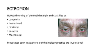
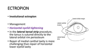
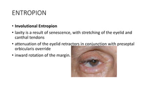
This document summarizes various periorbital malpositions and involutional changes including ectropion, entropion, and symblepharon. Ectropion is outward turning of the eyelid and has several types including involutional, cicatricial, and paralytic. Entropion is inward turning and can be involutional, acute spastic, or cicatricial. Symblepharon is an adhesion between conjunctival surfaces caused by inflammation, infection, or trauma. The document discusses examination, risk factors, pathophysiology, and management options for each condition including surgical procedures and medical treatment.
Approach to a patient with ectropion, entropion, symblepharon.pptxIddi Ndyabawe
This document discusses approaches to treating ectropion, entropion, and symblepharon. It describes the anatomy and causes of ectropion and entropion, including involutional, cicatricial, paralytis, and congenital types. Clinical features, grading scales, and surgical management techniques are outlined for different types and severities of ectropion and entropion. Symblepharon is defined as an adhesion between conjunctival surfaces, with treatment involving conjunctival grafts or flaps.
This document discusses various eyelid malpositions including entropion, ectropion, ptosis, and trichiasis. Entropion is when the eyelid rolls inward causing eyelashes to rub the eye, potentially damaging the cornea. Ectropion is when the eyelid rolls outward. Causes, symptoms, evaluations, and treatments are described for each condition. Conservative treatments include taping, lubricants, and botulinum toxin injections. Surgical treatments aim to tighten or reposition the eyelid depending on the specific malposition and its underlying cause. Prompt treatment is important to prevent corneal complications.
This document provides information on retinal detachment including:
1. It describes the three main types of retinal detachment - rhegmatogenous, tractional, and exudative - and their causes.
2. Risk factors, clinical manifestations, diagnosis, and surgical management options like cryopexy, laser photocoagulation, scleral buckling, pneumatic retinopexy, and vitrectomy are summarized.
3. Post-operative care involves positioning, monitoring for complications, restricting activities that raise intraocular pressure, and reporting any new visual issues or pain immediately.
This document provides information about ectropion, including:
1. It defines ectropion as eversion of the eyelid where the lid rolls away from the globe.
2. It classifies ectropion into categories such as congenital, involutional, cicatricial, paralytic, and mechanical.
3. It describes the evaluation, causes, clinical features and complications of different types of ectropion, as well as various surgical and non-surgical management approaches.
This lecture is about the different types of blunt trauma,presentation and management of the various forms of blunt trauma.Its complications and their approach.the Le Fort fractures
This document contains a series of multiple choice questions about various ophthalmic conditions and treatments. The questions cover topics like causes of blindness, types of entropion/ectropion, cranial nerve disorders, corneal diseases, conjunctivitis, pterygium, and treatments for conditions like ptosis, blepharitis and corneal ulcers. The questions are in a multiple choice format with 4 answer options for each.
The document provides information on eyelid anatomy and various eyelid conditions. It describes:
1. The anatomy of the eyelids including the anterior lamella with skin and orbicularis muscle, and posterior lamella with tarsal plate and conjunctiva. Common eyelid conditions are also summarized such as entropion, ectropion, lagophthalmos, ptosis, and trichiasis.
2. Causes, signs, and treatments for various types of ectropion and entropion are outlined, including involutional, cicatricial, paralytic, and congenital forms. Surgical procedures for correction including wedges resections and tightening of retractors are mentioned.
This document discusses various complications that can occur after cataract surgery, including operative complications such as posterior capsule rupture and postoperative complications like cystoid macular edema. It provides details on risk factors, signs and symptoms, diagnosis, and treatment approaches for each complication. The most common late complication is posterior capsular opacification, occurring in 20-25% of patients, which can be treated with Nd:YAG laser capsulotomy.
Eyelid Surgery for Ophthalmic Clinical Course UPPGHAlex Tan
This document provides an overview of surgical techniques for managing various eyelid conditions including entropion, ectropion, and ptosis. It discusses the underlying causes and pathologies of each condition and describes various surgical procedures for correction. For entropion, procedures described include Quickert sutures, lateral tarsal strips, and reattachment of retractors for involutional cases, and tarsotomy with or without grafts for cicatricial cases. For ectropion, techniques include tendon tightening and lazy-T procedures. For ptosis, factors in evaluation are outlined and techniques covered include levator resection, frontalis suspension, and aponeurotic surgery.
This document provides details on the preoperative evaluation and surgical techniques for blepharoplasty. The preoperative evaluation includes tests to assess for dry eyes, thyroid disease, visual function and anatomy of the eyelids and brows. Surgical techniques are described for upper and lower eyelid blepharoplasty including markings, incisions, fat and tissue removal and closure. Specific considerations are discussed for different ethnic groups and addressing brow ptosis or dry eyes. Photographs are taken preoperatively for reference and planning.
Cataract surgery can have several complications related to anesthesia, the surgery itself, or postoperative recovery. Intraoperative complications include issues with the incision, anterior chamber stability, iris handling, and posterior capsule rupture. Postoperative complications involve corneal edema, elevated pressure, inflammation, and capsule issues. Careful patient selection and surgical technique aim to prevent these complications from cataract surgery.
This document provides information on entropion, including anatomy of the eyelid, causes of entropion, classification, symptoms, assessment, differential diagnosis, and surgical and non-surgical management. Entropion is defined as inward rotation of the eyelid margin. It discusses various procedures for correcting entropion such as Quickert-Rathbun eversion sutures, lower lid retractor advancement combined with lateral tarsal strip or wedge resection, tarsal fracture/transverse tarsotomy, and use of posterior lamellar grafts for severe cicatricial entropion. Non-surgical management includes artificial tears, bandage contact lenses, lower lid taping, and botulinum
Retinal detachment occurs when the retina separates from its underlying layers, which can cause vision loss if not treated. It is often caused by breaks or tears in the retina that allow fluid to enter and detach the retina. Diagnosis involves examining the eye through various tests like ophthalmoscopy, ultrasound and angiography. Treatment options aim to reattach the retina through procedures like laser photocoagulation, cryotherapy, scleral buckling surgery, pneumatic retinopexy or vitrectomy. These work by sealing retinal breaks or reducing fluid flow to allow the retina to reattach.
This document discusses blunt eye trauma, including:
- It can cause closed or open globe injuries from mechanisms like coup, countercoup, or compression.
- Etiology includes direct blows, accidents, or increased intraocular pressure from force. Common causes are sports, work, or domestic violence.
- Pathophysiology involves increased pressure rupturing the sclera or retrobulbar hematoma compressing the optic nerve.
- Physical exam looks for vision loss, irregular globe contour, hyphema, or proptosis indicating rupture.
- Manifestations involve the orbit, nerves, anterior segment structures like the conjunctiva, cornea, anterior chamber, or uveitis. Management
1. The document discusses anatomy and physiology of the eye and ear, as well as various ocular and auditory conditions.
2. Assessment of vision and hearing involves testing visual acuity, pupillary response, and hearing through whisper tests and tuning forks.
3. Common eye conditions addressed include cataracts, glaucoma, retinal detachment, and macular degeneration. Common ear conditions include hearing loss, vertigo, tinnitus, and Meniere's disease.
The document discusses anophthalmia, which refers to the complete absence of the eye and orbital tissues. Anophthalmia can be congenital or acquired through procedures like enucleation, evisceration, and exenteration. These procedures require orbital implants, conformers, and prosthetics to rehabilitate the socket. Long-term complications can include socket contracture, eyelid malpositions, and implant exposure or migration. Proper surgical planning and techniques aim to minimize complications and provide stable, comfortable sockets that allow for good prosthetic motility and cosmesis.
EVISCERATION, ENUCLEATION, EXENTRATION, CYCLODESTRUCTIVE PROCEDURESReshma Peter
The document discusses four surgical procedures for removing an eye: evisceration, enucleation, exenteration, and cyclodestructive procedures. Evisceration involves removing the contents of the eye while leaving surrounding structures intact. Enucleation is the removal of the entire eye while leaving surrounding orbital contents intact. Exenteration is the removal of the entire orbital contents, including extraocular muscles. The document provides details on indications, techniques, advantages, and disadvantages of each procedure.
Glaucoma post cataract extraction, vitreoretinal surgery, keratoplasty and re...Haitham Al Mahrouqi
Glaucoma is a common complication of ocular surgeries such as cataract extraction, keratoplasty, vitreoretinal surgery, and refractive surgery. The mechanisms of glaucoma include retained viscoelastic, inflammation, pupillary block, and changes to the angle structure. Glaucoma can occur early due to these mechanisms or late due to factors like steroid response or inflammation. Careful case selection, prevention of complications, and close monitoring are important to manage glaucoma risk in postoperative patients.
Surgeries for open angle glaucoma were discussed, including trabeculectomy. Trabeculectomy involves making a conjunctival flap, applying antimetabolites, excising a block of tissue from the eye, and suturing the scleral flap. Potential complications include shallow anterior chamber, low or high eye pressure, infection, and visual loss. Careful postoperative monitoring and treatment are important to manage complications while lowering eye pressure to prevent further glaucoma damage.
ACCORDING TO AYURVEDA THE DISEASES OF SCLERA ARE CALLED AS SHUKLAGATA ROGAS. THE PROBABLE MODERN CORRELATION OF SHUKLAGATA ROGAS LIKE ARMA(PTERYGIUM) ETC. ARE EXPLAINED IN THIS PPT.
Traumatic chorioretinal pathologies can result from both penetrating and non-penetrating ocular injuries. Males under 40 are most commonly affected. Closed globe injuries may cause commotio retinae, while open globe injuries risk choroidal rupture. Indirect injuries like Valsalva retinopathy or Terson syndrome can also cause intraocular bleeding. Traumatic macular holes and retinal detachments require surgical repair like vitrectomy. Overall ocular trauma remains a major cause of blindness worldwide.
This document discusses the anatomy and management of the anophthalmic socket. It begins by defining an anophthalmic socket as an orbit lacking an eye globe, usually due to enucleation. True anophthalmia can be congenital or acquired. The most common causes of acquired anophthalmia are enucleation, exenteration, or evisceration performed to treat painful, blind eyes or remove tumors. The document outlines techniques for each procedure and discusses complications that can arise like socket contracture and management strategies like grafting. It emphasizes the importance of socket maintenance with conformers and prosthetics to prevent complications. In summary, this document provides an overview of anophthalmic socket anatomy,
The document summarizes management of ocular trauma from blunt injuries. It describes mechanisms of injury including kinetic energy transfer and oscillatory tissue damage. Treatment depends on injury severity and location, and may involve repair of open globe injuries, removal of intraocular foreign bodies, management of hyphema, lens subluxation or dislocation, and vitrectomy. The goal is to restore ocular integrity and address complications while minimizing further trauma from intervention.
1) Trabeculectomy is a glaucoma surgery that creates an opening in the eye to drain fluid from the anterior chamber and reduce intraocular pressure.
2) It involves making a partial thickness scleral flap, removing a block of tissue underneath, and suturing the flap loosely to allow fluid drainage.
3) Antifibrotic agents like mitomycin C or 5-fluorouracil are often applied to reduce scarring and improve surgical success rates.
This document discusses several age-related diseases that can affect the ocular adnexa. The eyelids are susceptible to ectropion, entropion, dermatochalasis, ptosis, and madarosis due to changes like laxity of tissues, loss of elasticity, and muscle weakening. The lacrimal apparatus can develop dry eye or dacryocystitis from reduced tear production and inflammation. The orbit may experience enophthalmos from shrinking fat pads or bulging fat into the eyelids. Other common age-related eye conditions discussed include presbyopia, cataracts, floaters, glaucoma, macular degeneration, retinal detachment, conjunctivitis,
Anophthalmia is the absence of the eyeball and can be congenital or acquired. The optimal anophthalmic socket has a well-positioned implant, healthy conjunctiva, symmetric eyelids, and a comfortable prosthesis. Complications after enucleation include enophthalmos, eyelid deformities, and socket contracture. Management involves orbital implants, dermis fat grafts, fornix deepening, and lid procedures to correct volume deficits and improve prosthesis retention.
Anophthalmia is the absence of the eyeball and can be congenital or acquired. The optimal management of an anophthalmic socket involves maintaining adequate volume with a well-positioned implant, healthy conjunctiva, and symmetric eyelids. Complications after enucleation like enophthalmos, eyelid deformities, and socket contracture can be addressed through procedures like dermis fat grafts, fornix deepening sutures, and implant replacement. Proper prosthesis fitting and care is also important for optimal cosmetic and functional results.
MICROBIAL AND PARASITIC INFECTIONS OF THE EYE.pptxBARNABASMUGABI
This document discusses various microbial and parasitic infections that can affect the eye. It covers lid infections like styes, hordeolums, and chalazions caused by staphylococcal or meibomian gland infections. It also discusses blepharitis, demodex, and parasitic infections like phthiriasis palpebrum. Orbital cellulitis and its complications are described. Infections of the lacrimal system like dacryocystitis and dacryoadenitis are covered. The document concludes with details on various types of conjunctivitis including bacterial, viral like adenovirus, and parasitic forms such as trachoma.
RADIOTHERAPY describes the various indicationsBARNABASMUGABI
Radiotherapy is the use of ionizing radiation to treat cancer and tumors. It can be classified as electromagnetic radiation like x-rays and gamma rays, or particulate radiation like protons, neutrons, and electrons. Radiotherapy is commonly used to treat retinoblastoma, uveal melanoma, and conjunctival tumors. For retinoblastoma, chemotherapy is the main treatment but radiotherapy techniques like plaque brachytherapy, proton beam therapy, and stereotactic radiotherapy may also be used. Uveal melanoma can be treated with plaque brachytherapy, proton beam therapy, or stereotactic radiotherapy. Conjunctival tumors are often treated with plaque brachytherapy or external beam
More Related Content
Similar to PERIOCULAR MALPOSITIONS AND INVOLUTIONAL CHANGES.pptx
Eyelid Surgery for Ophthalmic Clinical Course UPPGHAlex Tan
This document provides an overview of surgical techniques for managing various eyelid conditions including entropion, ectropion, and ptosis. It discusses the underlying causes and pathologies of each condition and describes various surgical procedures for correction. For entropion, procedures described include Quickert sutures, lateral tarsal strips, and reattachment of retractors for involutional cases, and tarsotomy with or without grafts for cicatricial cases. For ectropion, techniques include tendon tightening and lazy-T procedures. For ptosis, factors in evaluation are outlined and techniques covered include levator resection, frontalis suspension, and aponeurotic surgery.
This document provides details on the preoperative evaluation and surgical techniques for blepharoplasty. The preoperative evaluation includes tests to assess for dry eyes, thyroid disease, visual function and anatomy of the eyelids and brows. Surgical techniques are described for upper and lower eyelid blepharoplasty including markings, incisions, fat and tissue removal and closure. Specific considerations are discussed for different ethnic groups and addressing brow ptosis or dry eyes. Photographs are taken preoperatively for reference and planning.
Cataract surgery can have several complications related to anesthesia, the surgery itself, or postoperative recovery. Intraoperative complications include issues with the incision, anterior chamber stability, iris handling, and posterior capsule rupture. Postoperative complications involve corneal edema, elevated pressure, inflammation, and capsule issues. Careful patient selection and surgical technique aim to prevent these complications from cataract surgery.
This document provides information on entropion, including anatomy of the eyelid, causes of entropion, classification, symptoms, assessment, differential diagnosis, and surgical and non-surgical management. Entropion is defined as inward rotation of the eyelid margin. It discusses various procedures for correcting entropion such as Quickert-Rathbun eversion sutures, lower lid retractor advancement combined with lateral tarsal strip or wedge resection, tarsal fracture/transverse tarsotomy, and use of posterior lamellar grafts for severe cicatricial entropion. Non-surgical management includes artificial tears, bandage contact lenses, lower lid taping, and botulinum
Retinal detachment occurs when the retina separates from its underlying layers, which can cause vision loss if not treated. It is often caused by breaks or tears in the retina that allow fluid to enter and detach the retina. Diagnosis involves examining the eye through various tests like ophthalmoscopy, ultrasound and angiography. Treatment options aim to reattach the retina through procedures like laser photocoagulation, cryotherapy, scleral buckling surgery, pneumatic retinopexy or vitrectomy. These work by sealing retinal breaks or reducing fluid flow to allow the retina to reattach.
This document discusses blunt eye trauma, including:
- It can cause closed or open globe injuries from mechanisms like coup, countercoup, or compression.
- Etiology includes direct blows, accidents, or increased intraocular pressure from force. Common causes are sports, work, or domestic violence.
- Pathophysiology involves increased pressure rupturing the sclera or retrobulbar hematoma compressing the optic nerve.
- Physical exam looks for vision loss, irregular globe contour, hyphema, or proptosis indicating rupture.
- Manifestations involve the orbit, nerves, anterior segment structures like the conjunctiva, cornea, anterior chamber, or uveitis. Management
1. The document discusses anatomy and physiology of the eye and ear, as well as various ocular and auditory conditions.
2. Assessment of vision and hearing involves testing visual acuity, pupillary response, and hearing through whisper tests and tuning forks.
3. Common eye conditions addressed include cataracts, glaucoma, retinal detachment, and macular degeneration. Common ear conditions include hearing loss, vertigo, tinnitus, and Meniere's disease.
The document discusses anophthalmia, which refers to the complete absence of the eye and orbital tissues. Anophthalmia can be congenital or acquired through procedures like enucleation, evisceration, and exenteration. These procedures require orbital implants, conformers, and prosthetics to rehabilitate the socket. Long-term complications can include socket contracture, eyelid malpositions, and implant exposure or migration. Proper surgical planning and techniques aim to minimize complications and provide stable, comfortable sockets that allow for good prosthetic motility and cosmesis.
EVISCERATION, ENUCLEATION, EXENTRATION, CYCLODESTRUCTIVE PROCEDURESReshma Peter
The document discusses four surgical procedures for removing an eye: evisceration, enucleation, exenteration, and cyclodestructive procedures. Evisceration involves removing the contents of the eye while leaving surrounding structures intact. Enucleation is the removal of the entire eye while leaving surrounding orbital contents intact. Exenteration is the removal of the entire orbital contents, including extraocular muscles. The document provides details on indications, techniques, advantages, and disadvantages of each procedure.
Glaucoma post cataract extraction, vitreoretinal surgery, keratoplasty and re...Haitham Al Mahrouqi
Glaucoma is a common complication of ocular surgeries such as cataract extraction, keratoplasty, vitreoretinal surgery, and refractive surgery. The mechanisms of glaucoma include retained viscoelastic, inflammation, pupillary block, and changes to the angle structure. Glaucoma can occur early due to these mechanisms or late due to factors like steroid response or inflammation. Careful case selection, prevention of complications, and close monitoring are important to manage glaucoma risk in postoperative patients.
Surgeries for open angle glaucoma were discussed, including trabeculectomy. Trabeculectomy involves making a conjunctival flap, applying antimetabolites, excising a block of tissue from the eye, and suturing the scleral flap. Potential complications include shallow anterior chamber, low or high eye pressure, infection, and visual loss. Careful postoperative monitoring and treatment are important to manage complications while lowering eye pressure to prevent further glaucoma damage.
ACCORDING TO AYURVEDA THE DISEASES OF SCLERA ARE CALLED AS SHUKLAGATA ROGAS. THE PROBABLE MODERN CORRELATION OF SHUKLAGATA ROGAS LIKE ARMA(PTERYGIUM) ETC. ARE EXPLAINED IN THIS PPT.
Traumatic chorioretinal pathologies can result from both penetrating and non-penetrating ocular injuries. Males under 40 are most commonly affected. Closed globe injuries may cause commotio retinae, while open globe injuries risk choroidal rupture. Indirect injuries like Valsalva retinopathy or Terson syndrome can also cause intraocular bleeding. Traumatic macular holes and retinal detachments require surgical repair like vitrectomy. Overall ocular trauma remains a major cause of blindness worldwide.
This document discusses the anatomy and management of the anophthalmic socket. It begins by defining an anophthalmic socket as an orbit lacking an eye globe, usually due to enucleation. True anophthalmia can be congenital or acquired. The most common causes of acquired anophthalmia are enucleation, exenteration, or evisceration performed to treat painful, blind eyes or remove tumors. The document outlines techniques for each procedure and discusses complications that can arise like socket contracture and management strategies like grafting. It emphasizes the importance of socket maintenance with conformers and prosthetics to prevent complications. In summary, this document provides an overview of anophthalmic socket anatomy,
The document summarizes management of ocular trauma from blunt injuries. It describes mechanisms of injury including kinetic energy transfer and oscillatory tissue damage. Treatment depends on injury severity and location, and may involve repair of open globe injuries, removal of intraocular foreign bodies, management of hyphema, lens subluxation or dislocation, and vitrectomy. The goal is to restore ocular integrity and address complications while minimizing further trauma from intervention.
1) Trabeculectomy is a glaucoma surgery that creates an opening in the eye to drain fluid from the anterior chamber and reduce intraocular pressure.
2) It involves making a partial thickness scleral flap, removing a block of tissue underneath, and suturing the flap loosely to allow fluid drainage.
3) Antifibrotic agents like mitomycin C or 5-fluorouracil are often applied to reduce scarring and improve surgical success rates.
This document discusses several age-related diseases that can affect the ocular adnexa. The eyelids are susceptible to ectropion, entropion, dermatochalasis, ptosis, and madarosis due to changes like laxity of tissues, loss of elasticity, and muscle weakening. The lacrimal apparatus can develop dry eye or dacryocystitis from reduced tear production and inflammation. The orbit may experience enophthalmos from shrinking fat pads or bulging fat into the eyelids. Other common age-related eye conditions discussed include presbyopia, cataracts, floaters, glaucoma, macular degeneration, retinal detachment, conjunctivitis,
Anophthalmia is the absence of the eyeball and can be congenital or acquired. The optimal anophthalmic socket has a well-positioned implant, healthy conjunctiva, symmetric eyelids, and a comfortable prosthesis. Complications after enucleation include enophthalmos, eyelid deformities, and socket contracture. Management involves orbital implants, dermis fat grafts, fornix deepening, and lid procedures to correct volume deficits and improve prosthesis retention.
Anophthalmia is the absence of the eyeball and can be congenital or acquired. The optimal management of an anophthalmic socket involves maintaining adequate volume with a well-positioned implant, healthy conjunctiva, and symmetric eyelids. Complications after enucleation like enophthalmos, eyelid deformities, and socket contracture can be addressed through procedures like dermis fat grafts, fornix deepening sutures, and implant replacement. Proper prosthesis fitting and care is also important for optimal cosmetic and functional results.
MICROBIAL AND PARASITIC INFECTIONS OF THE EYE.pptxBARNABASMUGABI
This document discusses various microbial and parasitic infections that can affect the eye. It covers lid infections like styes, hordeolums, and chalazions caused by staphylococcal or meibomian gland infections. It also discusses blepharitis, demodex, and parasitic infections like phthiriasis palpebrum. Orbital cellulitis and its complications are described. Infections of the lacrimal system like dacryocystitis and dacryoadenitis are covered. The document concludes with details on various types of conjunctivitis including bacterial, viral like adenovirus, and parasitic forms such as trachoma.
RADIOTHERAPY describes the various indicationsBARNABASMUGABI
Radiotherapy is the use of ionizing radiation to treat cancer and tumors. It can be classified as electromagnetic radiation like x-rays and gamma rays, or particulate radiation like protons, neutrons, and electrons. Radiotherapy is commonly used to treat retinoblastoma, uveal melanoma, and conjunctival tumors. For retinoblastoma, chemotherapy is the main treatment but radiotherapy techniques like plaque brachytherapy, proton beam therapy, and stereotactic radiotherapy may also be used. Uveal melanoma can be treated with plaque brachytherapy, proton beam therapy, or stereotactic radiotherapy. Conjunctival tumors are often treated with plaque brachytherapy or external beam
Optic Disc Abnormalities and their presentationsBARNABASMUGABI
This document discusses various optic disc abnormalities including optic nerve hypoplasia, optic pits, and morning glory disc anomaly. Optic nerve hypoplasia is characterized by a decreased number of optic nerve fibers and can cause visual impairment. It has known risk factors such as young maternal age. Optic pits appear as depressions of the optic disc and can sometimes cause macular serous detachments. Morning glory disc anomaly is a funnel-shaped excavation of the optic disc that is typically unilateral and sometimes associated with retinal detachments or brain abnormalities.
OCULAR TOXOPLASMOSIS the blinding disease and is classified among the torch i...BARNABASMUGABI
the disease spreads from the cats and causes retinochoroiditis and its a blinding disease aswell.its managed with spiromyscin,folinic acid and incase of retinal detachment,vitrectomy is done
OCULAR VIRAL ILLNESSES with different ocular manifestationsBARNABASMUGABI
it entails a description of the ocular manifestations of various viral infections and their management aswell as their complications and how to go about the complications
LEUKEMIC DISEASES AND THE EYE.pptx. This talks about the ocular manifestation...BARNABASMUGABI
This document provides an overview of leukemic diseases and their ocular manifestations. It discusses the different types of leukemia including acute myeloid leukemia, acute lymphoblastic leukemia, chronic myeloid leukemia, and chronic lymphocytic leukemia. It covers the pathogenesis, classification, signs and symptoms, diagnostic testing including blood counts, bone marrow examination, and cytochemical staining. It also describes the various ocular changes that can occur due to anemia, thrombocytopenia, hyperviscosity, thrombosis, infiltration of tissues, and metabolic abnormalities associated with leukemias.
HYPERTENSIVE RETINOPATHY. The effects of hypertension on the eyeBARNABASMUGABI
Hypertension can cause narrowing of retinal arterioles and pre-existing arteriosclerosis. Hypertensive retinopathy is characterized by vasoconstriction of retinal arterioles, leakage from these vessels, and progression of arteriosclerosis in the retina.
1.Ocular headache and the causes of raised ocular pressureBARNABASMUGABI
This document provides an overview of headache evaluation and management for ophthalmologists. It begins by classifying headaches as either primary or secondary, with the primary types including migraine, tension-type headache, and trigeminal autonomic cephalgias. For evaluation, it recommends taking a thorough history and performing ocular, neurological, and laboratory exams. It describes features that warrant neuroimaging using the "SNOOP" mnemonic. Treatment approaches for migraine and tension-type headaches include acute symptomatic relief as well as prophylactic management. Specific headache syndromes like cluster headache and SUNCT are also outlined. Secondary headaches can result from ocular, orbital, vascular, or intracranial pathology.
This document provides an overview of diabetic retinopathy. It discusses the etiology, anatomy, epidemiology, pathophysiology, risk factors, classification, imaging, and treatment of diabetic retinopathy. Some key points include:
- Diabetic retinopathy is caused by prolonged hyperglycemia and is a leading cause of blindness. It affects the retinal microvasculature.
- Risk factors include duration of diabetes, glycemic control, hypertension, and pregnancy.
- Stages include mild, moderate, and severe non-proliferative diabetic retinopathy and proliferative diabetic retinopathy.
- Diabetic macular edema can occur at any stage and is a major cause of
This document provides an overview of several retinal vascular diseases including sickle cell disease, vasculitis, cystoid macular edema, Coats disease, macular telangiectasia, phakomatoses, radiation retinopathy, Valsalva retinopathy, and Purtscher retinopathy. Key details are provided on the pathogenesis, clinical presentation, diagnosis, and management of each condition. The document is intended as an educational guide for retinal specialists and contains technical terminology related to retinal anatomy and diseases.
A 36-year-old man presented with complaints of blurry vision when reading or doing near tasks. Examination found compound hyperopic astigmatism. Refraction found +2.00/-1.00X100° in the right eye and +2.25/-1.00X100° in the left eye. Astigmatism is caused by an irregular curvature of the cornea or lens that results in unequal refractive power in different meridians. It is typically treated with spectacles containing cylindrical lenses.
This document presents a case of orbital Burkitt lymphoma in a 14-year-old male from Uganda who initially presented with abdominal symptoms and was diagnosed with Burkitt lymphoma. He received chemotherapy but later developed right eye swelling and proptosis. Examination found a large orbital mass and biopsy confirmed recurrent Burkitt lymphoma. The key points discussed include the clinical features of Burkitt lymphoma, the diagnostic approach involving imaging and biopsy, the characteristic pathology findings, and management with chemotherapy and possible radiation therapy.
This document provides information on evaluating patients for refractive surgery. It discusses examining the patient's medical, ocular, and refractive history. Important tests include visual acuity, refraction, corneal topography and tomography to check for ectasia risk, wavefront analysis, and evaluating dry eye and ocular surface disease. Key considerations are patient expectations, corneal health, stability of refractive error, and identifying contraindications.
This document presents a case of orbital Burkitt lymphoma in a 14-year-old male from Uganda who originally presented with abdominal symptoms and was diagnosed with Burkitt lymphoma. He received chemotherapy but later developed right eye swelling and proptosis. Examination found a large orbital mass and biopsy confirmed recurrent Burkitt lymphoma. The key points discussed include the clinical features of Burkitt lymphoma, the diagnostic approach involving imaging and biopsy, the characteristic pathology findings, and management with chemotherapy and possible radiation therapy.
The document discusses the anatomy, development and physiology of the lacrimal secretory and drainage systems. It describes the embryological development of the lacrimal system from ectodermal tissue. It details the anatomy of the lacrimal gland, puncta, canaliculi, sac, duct and nasolacrimal drainage system. It explains the physiology of tear secretion, composition of the tear film layers, and the role of nerves, blood vessels and muscles in lacrimal secretion and drainage.
Clinical refraction involves determining the eye's best corrected vision and refractive error using objective and subjective techniques. Retinoscopy is an objective technique that uses the characteristics of the retinal reflex seen through a retinoscope to neutralize the reflex and determine the refractive error. The power of the correcting lens at neutrality minus the working distance provides the actual refractive error. Subjective techniques refine the result by having the patient respond to lenses placed before their eyes.
A 24-year-old male presented with blunt trauma to the right eye after being punched 1 week prior without eye protection. On examination, he had a hyphema, traumatic cataract, corneal edema, and uveitis in the right eye. He was managed conservatively with medications, close monitoring, and later referred to a vitreoretinal surgeon. Over subsequent follow ups his condition improved, though he was still advised to limit activity and continue regular monitoring. Blunt ocular trauma can cause various injuries like hyphema, cataract, uveitis, retinal tears or detachment depending on the force and location of impact. Evaluation involves examination to identify injuries and rule out perforating injuries, with
Primary health care is a vital component of eye care that includes promotion of eye health, prevention and treatment of conditions that may lead to visual loss. Primary eye care aims to make eye care services universally accessible through community-based programs. A primary eye care worker is trained to manage some eye conditions, diagnose others, and refer patients appropriately. Essential components of primary eye care include health promotion, disease prevention, treatment of common diseases, and rehabilitation. Regular training, supervision, and interaction between full-time eye care workers and volunteer community workers is important for a successful primary eye care program.
Muktapishti is a traditional Ayurvedic preparation made from Shoditha Mukta (Purified Pearl), is believed to help regulate thyroid function and reduce symptoms of hyperthyroidism due to its cooling and balancing properties. Clinical evidence on its efficacy remains limited, necessitating further research to validate its therapeutic benefits.
Osteoporosis - Definition , Evaluation and Management .pdfJim Jacob Roy
Osteoporosis is an increasing cause of morbidity among the elderly.
In this document , a brief outline of osteoporosis is given , including the risk factors of osteoporosis fractures , the indications for testing bone mineral density and the management of osteoporosis
Promoting Wellbeing - Applied Social Psychology - Psychology SuperNotesPsychoTech Services
A proprietary approach developed by bringing together the best of learning theories from Psychology, design principles from the world of visualization, and pedagogical methods from over a decade of training experience, that enables you to: Learn better, faster!
TEST BANK For Community Health Nursing A Canadian Perspective, 5th Edition by...Donc Test
TEST BANK For Community Health Nursing A Canadian Perspective, 5th Edition by Stamler, Verified Chapters 1 - 33, Complete Newest Version Community Health Nursing A Canadian Perspective, 5th Edition by Stamler, Verified Chapters 1 - 33, Complete Newest Version Community Health Nursing A Canadian Perspective, 5th Edition by Stamler Community Health Nursing A Canadian Perspective, 5th Edition TEST BANK by Stamler Test Bank For Community Health Nursing A Canadian Perspective, 5th Edition Pdf Chapters Download Test Bank For Community Health Nursing A Canadian Perspective, 5th Edition Pdf Download Stuvia Test Bank For Community Health Nursing A Canadian Perspective, 5th Edition Study Guide Test Bank For Community Health Nursing A Canadian Perspective, 5th Edition Ebook Download Stuvia Test Bank For Community Health Nursing A Canadian Perspective, 5th Edition Questions and Answers Quizlet Test Bank For Community Health Nursing A Canadian Perspective, 5th Edition Studocu Test Bank For Community Health Nursing A Canadian Perspective, 5th Edition Quizlet Test Bank For Community Health Nursing A Canadian Perspective, 5th Edition Stuvia Community Health Nursing A Canadian Perspective, 5th Edition Pdf Chapters Download Community Health Nursing A Canadian Perspective, 5th Edition Pdf Download Course Hero Community Health Nursing A Canadian Perspective, 5th Edition Answers Quizlet Community Health Nursing A Canadian Perspective, 5th Edition Ebook Download Course hero Community Health Nursing A Canadian Perspective, 5th Edition Questions and Answers Community Health Nursing A Canadian Perspective, 5th Edition Studocu Community Health Nursing A Canadian Perspective, 5th Edition Quizlet Community Health Nursing A Canadian Perspective, 5th Edition Stuvia Community Health Nursing A Canadian Perspective, 5th Edition Test Bank Pdf Chapters Download Community Health Nursing A Canadian Perspective, 5th Edition Test Bank Pdf Download Stuvia Community Health Nursing A Canadian Perspective, 5th Edition Test Bank Study Guide Questions and Answers Community Health Nursing A Canadian Perspective, 5th Edition Test Bank Ebook Download Stuvia Community Health Nursing A Canadian Perspective, 5th Edition Test Bank Questions Quizlet Community Health Nursing A Canadian Perspective, 5th Edition Test Bank Studocu Community Health Nursing A Canadian Perspective, 5th Edition Test Bank Quizlet Community Health Nursing A Canadian Perspective, 5th Edition Test Bank Stuvia
Clinic ^%[+27633867063*Abortion Pills For Sale In Tembisa Central19various
Clinic ^%[+27633867063*Abortion Pills For Sale In Tembisa Central Clinic ^%[+27633867063*Abortion Pills For Sale In Tembisa CentralClinic ^%[+27633867063*Abortion Pills For Sale In Tembisa CentralClinic ^%[+27633867063*Abortion Pills For Sale In Tembisa CentralClinic ^%[+27633867063*Abortion Pills For Sale In Tembisa Central
Here is the updated list of Top Best Ayurvedic medicine for Gas and Indigestion and those are Gas-O-Go Syp for Dyspepsia | Lavizyme Syrup for Acidity | Yumzyme Hepatoprotective Capsules etc
Adhd Medication Shortage Uk - trinexpharmacy.comreignlana06
The UK is currently facing a Adhd Medication Shortage Uk, which has left many patients and their families grappling with uncertainty and frustration. ADHD, or Attention Deficit Hyperactivity Disorder, is a chronic condition that requires consistent medication to manage effectively. This shortage has highlighted the critical role these medications play in the daily lives of those affected by ADHD. Contact : +1 (747) 209 – 3649 E-mail : sales@trinexpharmacy.com
2. OUTLINE
• ECTROPION
• History and examination
• Risk factors
• Pathophysiology
• Types of ectropion
• ENTROPION
• Special tests
• Types of ectropion
• SYMBLEPHARON
• Definitoin
• Etiology
• Pathophysiology
• Management
• Complications
• prognosis
3. ECTROPION
Outward turning of the eyelid margin and classified as
• congenital
• Involutional
• cicatricial
• paralytic
• Mechanical
Most cases seen in a general ophthalmology practice are involutional
4. ECTROPION
• History and Examination
• History of the presenting condition
• General medical history.
• A detailed ocular exam including:
₋ visual acuity
₋ ocular motility
₋ slit-lamp examination
₋ testing of tearing and protective mechanisms should be performed
6. ECTROPION
• Risk factors
• Age (gravity, loss of elasticity)
• Eyelid rubbing
• Repeated eyelid pulling (ex. contact-lens use)
• Floppy eyelid syndrome
• Long term use of eye drops
• Skin conditions which involve the eyelid
• Trauma
• Prior Eyelid Surgery
7. ECTROPION
• Pathophysiology
• In involutional ectropion,
the tarsoligamentous sling
becomes lax.
• In paralytic ectropion, orbicularis
muscle tone is weak or absent due
to facial nerve palsy.
8. ECTROPION
• Pathophysiology
• In cicatricial ectropion, the anterior
or middle lamellae are shortened due
to scarring.
• Midfacial hypoplasia results in both
decreased lower eyelid support and
increased propensity for lower eyelid
retraction
9. ECTROPION
• Involutional ectropion
• Results from horizontal eyelid laxity
in the medial or lateral canthal tendons
or both.
• Leads to loss of eyelid apposition to the
globe and eversion of the eyelid margin
• Chronic conjunctival inflammation with
hypertrophy and keratinization
• usually occurs in the lower eyelid
10. ECTROPION
• Involutional ectropion
• Management
• Horizontal eyelid tightening
• In the lateral tarsal strip procedure,
the tarsus is sutured directly to the
lateral orbital rim periosteum
• Repair of medial canthal laxity is more
challenging than repair of horizontal
lower eyelid laxity
11. ECTROPION
• Involutional ectropion
• Management
• Medial spindle procedure
• In cases of mild medial ectropion
with punctal eversion
• The procedure involves a horizontal
fusiform excision of conjunctiva and
eyelid retractors 4 mm inferior to the
puncta, followed by inverting sutures
for closure
13. ECTROPION
• Cicatricial Ectropion
• Cicatricial ectropion of the upper or
lower eyelid occurs when there is a
deficiency of skin
• chronic inflammation of the eyelid
from dermatologic conditions such
as rosacea, atopic dermatitis,…
• Management consists of:
• addressing the under lying cause
• conservative medical protection of the cornea.
14. ECTROPION
• Cicatricial Ectropion
• Management…
• 3-step procedure:
Vertical cicatricial traction is surgically released through an anterior
approach.
The eyelid is horizontally tightened.
The anterior lamella is vertically augmented by means of a midface-l
ift, full-t hickness skin graft, or adjacent tissue transfer, and the eyelid
is placed on superior traction with a suture.
15. ECTROPION
• Cicatricial Ectropion
• Management
• Treatment of cicatricial ectropion
or retraction of the upper eyelid
usually requires only release of
traction and augmentation of the
vertically shortened anterior lamella
with a full- thickness skin graft.
• The postauricular, preauricular, supraclavicular, and medial upper arm
areas are potential donor sites,upper eyelid skin from the fellow eye
16. ECTROPION
• Mechanical Ectropion
• Mechanical ectropion is usually
caused by the gravitational effect
of a bulky eyelid mass.
• Other causes include:
• fluid accumulation
• herniated orbital fat
• poorly fitted spectacles.
• Treatment is focused on addressing the underlying etiology.
17. ECTROPION
• Paralytic Ectropion
• Paralytic ectropion usually follows CN VII paralysis or palsy
• concomitant upper eyelid lagophthalmos
• Poor blinking and eyelid closure
• frequent reports of tearing in these patients.
• Neurologic evaluation may be needed to determine the cause of the
CN VII paralysis.
18. ECTROPION
• Paralytic Ectropion
• In cases resulting from stroke or intracranial surgery, clinical
evaluation of corneal sensation is indicated
• Management
• Lubricating drops
• viscous tear supplementation
• ointments
• taping of the temporal half of the lower eyelid
• moisture chambers can be used.
19. ECTROPION
• Paralytic Ectropion
• Long- term or permanent paralysis
• tarsorrhaphy
• medial or lateral canthoplasty
• suspension procedures
• horizontal tightening procedures are useful.
20. ECTROPION
• Paralytic Ectropion
• A “temporary tarsorrhaphy”:
• Nonabsorbable sutures between the upper and lower eyelid margins.
• injection of botulinum toxin into the levator muscle
• Permanent tarsorrhaphy involves de-epithelialization of the upper
and lower eyelid margins, avoiding the lash follicles.
21. ECTROPION
• Paralytic Ectropion
• Occasionally, a fascia lata or silicone suspension sling of the lower
eyelid may be indicated.
• Vertical elevation of the lower eyelid is useful in reducing exposure of
the inferior cornea.
• recession of the lower eyelid retractors
• spacer graft.
• Surgical midface elevation
22. ENTROPION
• Introduction
• Entropion is an inversion of the eyelid margin.
• Lower eyelid entropion is much more common than upper eyelid
entropion
• unilateral or bilateral
24. ENTROPION
• Special Tests
• Horizontal lid laxity can be evaluated
with the snap back test.
• Lower lid retractor function is evaluated
by measuring lower lid excursion in downgaze.
• Spastic entropion can be elicited by forcefully
closing the eyelids, causing override of the
pretarsal orbicularis oculi by the preseptal orbicularis.
25. ENTROPION
• Involutional Entropion
• occurs in the lower eyelids
• Causative factors include:
• horizontal laxity of the eyelid
• attenuation or disinsertion of eyelid retractors
• and overriding by the preseptal orbicularis oculi muscle.
• Histologic examination of tarsal plates showed degenerated and
disorganized collagen fibers with abnormal elastogenesis.
• snapback and distraction testing
26. ENTROPION
• Involutional Entropion
• laxity is a result of senescence, with stretching of the eyelid and
canthal tendons
• attenuation of the eyelid retractors in conjunction with preseptal
orbicularis override
• inward rotation of the margin.
27. ENTROPION
• Involutional Entropion
• Several clinical clues may suggest disinsertion of the retractors:
• a white subconjunctival line several millimeters below the inferior
tarsal border
• a deeper- than- normal inferior fornix
• elevation of the lower eyelid
• minimal movement of the lower eyelid on downgaze
28. ENTROPION
• Management:
• a combination of procedures is necessary
• Procedures to repair involutional entropion of the lower eyelid:
• Temporizing measures
• Lubrication and a bandage contact lens
• Rotational suture techniques
• Recurrance if used in isolation
29. ENTROPION
• Management:
• Surgical repair
• Direct repair of lower eyelid retractor defects through a skin incision
or a transconjunctival approach
• a small amount of preseptal orbicularis oculi muscle can be removed
• Reinsertion of the eyelid retractors and limited myectomy of the
orbicularis in conjunction with a lower eyelid shortening procedure
30. ENTROPION
• Acute Spastic Entropion
• Acute spastic entropion arises from ocular irritation or inflammation
• Sustained contraction of the orbicularis oculi muscle leads to inward
rotation of the eyelid margin
• Taping of the entropic eyelid to evert the margin, cautery, or various
rotational suture techniques
• botulinum toxin injection
31. ENTROPION
• Cicatricial Entropion
• cicatricial entropion is caused by vertical tarsoconjunctival
contracture and internal rotation of the eyelid margin
• Conditions that may lead to cicatricial entropion include:
• autoimmune
• inflammatory
• infectious
• surgical
• traumatic
32. ENTROPION
Cicatricial Entropion
• The long-term use of topical glaucoma medications, especially miotics
and prostaglandins
• Distinguishing cicatricial entropion from involutional entropion:
• digital eversion
• inspection of the posterior lamella may reveal scarring of the tarsal
conjunctiva in cases of cicatricial entropion.
33. ENTROPION
• Cicatricial Entropion
• Management
• careful preoperative evaluation
• lubricating drops and ointments, barriers to symblepharon formation,
and eyelash ablation with lash cautery
• topical and systemic medications until a state of stability is attained.
• The tarsal fracture operation is useful in cases of mild to moderate
cicatricial entropion (marginal entropion)
34. ENTROPION
• Cicatricial Entropion
• Management
• A posterior horizontal tarsal incision is made 2 mm distal to the eyelid
margin.
• Eyelid margin is rotated away from the globe.
• The eyelid position is stabilized with everting sutures.
35. ENTROPION
• Cicatricial Entropion
• Management
• Incase of tarsal scarring:
• In the upper eyelid, tarsoconjunctival and other mucosal grafts are
useful tarsal substitutes.
• In the lower eyelid, autogenous ear cartilage, preserved scleral grafts,
and hard- palate mucosa have been used.
36. SYMBLEPHARON
• Symblepharon is an adhesion between conjunctival surfaces.
• It can occur as a result of inflammation, infection, trauma, or previous
surgery
• Conjunctival Z- plasties
• More extensive symblepharon formation requires a full-t hickness
conjunctival graft or flap, a partial-t hickness buccal mucous
membrane graft, or an amniotic membrane graft.
40. SYMBLEPHARON
• Pathophysiology
• Symblepharon occurs from an
abnormal healing process after
injury to the conjunctiva.
• loss of epithelial cells from both
the bulbar and palpebral conjunctiva
• an abnormal adhesion between the
bulbar and palpebral conjunctiva.
42. SYMBLEPHARON
• Diagnosis
• Physical examination
• small adhesion between the two layers of conjunctiva
• fornix of the eye may become obliterated
• cicatricial entropion
• permanent lagophthalmos
• exposure of the cornea.
43. SYMBLEPHARON
• Management
• Medical therapy
• Aims to prevent or decrease symblepharon formation
• to treat the underlying pathology.
• immune modulating therapy
• Steroids and other immuno-suppressive drugs
• preservative free artificial tears and eye lubricants.
44. SYMBLEPHARON
• Management
• Surgery
• tissue grafting to the affected areas, and reconstruction of a normal fornix.
• cicatrix lysis and intraoperative mitomycin C (MMC)
• reconstruction using tissue grafts from either oral mucosal transplantation
and autografting
• Cultivated limbal stem cell transplantation
46. SYMBLEPHARON
• Complications
• The adhesion can encroach on the limbus and grow over the cornea,
leading to vision loss.
• The adhesions can also decrease eye movement
• Diplopia
• prevent the normal functioning of the eyelids through mechanical
forces
47. SYMBLEPHARON
• Prognosis
• mild and cause no symptoms or damage to the eye.
• permanent blindness, in others there may be full resolution with
good visual outcome.