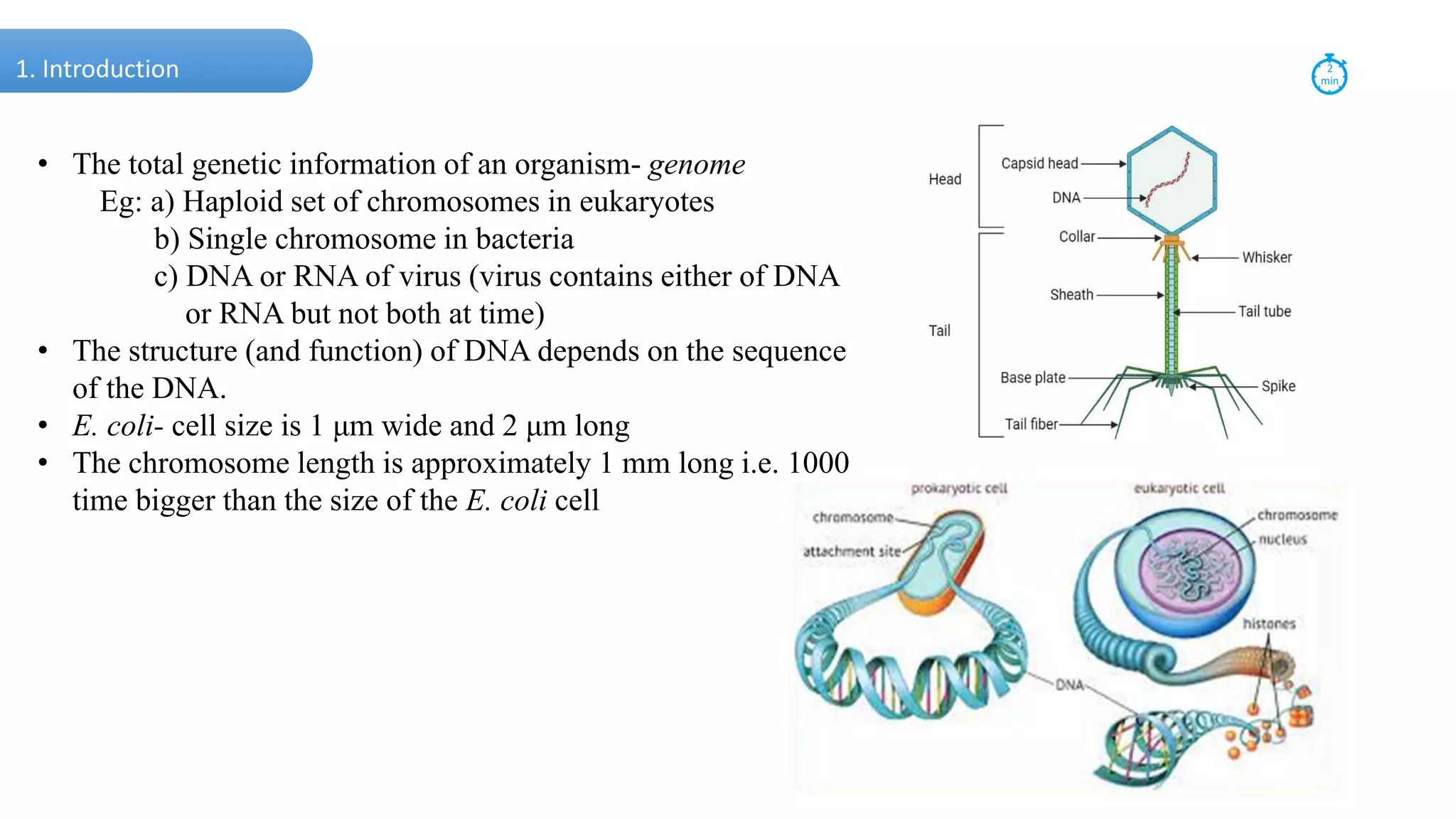
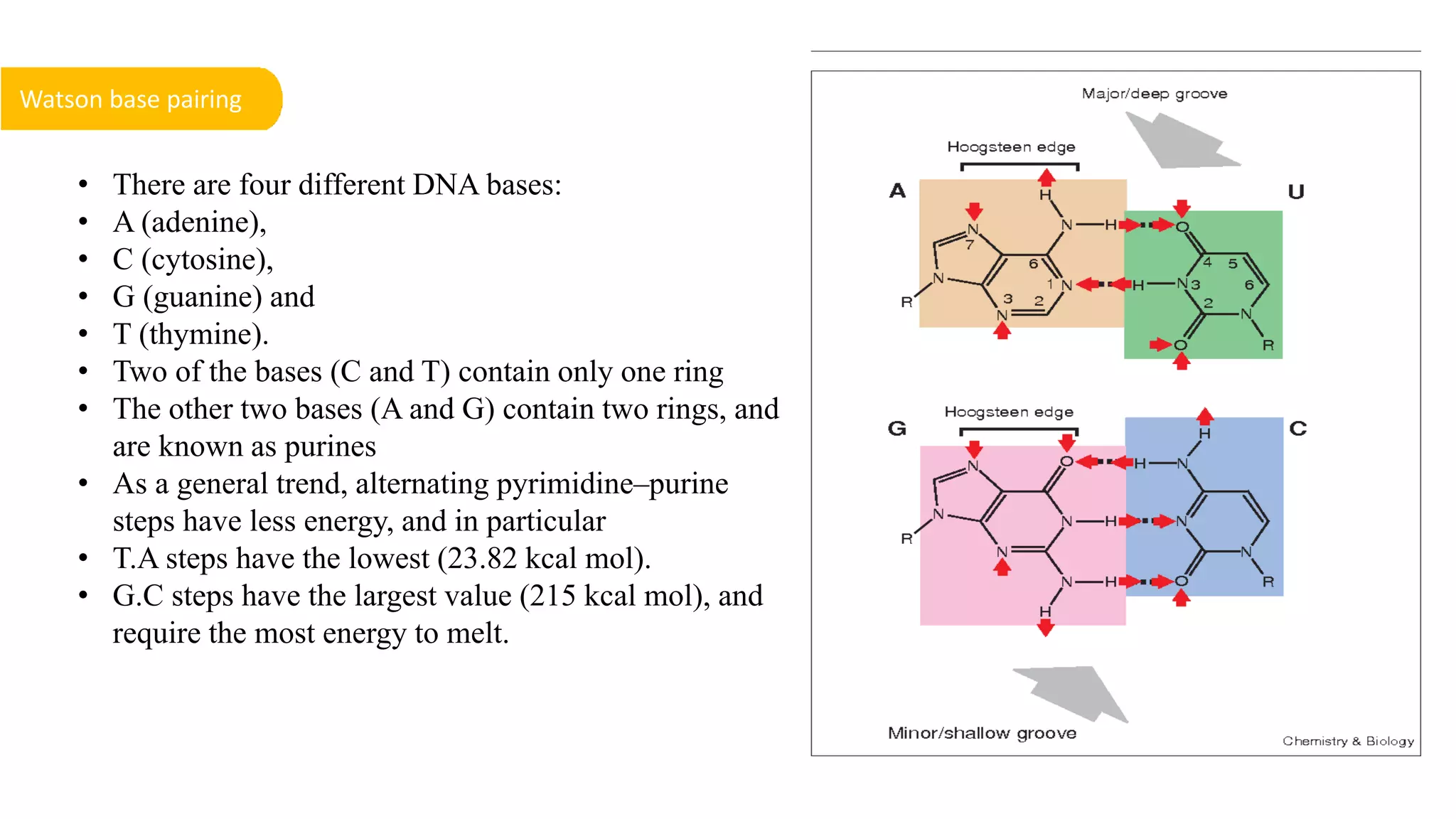
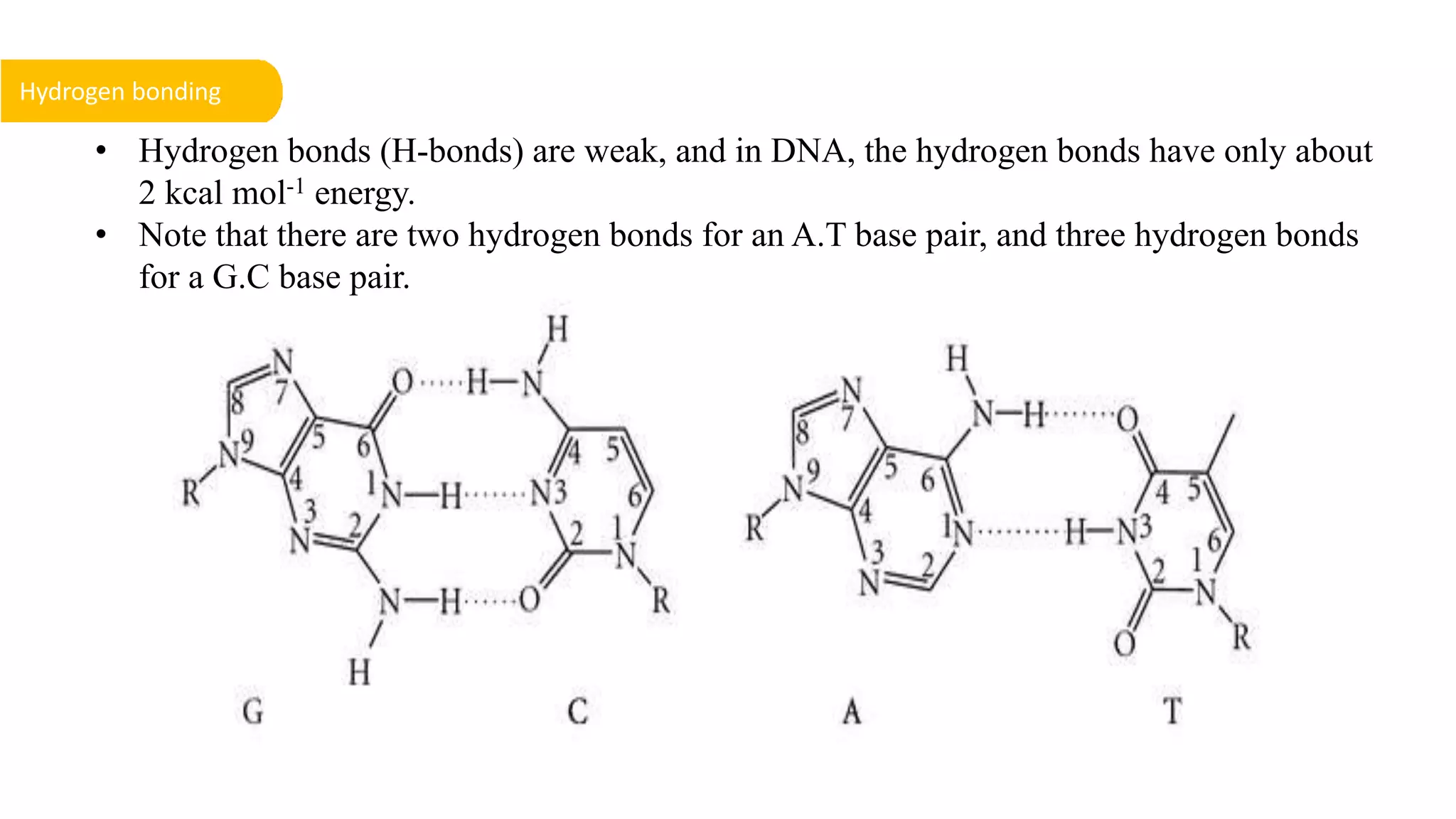
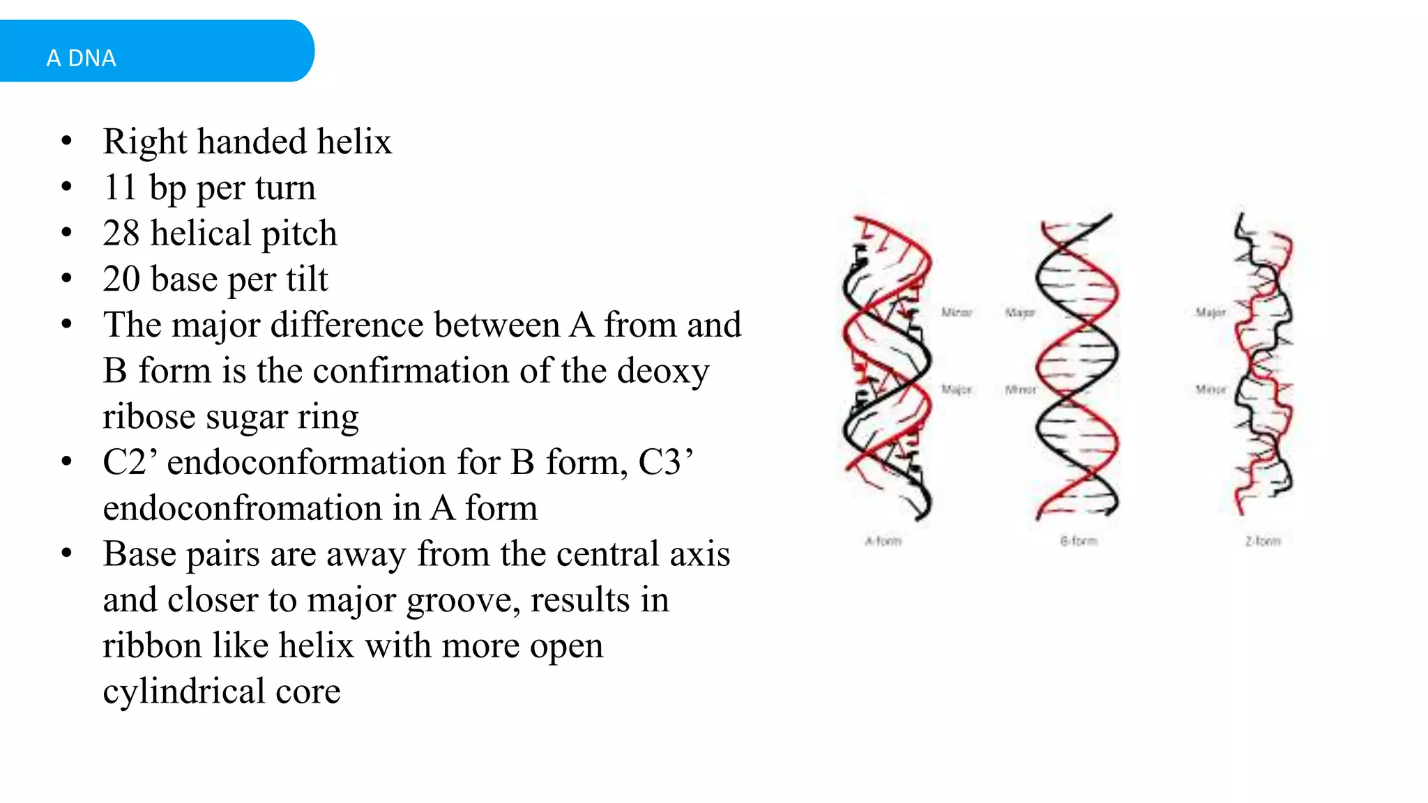
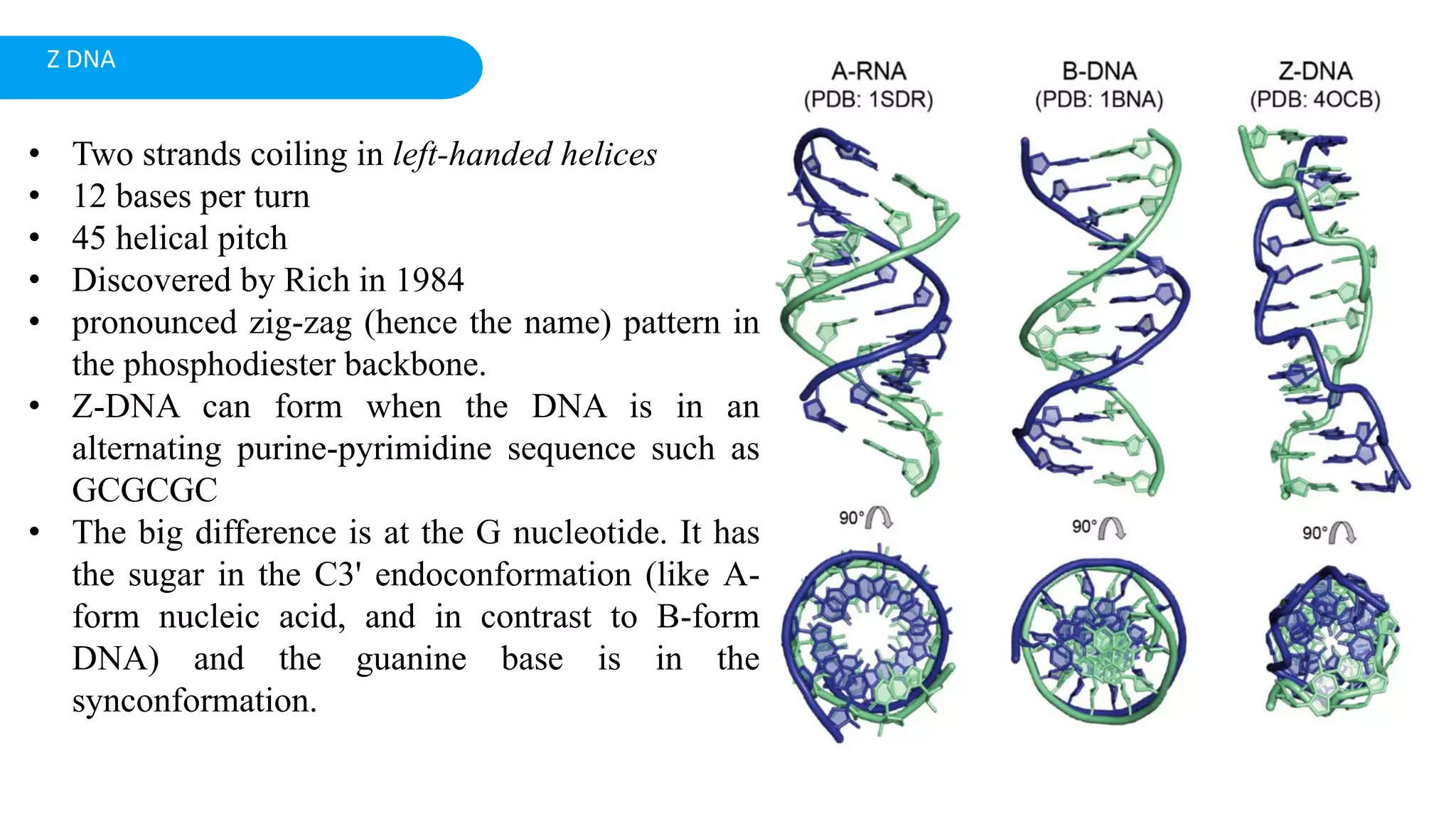
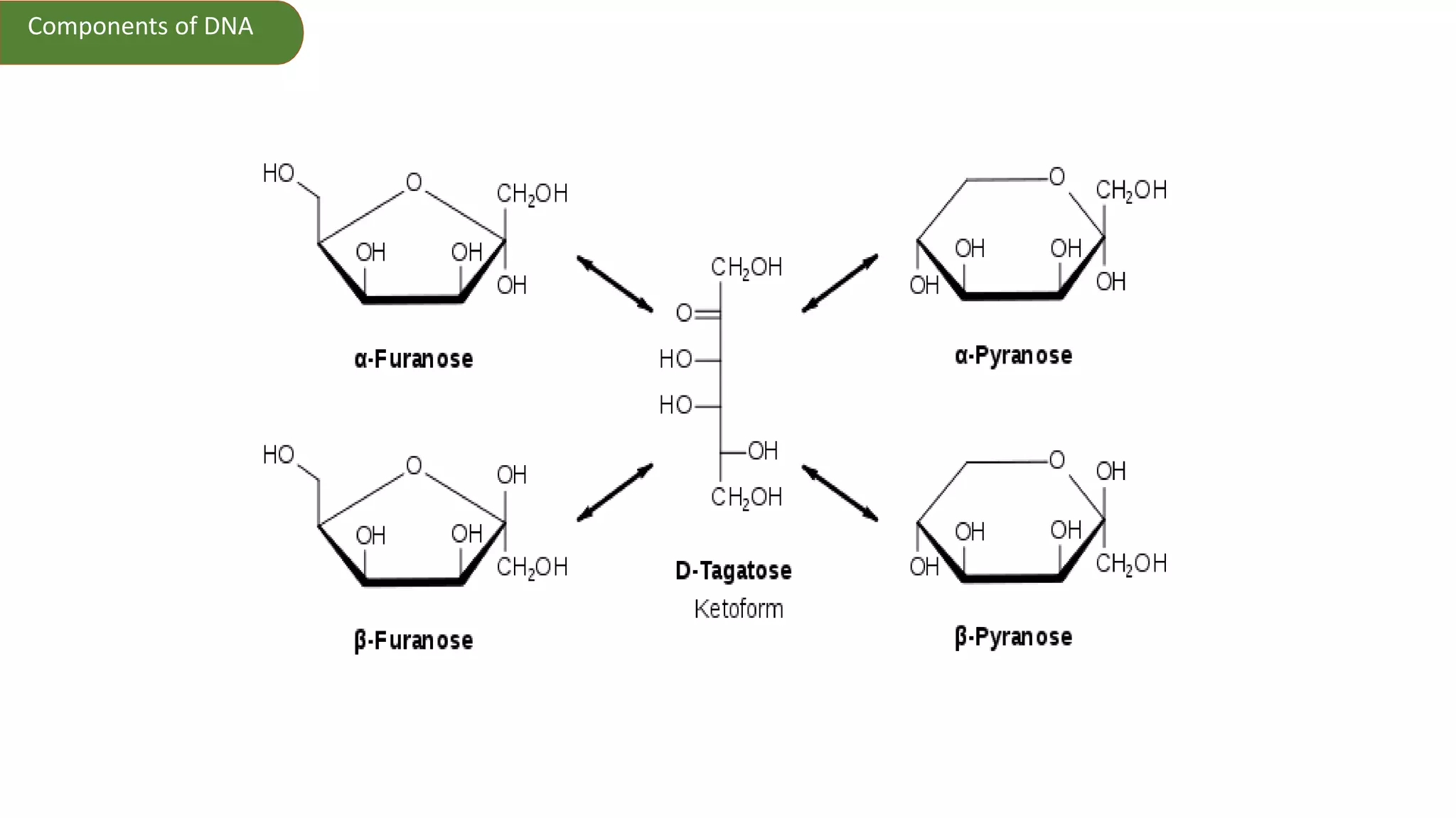
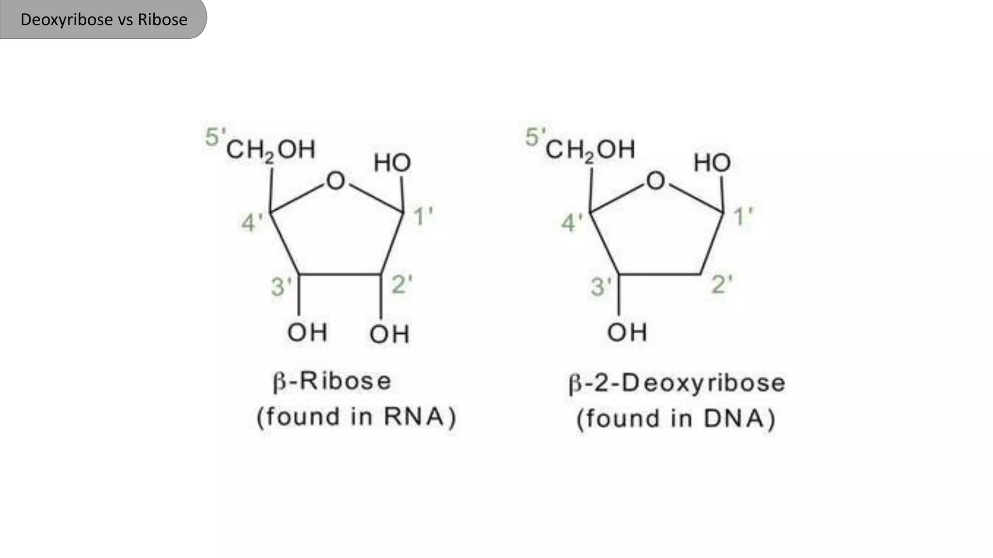
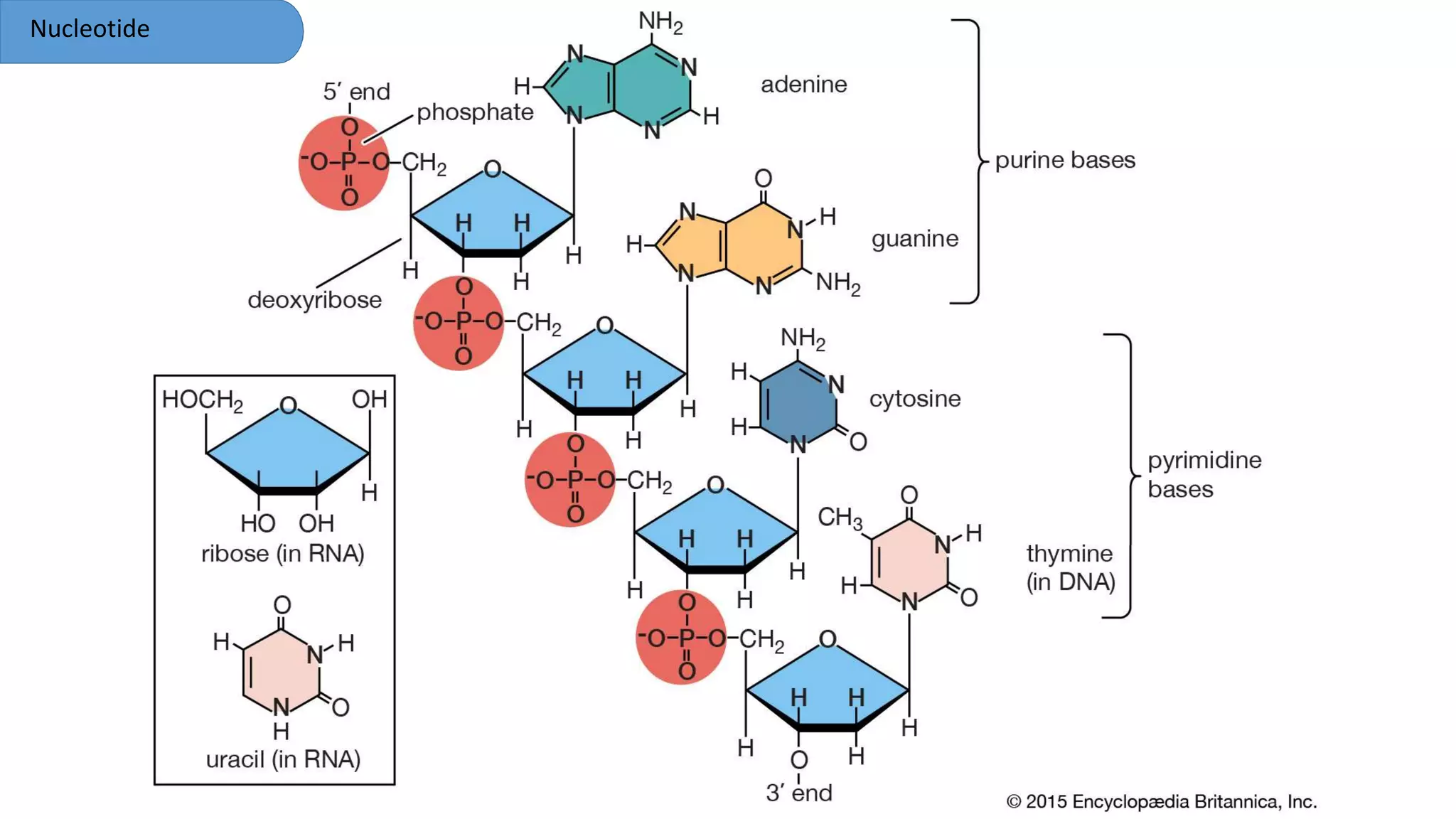
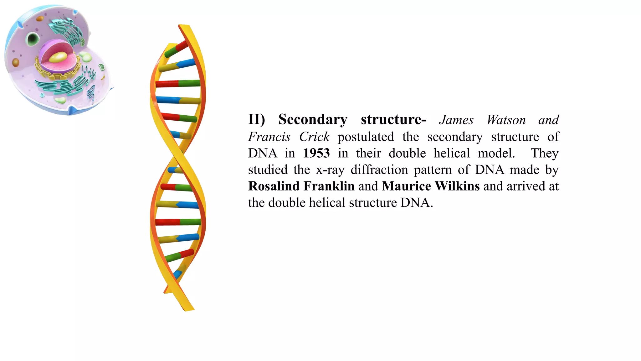
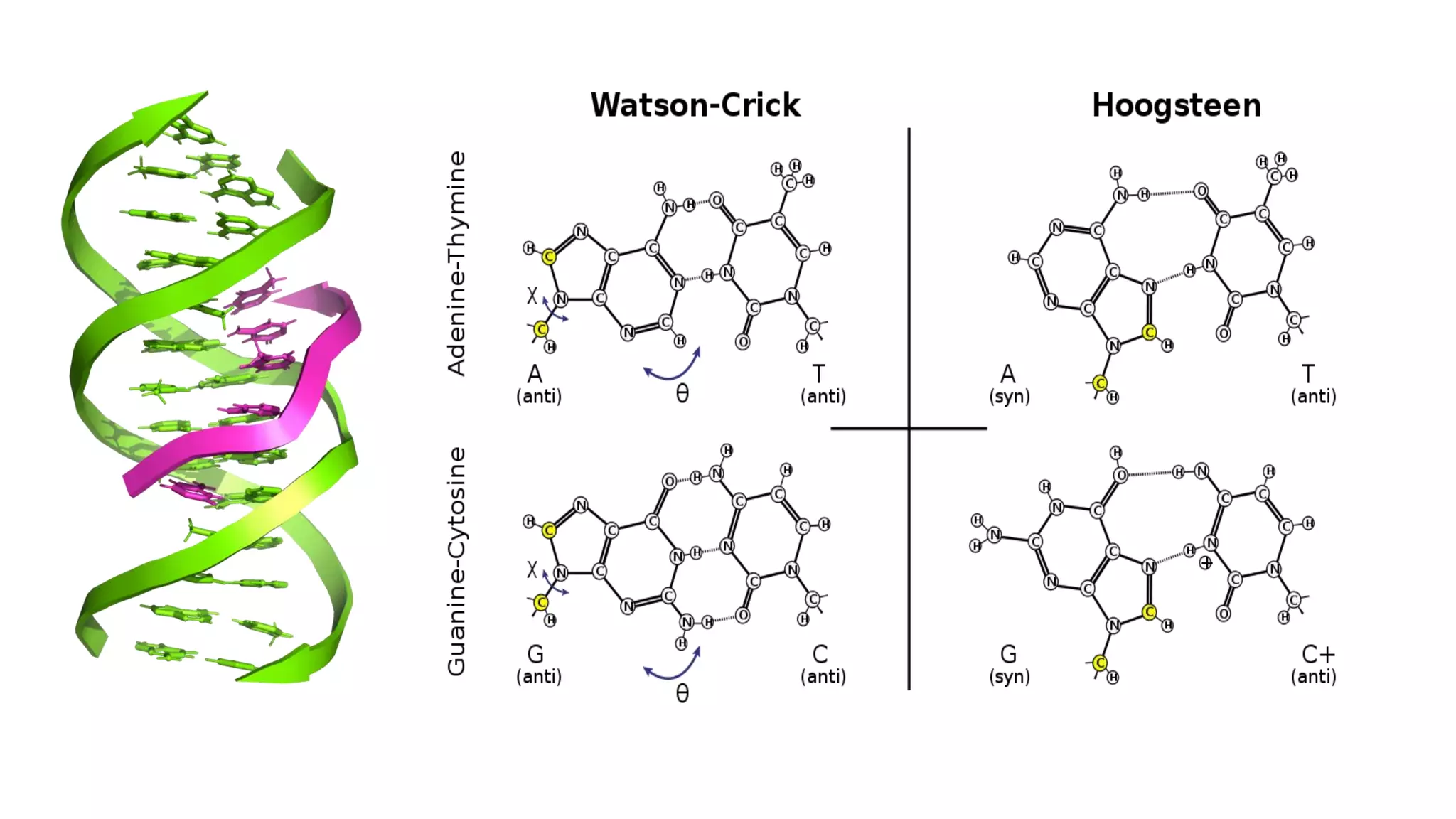
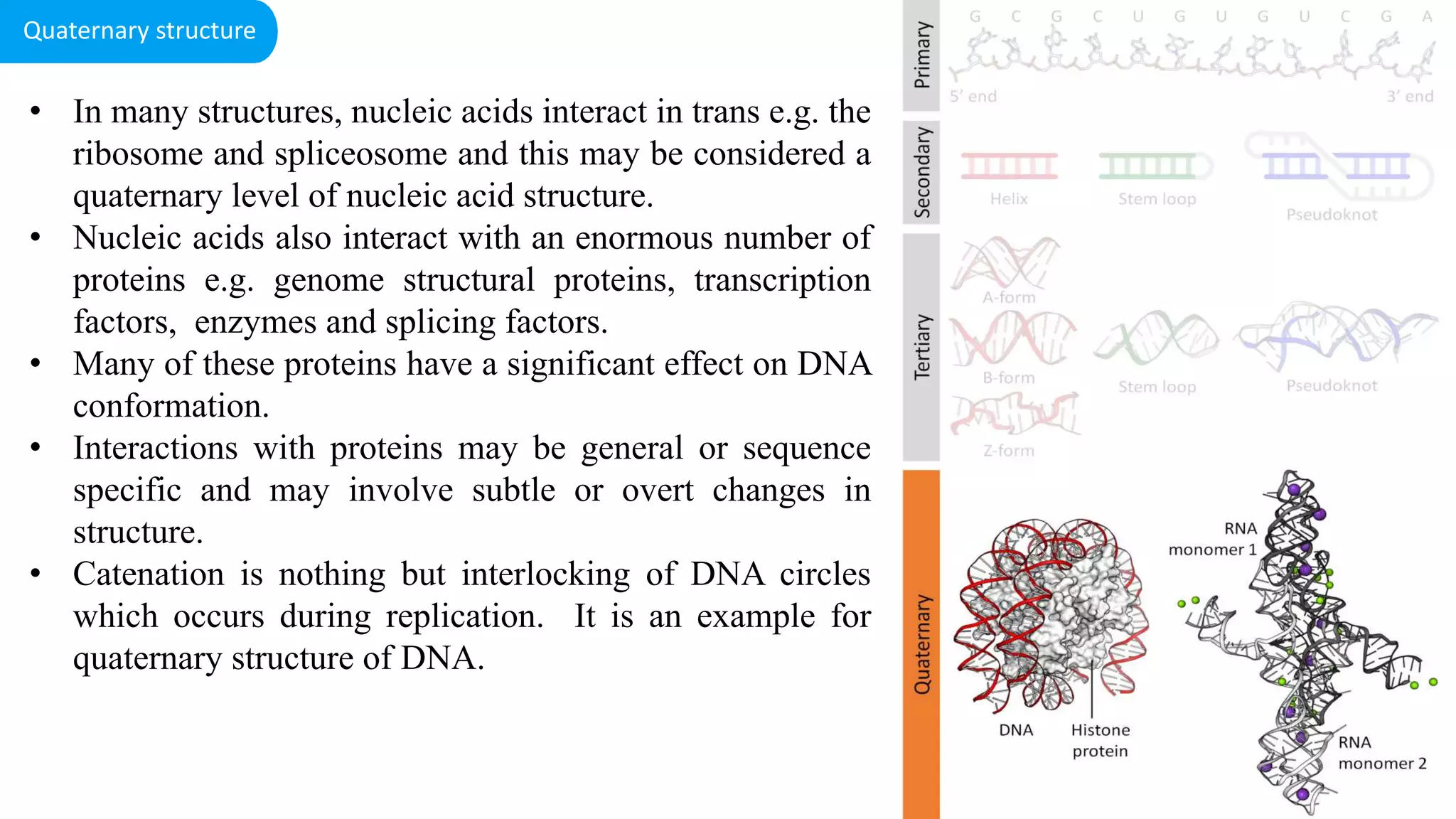
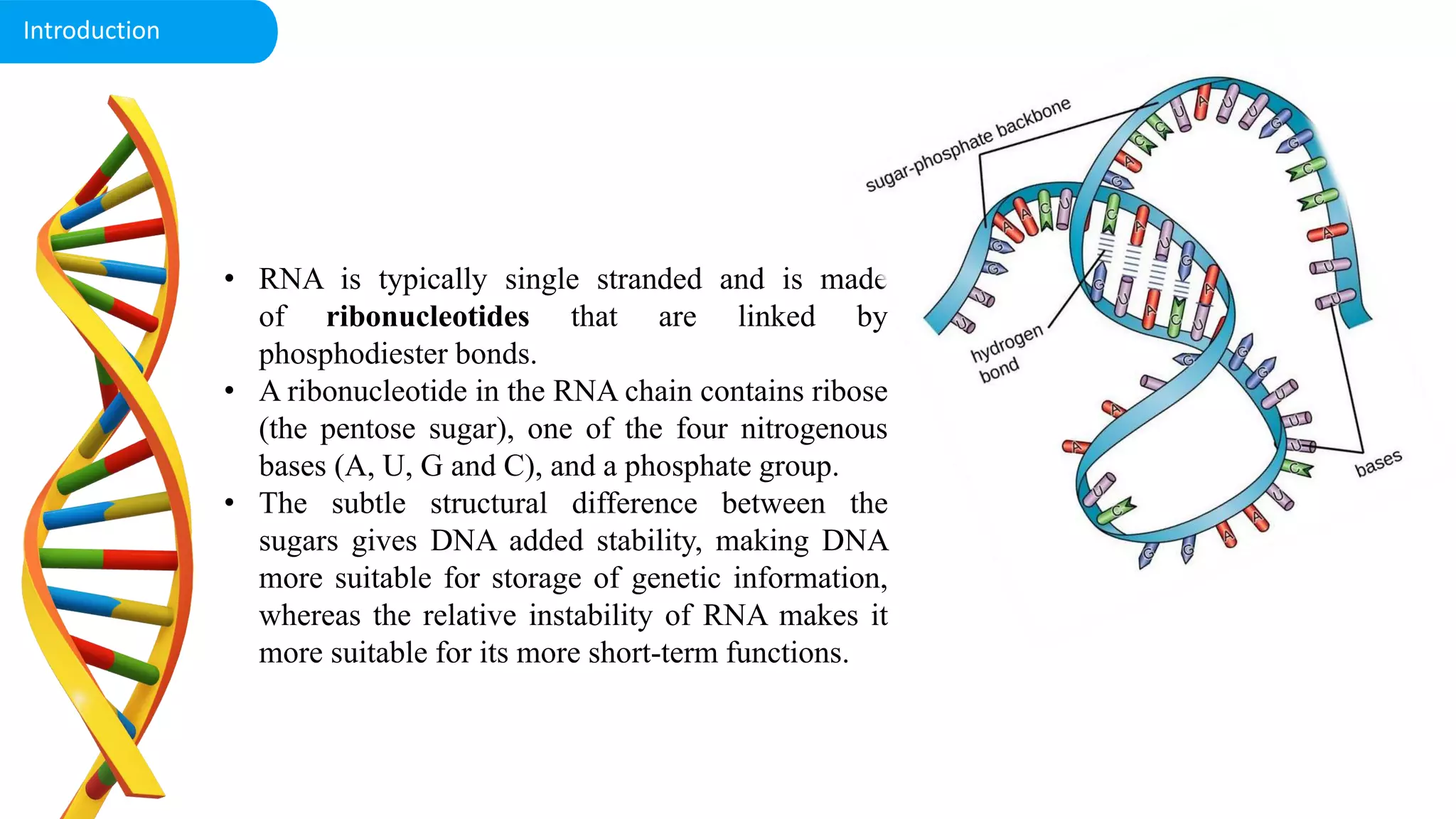
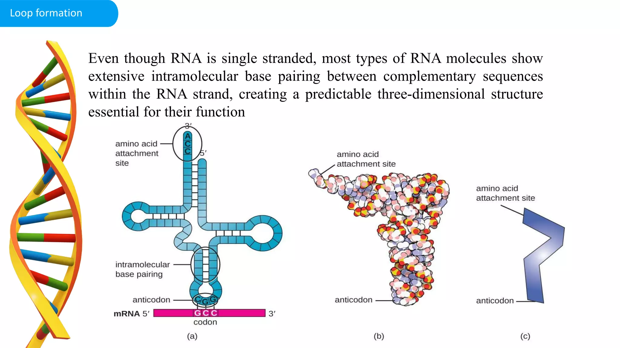
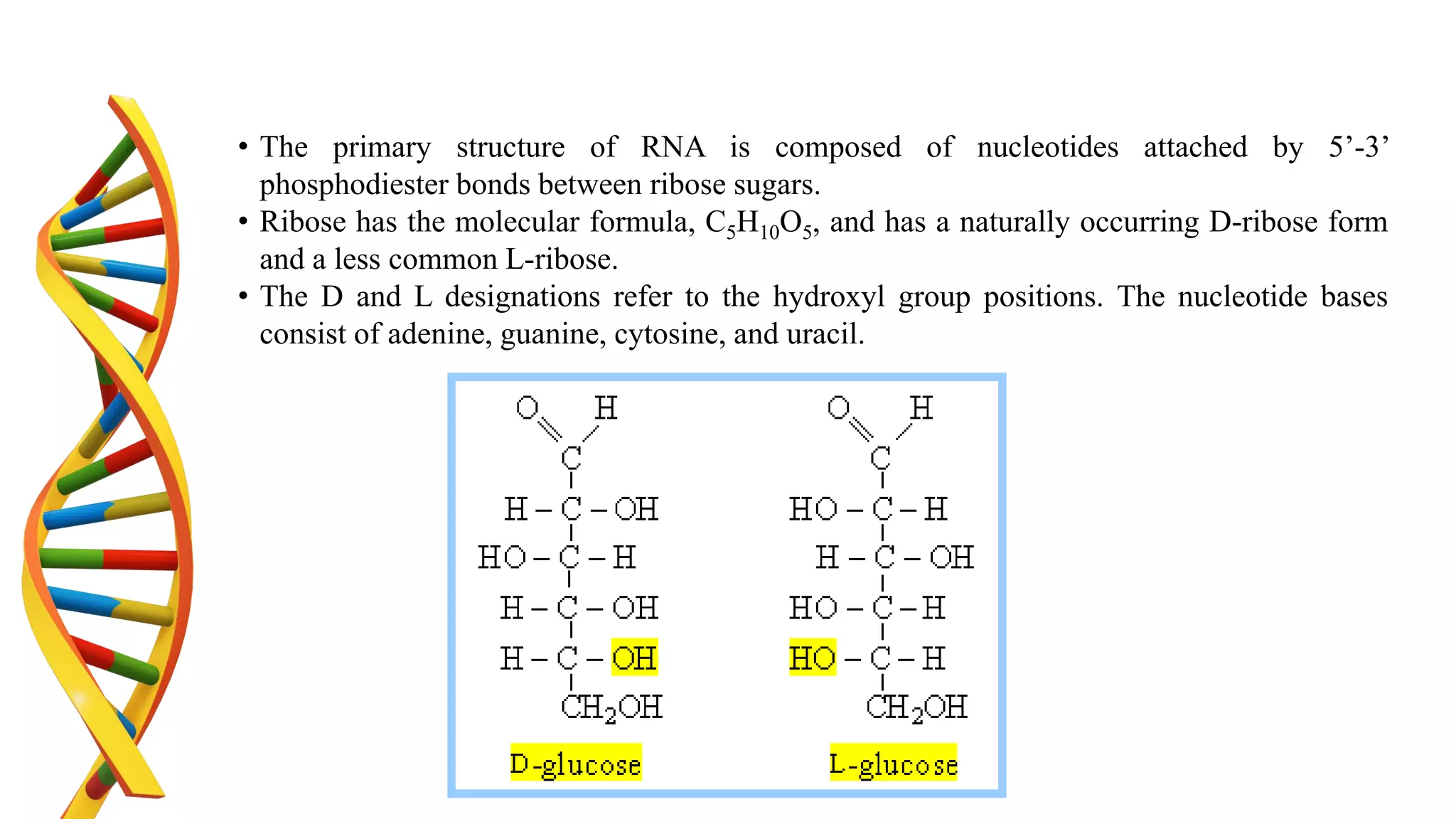
Duration: 60 min Post Graduate Grades: PG PJTSAU
DNA structure
Microbial Genetics 3(2+1)
Background information
• DNA is genetic material of living things except: RNA viruses
• Any living organism may contain only one type of cell either
A. Prokaryotic cells (Pro =before; karyon= nut or kernel)
B. Eukaryotic cells.
• The terms prokaryotic and eukaryotic were suggested by Hans Ris in the 1960’s.
• The DNA/nuclear material is tightly packed in the nucleoid. But why?
• Recognize and characterize the types of DNA
• Identify the parts which differ between eukaryotic cells and prokaryotic cells.
• Define different DNA helices
Learning objectives
1. Introduction 2
min
• The total genetic information of an organism- genome
Eg: a) Haploid set of chromosomes in eukaryotes
b) Single chromosome in bacteria
c) DNA or RNA of virus (virus contains either of DNA
or RNA but not both at time)
• The structure (and function) of DNA depends on the sequence
of the DNA.
• E. coli- cell size is 1 μm wide and 2 μm long
• The chromosome length is approximately 1 mm long i.e. 1000
time bigger than the size of the E. coli cell
What does a DNA look like?
2
min
• There are four different DNA bases:
• A (adenine),
• C (cytosine),
• G (guanine) and
• T (thymine).
• Two of the bases (C and T) contain only one ring
• The other two bases (A and G) contain two rings, and
are known as purines
• As a general trend, alternating pyrimidine–purine
steps have less energy, and in particular
• T.A steps have the lowest (23.82 kcal mol).
• G.C steps have the largest value (215 kcal mol), and
require the most energy to melt.
Watson base pairing
• Hydrogen bonds (H-bonds) are weak, and in DNA, the hydrogen bonds have only about
2 kcal mol-1 energy.
• Note that there are two hydrogen bonds for an A.T base pair, and three hydrogen bonds
for a G.C base pair.
Hydrogen bonding
4. Eukaryotic vs. Prokaryotic: Is there any difference? 5
min
Prokaryotic cell
• A single cellular DNA but recent research has shown
the presence of even multiple and linear forms of
DNA
• Small in size lacks membrane bound nucleus
• No problem of end replication
• Plasmids are extrachromosomal, covalently closed
circular (CCC)
• No histone proteins
• Have single origin of replication
• Chromosomes lack centromere, telomere
• Replication is relatively simple, one replication fork
• DNA replication speed is 2000 bp/sec
• Two proteins are required for the initiation of DNA
replication
• Genes are organized as operons, high percentage of
genes code for proteins
4. Eukaryotic genome
eukaryotic cell
• Multiple copies of chromosome
• Multiple origin of replication
• Replication speed is 100 bp/sec
• Multiple protein component is required to initiate the
replication
• Each gene code for single protein, only 5 % of the
DNA codes for protein
• Genes have two versions
DNA packing
• The DNA molecule consists of two strands
that wind around one another to form a
shape known as a double helix. Each strand
has a backbone made of alternating sugar
(deoxyribose) and phosphate groups.
Attached to each sugar is one of four bases-
-adenine (A), cytosine (C), guanine (G),
and thymine (T).
• Phoshodiester back bone: The phosphate
backbone consists of deoxyribose sugar
molecules linked together by phosphate
groups
• The backbone continues on in a simple
repetitive pattern
• The phosphate groups have a negative
charge, giving the outside of the DNA an
overall negative charge.
DNA Structure
There are three families of DNA helices:
• A-DNA :- which can readily form
within certain stretches of purines
(e.g.GAGGGA);
• B-DNA :- which is favored by mixed
sequences (although the exact
conformation depends on the particular
nucleotide sequence)
• Z-DNA :- which is favored by
alternating pyrimidine– purine steps
(e.g. CGCGCG).
• The A-and B-DNA families are right-
handed helices, while the Z-DNA
family has a left-handed orientation of
the helix
Types of DNA helices
• Also known as whattson-Crick form
• Two strands of DNA wound around the same axis
• The two strands are held together with Hydrogen
bonding
• Two strands are antiparlel
• The nucleotides arrayed in 5’ to 3’ orientation on
one strand which align with complementary
nucleotides in 3’ to 5’ orientation
• Based on chargaffs rule A binds with T with two
hydrogen bonds and G binds with C with three
hydrogen bonds
• 34 nm between bp
• 3.4 nm per turn
• 10 bp per turn
B DNA
A DNA
• Right handed helix
• 11 bp per turn
• 28 helical pitch
• 20 base per tilt
• The major difference between A from and
B form is the confirmation of the deoxy
ribose sugar ring
• C2’ endoconformation for B form, C3’
endoconfromation in A form
• Base pairs are away from the central axis
and closer to major groove, results in
ribbon like helix with more open
cylindrical core
• Two strands coiling in left-handed helices
• 12 bases per turn
• 45 helical pitch
• Discovered by Rich in 1984
• pronounced zig-zag (hence the name) pattern in
the phosphodiester backbone.
• Z-DNA can form when the DNA is in an
alternating purine-pyrimidine sequence such as
GCGCGC
• The big difference is at the G nucleotide. It has
the sugar in the C3' endoconformation (like A-
form nucleic acid, and in contrast to B-form
DNA) and the guanine base is in the
synconformation.
Z DNA
• The first is the ionic or hydration environment, which can facilitate conversion between
different helical forms.
• A-DNA is favored by low hydration, whereas Z-DNA can be favored by high salt.
• The second condition is the DNA sequence: A-DNA is favored by certain stretches of
purines (or pyrimidines), whereas Z-DNA can be most readily formed by alternating
purine-pyrimidine steps.
• The third condition is the presence of proteins that can bind to DNA in one helical
conformation and force the DNA to adopt a different conformation, such as proteins
which bind to B-DNA and can drive it to either A-or Z forms.
• In living cells, most of the DNA is in a mixture of Aand B-DNA conformations, with a
few small regions capable of forming Z-DNA.
5. Activity – Every cell is unique
C-DNA
• Formed at 66% relative humidity and in presence of Li+ and Mg2+ ions.
• Right-handed with the axial rise of 3.32 A° per base pair
• 33 base pairs per turn
• Helical pitch 3.32A°×9.33°A=30.97A°.
• Base pair rotation=38.58°.
• Has a diameter of 19 A°, smaller than that of A-&B- DNA.
• The tilt of base is 7.8°
D-DNA
• Rare variant with 8 base pairs per helical turn
• These forms of DNA found in some DNA molecules devoid of guanine.
• The axial rise of 3.03 A°per base pairs
• The tilt of 16.7° from the axis of the helix.
E- DNA
• Extended or eccentric DNA.
• E-DNA has a long helical axis rise and base perpendicular to the helical axis.
• Deep major groove and the shallow minor groove.
• E-DNA allowed to crystallize for a period time longer, the methylated sequence forms
standard A-DNA.
• E-DNA is the intermediate in the crystallographic pathway from B-DNA to A-DNA.
Components of DNA
Ribose isomers
Furanose vs Pyranose
Deoxyribose vs Ribose
Nitrogen bases
Nucleotide
Nucleotide
Bhor’s model
Bhor’s model of Phosphorous
Valency of Phosphorous
Bhor’s Model of oxygen
Nucleotide
RNA
There are four different structures namely primary secondary, tertiary
and quaternary
i) Primary structure - deoxyribo nucleotides linked to each other
by phosphodiester bonds.
• The substrates for polymerization are nucleoside triphosphates,
but the repeating unit or monomer, of a nucleic acid is a
monophosphate.
• During polymerization, the 3’-OH group of the terminal
nucleotide residue in the existing chain makes a nucleophilic
attack upon the alpha phosphate of the incoming nucleoside
triphosphate to form 5’ to 3’ phosphodiester bond. This reaction is
catalyzed by DNA polymerase.
• Serial polymerization generates long polymers variously called
chains or strands, containing an invariant sugar-phosphate
backbone with 5’ to 3’ polarity and projecting nitrogenous
base.
Structure of DNA
II) Secondary structure- James Watson and
Francis Crick postulated the secondary structure of
DNA in 1953 in their double helical model. They
studied the x-ray diffraction pattern of DNA made by
Rosalind Franklin and Maurice Wilkins and arrived at
the double helical structure DNA.
• Nucleic acid tertiary structures reflect interactions which contribute to overall three dimensional
shape.
• This includes interactions between different secondary structure elements, interactions between single
strands and secondary structures and topological properties of nucleic acids.
• Examples for tertiary include cruciform, triple helices and super coils.
III) Tertiary structure
• Cruciform are cross-links structures.
• They are likely to form when a DNA sequence contains a palindrome or inverted repeats
• palindrome is defined as a sequence that provides the same information whether it is read forward or
backward e.g. MADAM I’M ADAM.
• In one proposed mechanism, cruciform formation begins with a small bubble, or proto cruciform and
progresses as intra-strand base pairing occurs.
• The mechanism, which bubble initiates formation, is unknown.
• Cruciform DNA is found in both prokaryotes and eukaryotes and has a role in DNA transcription and DNA
replication, double strand repair, DNA translocation and recombination.
• There are two mechanisms of formation of cruciform DNA
a) C- type (temperature dependent)
b) S- type (salt extrusion)
a) Cruciform
b) triple /quadruple helix:
• In DNA, tertiary interactions involve single stands
interacting with duplexes or duplexes interacting with
duplexes, resulting in the formation of triple and quadruple
strand structures.
• In certain circumstances e.g. low pH, a DNA sequence
containing a long segments consisting of a polypurine
stand hydrogen-bonded to a polypyrimidine strand can
form a triple helix.
• The formation of the triple helix, also referred to a H-
DNA, depends on the formation of nonconventional base
pairing (HOOGSTEEN Base pairing), which occurs
without disruption the Watson-Crick base pairs.
• The significance of H-DNA is unclear, although it is
implicated in the regulation of some genes.
• Quadruple structure is one of the tertiary structure of
DNA. Guanosine repeats like in telomere ex: (T4G2)2 in
Oxytrichia can associate to form cyclic tetramers known as
G-quartets.
b) Triple helix
• In many structures, nucleic acids interact in trans e.g. the
ribosome and spliceosome and this may be considered a
quaternary level of nucleic acid structure.
• Nucleic acids also interact with an enormous number of
proteins e.g. genome structural proteins, transcription
factors, enzymes and splicing factors.
• Many of these proteins have a significant effect on DNA
conformation.
• Interactions with proteins may be general or sequence
specific and may involve subtle or overt changes in
structure.
• Catenation is nothing but interlocking of DNA circles
which occurs during replication. It is an example for
quaternary structure of DNA.
Quaternary structure
Ribonucleic acid
(RNA)
• RNA is typically single stranded and is made
of ribonucleotides that are linked by
phosphodiester bonds.
• A ribonucleotide in the RNA chain contains ribose
(the pentose sugar), one of the four nitrogenous
bases (A, U, G and C), and a phosphate group.
• The subtle structural difference between the
sugars gives DNA added stability, making DNA
more suitable for storage of genetic information,
whereas the relative instability of RNA makes it
more suitable for its more short-term functions.
Introduction
The RNA specific pyrimidine uracil forms a
complementary base pair with adenine and is used
instead of the thymine used in DNA.
Uracil
Even though RNA is single stranded, most types of RNA molecules show
extensive intramolecular base pairing between complementary sequences
within the RNA strand, creating a predictable three-dimensional structure
essential for their function
Loop formation
• The primary structure of RNA is composed of nucleotides attached by 5’-3’
phosphodiester bonds between ribose sugars.
• Ribose has the molecular formula, C5H10O5, and has a naturally occurring D-ribose form
and a less common L-ribose.
• The D and L designations refer to the hydroxyl group positions. The nucleotide bases
consist of adenine, guanine, cytosine, and uracil.
• Two hydrogen bonds form between adenine and uracil, while three bonds form
between cytosine and guanine.
• The base pairing via hydrogen bonds is the basis of RNA secondary structure.
• The RNA tertiary structure is the result of RNA folding, which creates a three-
dimensional shape consisting of helices and grooves.
• RNA differs from DNA in that it contains a uracil nucleotide instead of thymine and
carries a 2’ hydroxyl group rather than a 2’ hydrogen.
• Due to its interaction with the solvent environment, the 2’ hydroxyl group
contributes to RNA conformation.
• mRNA is transcribed from DNA and contains the genetic blueprint to make proteins.
• Prokaryotic mRNA does not need to be processed and can proceed to synthesize proteins
immediately.
• In eukaryotes, a freshly transcribed RNA transcript is considered a pre-mRNA and needs to
undergo maturation to form mRNA.
• A pre-mRNA contains non-coding and coding regions known as introns and exons,
respectively.
• During pre-mRNA processing, the introns are spliced, and the exons are joined together.
• A 5’ cap known as 7-methylguanosine is added to the 5’ end of the RNA transcript and the
3’ end is polyadenylated.
• Polyadenylation refers to the process where a poly(A) tail, which is a sequence of adenine
nucleotides, is added to the transcript.
• The 5’ cap protects the mRNA from degradation, and the 3’ poly(A) tail contributes to the
stability of mRNA and aids it in transport.
• Researchers are also studying mRNA as an anti-cancer treatment due to its ability to modify
cells.

DNA structure

  • 1.
    Duration: 60 minPost Graduate Grades: PG PJTSAU DNA structure Microbial Genetics 3(2+1)
  • 2.
    Background information • DNAis genetic material of living things except: RNA viruses • Any living organism may contain only one type of cell either A. Prokaryotic cells (Pro =before; karyon= nut or kernel) B. Eukaryotic cells. • The terms prokaryotic and eukaryotic were suggested by Hans Ris in the 1960’s. • The DNA/nuclear material is tightly packed in the nucleoid. But why? • Recognize and characterize the types of DNA • Identify the parts which differ between eukaryotic cells and prokaryotic cells. • Define different DNA helices Learning objectives
  • 3.
    1. Introduction 2 min •The total genetic information of an organism- genome Eg: a) Haploid set of chromosomes in eukaryotes b) Single chromosome in bacteria c) DNA or RNA of virus (virus contains either of DNA or RNA but not both at time) • The structure (and function) of DNA depends on the sequence of the DNA. • E. coli- cell size is 1 μm wide and 2 μm long • The chromosome length is approximately 1 mm long i.e. 1000 time bigger than the size of the E. coli cell
  • 4.
    What does aDNA look like? 2 min
  • 5.
    • There arefour different DNA bases: • A (adenine), • C (cytosine), • G (guanine) and • T (thymine). • Two of the bases (C and T) contain only one ring • The other two bases (A and G) contain two rings, and are known as purines • As a general trend, alternating pyrimidine–purine steps have less energy, and in particular • T.A steps have the lowest (23.82 kcal mol). • G.C steps have the largest value (215 kcal mol), and require the most energy to melt. Watson base pairing
  • 6.
    • Hydrogen bonds(H-bonds) are weak, and in DNA, the hydrogen bonds have only about 2 kcal mol-1 energy. • Note that there are two hydrogen bonds for an A.T base pair, and three hydrogen bonds for a G.C base pair. Hydrogen bonding
  • 8.
    4. Eukaryotic vs.Prokaryotic: Is there any difference? 5 min Prokaryotic cell • A single cellular DNA but recent research has shown the presence of even multiple and linear forms of DNA • Small in size lacks membrane bound nucleus • No problem of end replication • Plasmids are extrachromosomal, covalently closed circular (CCC) • No histone proteins • Have single origin of replication • Chromosomes lack centromere, telomere • Replication is relatively simple, one replication fork • DNA replication speed is 2000 bp/sec • Two proteins are required for the initiation of DNA replication • Genes are organized as operons, high percentage of genes code for proteins
  • 9.
    4. Eukaryotic genome eukaryoticcell • Multiple copies of chromosome • Multiple origin of replication • Replication speed is 100 bp/sec • Multiple protein component is required to initiate the replication • Each gene code for single protein, only 5 % of the DNA codes for protein • Genes have two versions DNA packing
  • 10.
    • The DNAmolecule consists of two strands that wind around one another to form a shape known as a double helix. Each strand has a backbone made of alternating sugar (deoxyribose) and phosphate groups. Attached to each sugar is one of four bases- -adenine (A), cytosine (C), guanine (G), and thymine (T). • Phoshodiester back bone: The phosphate backbone consists of deoxyribose sugar molecules linked together by phosphate groups • The backbone continues on in a simple repetitive pattern • The phosphate groups have a negative charge, giving the outside of the DNA an overall negative charge. DNA Structure
  • 11.
    There are threefamilies of DNA helices: • A-DNA :- which can readily form within certain stretches of purines (e.g.GAGGGA); • B-DNA :- which is favored by mixed sequences (although the exact conformation depends on the particular nucleotide sequence) • Z-DNA :- which is favored by alternating pyrimidine– purine steps (e.g. CGCGCG). • The A-and B-DNA families are right- handed helices, while the Z-DNA family has a left-handed orientation of the helix Types of DNA helices
  • 12.
    • Also knownas whattson-Crick form • Two strands of DNA wound around the same axis • The two strands are held together with Hydrogen bonding • Two strands are antiparlel • The nucleotides arrayed in 5’ to 3’ orientation on one strand which align with complementary nucleotides in 3’ to 5’ orientation • Based on chargaffs rule A binds with T with two hydrogen bonds and G binds with C with three hydrogen bonds • 34 nm between bp • 3.4 nm per turn • 10 bp per turn B DNA
  • 13.
    A DNA • Righthanded helix • 11 bp per turn • 28 helical pitch • 20 base per tilt • The major difference between A from and B form is the confirmation of the deoxy ribose sugar ring • C2’ endoconformation for B form, C3’ endoconfromation in A form • Base pairs are away from the central axis and closer to major groove, results in ribbon like helix with more open cylindrical core
  • 14.
    • Two strandscoiling in left-handed helices • 12 bases per turn • 45 helical pitch • Discovered by Rich in 1984 • pronounced zig-zag (hence the name) pattern in the phosphodiester backbone. • Z-DNA can form when the DNA is in an alternating purine-pyrimidine sequence such as GCGCGC • The big difference is at the G nucleotide. It has the sugar in the C3' endoconformation (like A- form nucleic acid, and in contrast to B-form DNA) and the guanine base is in the synconformation. Z DNA
  • 15.
    • The firstis the ionic or hydration environment, which can facilitate conversion between different helical forms. • A-DNA is favored by low hydration, whereas Z-DNA can be favored by high salt. • The second condition is the DNA sequence: A-DNA is favored by certain stretches of purines (or pyrimidines), whereas Z-DNA can be most readily formed by alternating purine-pyrimidine steps. • The third condition is the presence of proteins that can bind to DNA in one helical conformation and force the DNA to adopt a different conformation, such as proteins which bind to B-DNA and can drive it to either A-or Z forms. • In living cells, most of the DNA is in a mixture of Aand B-DNA conformations, with a few small regions capable of forming Z-DNA. 5. Activity – Every cell is unique
  • 16.
    C-DNA • Formed at66% relative humidity and in presence of Li+ and Mg2+ ions. • Right-handed with the axial rise of 3.32 A° per base pair • 33 base pairs per turn • Helical pitch 3.32A°×9.33°A=30.97A°. • Base pair rotation=38.58°. • Has a diameter of 19 A°, smaller than that of A-&B- DNA. • The tilt of base is 7.8° D-DNA • Rare variant with 8 base pairs per helical turn • These forms of DNA found in some DNA molecules devoid of guanine. • The axial rise of 3.03 A°per base pairs • The tilt of 16.7° from the axis of the helix. E- DNA • Extended or eccentric DNA. • E-DNA has a long helical axis rise and base perpendicular to the helical axis. • Deep major groove and the shallow minor groove. • E-DNA allowed to crystallize for a period time longer, the methylated sequence forms standard A-DNA. • E-DNA is the intermediate in the crystallographic pathway from B-DNA to A-DNA.
  • 17.
  • 18.
  • 19.
  • 20.
  • 21.
  • 22.
  • 23.
  • 24.
  • 25.
    Bhor’s model ofPhosphorous
  • 26.
  • 27.
  • 28.
  • 29.
  • 30.
    There are fourdifferent structures namely primary secondary, tertiary and quaternary i) Primary structure - deoxyribo nucleotides linked to each other by phosphodiester bonds. • The substrates for polymerization are nucleoside triphosphates, but the repeating unit or monomer, of a nucleic acid is a monophosphate. • During polymerization, the 3’-OH group of the terminal nucleotide residue in the existing chain makes a nucleophilic attack upon the alpha phosphate of the incoming nucleoside triphosphate to form 5’ to 3’ phosphodiester bond. This reaction is catalyzed by DNA polymerase. • Serial polymerization generates long polymers variously called chains or strands, containing an invariant sugar-phosphate backbone with 5’ to 3’ polarity and projecting nitrogenous base. Structure of DNA
  • 31.
    II) Secondary structure-James Watson and Francis Crick postulated the secondary structure of DNA in 1953 in their double helical model. They studied the x-ray diffraction pattern of DNA made by Rosalind Franklin and Maurice Wilkins and arrived at the double helical structure DNA.
  • 32.
    • Nucleic acidtertiary structures reflect interactions which contribute to overall three dimensional shape. • This includes interactions between different secondary structure elements, interactions between single strands and secondary structures and topological properties of nucleic acids. • Examples for tertiary include cruciform, triple helices and super coils. III) Tertiary structure
  • 33.
    • Cruciform arecross-links structures. • They are likely to form when a DNA sequence contains a palindrome or inverted repeats • palindrome is defined as a sequence that provides the same information whether it is read forward or backward e.g. MADAM I’M ADAM. • In one proposed mechanism, cruciform formation begins with a small bubble, or proto cruciform and progresses as intra-strand base pairing occurs. • The mechanism, which bubble initiates formation, is unknown. • Cruciform DNA is found in both prokaryotes and eukaryotes and has a role in DNA transcription and DNA replication, double strand repair, DNA translocation and recombination. • There are two mechanisms of formation of cruciform DNA a) C- type (temperature dependent) b) S- type (salt extrusion) a) Cruciform
  • 34.
    b) triple /quadruplehelix: • In DNA, tertiary interactions involve single stands interacting with duplexes or duplexes interacting with duplexes, resulting in the formation of triple and quadruple strand structures. • In certain circumstances e.g. low pH, a DNA sequence containing a long segments consisting of a polypurine stand hydrogen-bonded to a polypyrimidine strand can form a triple helix. • The formation of the triple helix, also referred to a H- DNA, depends on the formation of nonconventional base pairing (HOOGSTEEN Base pairing), which occurs without disruption the Watson-Crick base pairs. • The significance of H-DNA is unclear, although it is implicated in the regulation of some genes. • Quadruple structure is one of the tertiary structure of DNA. Guanosine repeats like in telomere ex: (T4G2)2 in Oxytrichia can associate to form cyclic tetramers known as G-quartets. b) Triple helix
  • 36.
    • In manystructures, nucleic acids interact in trans e.g. the ribosome and spliceosome and this may be considered a quaternary level of nucleic acid structure. • Nucleic acids also interact with an enormous number of proteins e.g. genome structural proteins, transcription factors, enzymes and splicing factors. • Many of these proteins have a significant effect on DNA conformation. • Interactions with proteins may be general or sequence specific and may involve subtle or overt changes in structure. • Catenation is nothing but interlocking of DNA circles which occurs during replication. It is an example for quaternary structure of DNA. Quaternary structure
  • 37.
  • 38.
    • RNA istypically single stranded and is made of ribonucleotides that are linked by phosphodiester bonds. • A ribonucleotide in the RNA chain contains ribose (the pentose sugar), one of the four nitrogenous bases (A, U, G and C), and a phosphate group. • The subtle structural difference between the sugars gives DNA added stability, making DNA more suitable for storage of genetic information, whereas the relative instability of RNA makes it more suitable for its more short-term functions. Introduction
  • 39.
    The RNA specificpyrimidine uracil forms a complementary base pair with adenine and is used instead of the thymine used in DNA. Uracil
  • 40.
    Even though RNAis single stranded, most types of RNA molecules show extensive intramolecular base pairing between complementary sequences within the RNA strand, creating a predictable three-dimensional structure essential for their function Loop formation
  • 41.
    • The primarystructure of RNA is composed of nucleotides attached by 5’-3’ phosphodiester bonds between ribose sugars. • Ribose has the molecular formula, C5H10O5, and has a naturally occurring D-ribose form and a less common L-ribose. • The D and L designations refer to the hydroxyl group positions. The nucleotide bases consist of adenine, guanine, cytosine, and uracil.
  • 42.
    • Two hydrogenbonds form between adenine and uracil, while three bonds form between cytosine and guanine. • The base pairing via hydrogen bonds is the basis of RNA secondary structure. • The RNA tertiary structure is the result of RNA folding, which creates a three- dimensional shape consisting of helices and grooves. • RNA differs from DNA in that it contains a uracil nucleotide instead of thymine and carries a 2’ hydroxyl group rather than a 2’ hydrogen. • Due to its interaction with the solvent environment, the 2’ hydroxyl group contributes to RNA conformation.
  • 43.
    • mRNA istranscribed from DNA and contains the genetic blueprint to make proteins. • Prokaryotic mRNA does not need to be processed and can proceed to synthesize proteins immediately. • In eukaryotes, a freshly transcribed RNA transcript is considered a pre-mRNA and needs to undergo maturation to form mRNA. • A pre-mRNA contains non-coding and coding regions known as introns and exons, respectively. • During pre-mRNA processing, the introns are spliced, and the exons are joined together. • A 5’ cap known as 7-methylguanosine is added to the 5’ end of the RNA transcript and the 3’ end is polyadenylated. • Polyadenylation refers to the process where a poly(A) tail, which is a sequence of adenine nucleotides, is added to the transcript. • The 5’ cap protects the mRNA from degradation, and the 3’ poly(A) tail contributes to the stability of mRNA and aids it in transport. • Researchers are also studying mRNA as an anti-cancer treatment due to its ability to modify cells.