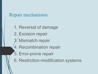
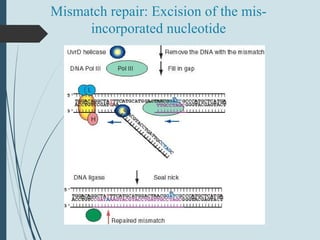
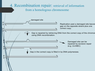
welcome
University of agricUltUral and
horticUltUral and horticUltUral
shivamogga
COLLEGE OF HORTICULTURE, MUDIGERE
Topic : DNA damage repair.
By,
Mahammed Faizan
Jr. M.Sc(HORT) GPB
Introduction
 Each cell contains only one or two copies of its DNA, the DNA
sequence is highly protected from harm.
 DNA is a relatively stable molecule, but Earth’s natural
environment is quite toxic, and damage to DNA is inevitable.
 DNA can also be altered by mistakes made during its own
replication or recombination.
 Damage and sequence alterations to DNA are often quickly
repaired, but when they are not, the DNA becomes
permanently altered and harbors a mutation.
 Mutations are changes in DNA sequence, and when mutations
occur in germ-line cells, these changes are inheritable.
 A cancer cell has mutations that prevent cell death,
resulting in loss of cell cycle control and unregulated cell
division, which leads to malignant tumors that can end
the life of the entire organism.
 The cell has a limited amount of time to fix the initial
alteration and restore the DNA to its normal sequence,
before replication converts the alteration into a mutation
that will be passed on to the next generation.
Co ntinue..
Co ntinue..
 Mutation
 Somatic mutations : occur in somatic cells and only affect
the individual in which the mutation arises.
 Germ-line mutations: alter gametes and passed to the next
generation.
 Mutations are quantified in two ways:
1. Mutation rate = probability of a particular type of mutation
per unit time (or generation).
2. Mutation frequency = number of times a particular
mutation occurs in a population of cells or individuals.
Typesof DNA mutations
 A mutation is a change in a DNA sequence that is
propagated through cellular generations. Mutations can be
as small as a single base pair or can range from a few base
pairs to thousands.
 Mutations of one or a few base pairs usually result from
errors in replication or damaged nucleotides.
 Mutation can have different effects on gene function.
A Point Mutation CanAlter One
AminoAcid
 A change in a single base pair is often
referred to as a point mutation.
 Point mutations fall into two categories:
i. A transition mutation is the exchange of
a purine-pyrimidine base pair for the other
purine-pyrimidine base pair: C G≡ becomes
T=A, or T=A becomes C G.≡
ii. A transversion mutation is the
replacement of a purine-pyrimidine base
pair with a pyrimidine-purine base pair, or
vice versa.
For example, C G≡ becomes either G C≡
or A=T.
 Transition mutations are nearly 10 times more
frequent than transversions.
 A point mutation in the protein-coding region of a
gene can result in an altered protein with partial or
complete loss of function.
 If the protein is central to cell viability, the cell
could die.
Point mutationsin aprotein-coding region can be
classified by their effect on theprotein sequence.
 The DNA sequence encoding a protein is read in codons. Each
codon corresponds to an amino acid .
 A silent mutation is a nucleotide change that produces a
codon for the same amino acid. For example, GAA and GAG
both code for glutamate.
 A missense mutation is a nucleotide change that results in a
different amino acid, such as a change from glutamate (GAA)
to glutamine (CAA).
 A nonsense mutation changes the nucleotide sequence so
that instead of encoding an amino acid, the triplet functions as
a stop codon, terminating the protein.
Small Insertion and Deletion
MutationsChangeProtein Length
 Another type of mutation is the gain or loss of
one or more base pairs.
i. Insertion mutations occur when one or
more base pairs are added to the wild-type
sequence.
ii. Deletion mutations are due to the loss of
one or more base pairs.
 Insertion and deletion mutations are
collectively referred to as indels.
 The DNA sequence from the start codon to
the stop codon is referred to as a reading
frame.
 Because nucleotides are decoded in triplets, an
indel mutation of only one or two base pairs in
the coding sequence of a protein throws off the
reading frame after the mutation, resulting in a
frameshift mutation.
Typesof mutationsin ORFs
1- Nonsynonymous/missense mutation
 Base pair substitution results in substitution of a different amino
acid.
2- Nonsense mutation
 Base pair substitution results in a stop codon (and shorter
polypeptide).
Co ntinue..
3- Neutral nonsynonymous mutation
 Base pair substitution results in substitution of an amino
acid with similar chemical properties (protein function is
not altered).
4- Synonymous/silent mutation
 Base pair substitution results in the same amino acid.
5- Frameshift mutations:
 Deletions or insertions (not divisible by 3) result in
translation of incorrect amino acids, stops codons
(shorter polypeptides), or read-through of stop codons
(longer polypeptides).
Co ntinue..
Repair mechanisms
1. Reversal of damage
2. Excision repair
3. Mismatch repair
4. Recombination repair
5. Error-prone repair
6. Restriction-modification systems
1. Reversal of damage
Enzymatically un-do the damage
a) Photoreactivation
b) Removal of methyl groups
Photolyase breaks apart pyrimidine dimers
N
HN
H
O
O
CH3
Thymine
N
HN
H
O
O
CH3
Thymine
d-ribose d-ribose
55
6 6 Photolyase breaks the
bonds between the dTs
1a. Photoreactivation
 Photolyase: binds a pyrimidine dimers and catalyzes
a photochemical reaction
 Breaks the cyclobutane ring and reforms two
adjacent T’s
 2 subunits, encoded by phrA and phrB.
N
N
N
N
N
O
N
N
O
O CH3
O
OH
CH2HO
H
O
OH
H2C OH
H
H
H
|||||
|||||
6-O-methyl-dG dT
H3C
H
6-O-methylguanine methyltransferase: removes the
mutagenic methyl group
Conversion of 6-O-methyl-dG back to dG
1b. Removal of methyl groups
 6-O-methylguanine methyltransferase
 Recognizes 6-O-methylguanine in DNA,
removes the methyl group
 Transfers the methyl group to an amino
acid of the enzyme
 “suicide” mechanism
 Encoded by ada gene in E. coli
2. Excision repair
 General Process:
 remove damage (base or DNA backbone)
 ss nick/gap provides 3’OH for DNA Pol I initiation
 DNA ligase seals nick
 Nucleotide excision repair:
 Cut out a segment of DNA around a damaged
base.
 Base excision repair:
 Cut out the base, then cut next to the
apurinic/apyrimidinic site, and let DNA Pol I
repair
1. Nucleotide excision repair
(NER)
targets large, bulky lesions and
removes DNA on either side of
them. In contrast to base
excision repair, NER does not
require specific recognition of a
damaged nucleotide and thus it
can remove DNA lesions.
Discovery of mutants defective in DNA
repair
1 0 0 -
w t
5 0 -
u vr -
% Su r vivo r s
1 0 -
D o se o f U V
UvrABC excision repair
5'
3'
damaged site
5'
3'
A A
B
5'
3'
A A
B
+
(UvrA) UvrB recognizes the damaged site2
(UvrA) dissociates
2
ATP
Cleavage and helicase
5'
3'
B
C
5'
3'
B
C
5'
3'
B
C5'
3'
B+
UvrC binds UvrB at the damaged site
UvrBC nicks both 5' and 3' to the damaged site
UvrD (helicase II) unwinds and liberates the damaged fragmentATP
ATP
Fill in with polymerase and ligate
5'
3'
5'
3'
5'
3'
DNA polymerase I fills in the gapdNTPs
NAD DNA ligase
2b. Base excision repair
1. Base excision repair (BER)
functions at the level of a single
damaged nucleotide that
distorts DNA very little. It is
also the main pathway for the
repair of single-strand DNA
breaks that lack a ligatable
junction and therefore require
“cleaning” of the 3 or 5′ ′
terminus for ligation.
3. Mismatch repair
 Action of DNA polymerase III (including
proofreading exonuclease) results in 1
misincorporation per 108
bases synthesized.
 Mismatch repair reduces this rate to 1 change
in every 1010
or 1011
bases.
 Recognize mispaired bases in DNA, e.g. G-T
or A-C base pairs
 These do not cause large distortions in the
helix: the mismatch repair system apparently
reads the sequence of bases in the DNA.
Role of methylation in discriminating
parental and progeny strands
 dam methylase acts on the A of GATC (note that
this sequence is symmetrical or pseudo
palindromic).
 Methylation is delayed for several minutes after
replication.
 Mismatch repair works on the un-methylated
strand (which is newly replicated) so that
replication errors are removed preferentially.
Action of MutS, MutL, MutH
 MutS: recognizes the mismatch (heteroduplex)
 MutL: a dimer; in presence of ATP, binds to MutS-
heteroduplex complex to activate MutH
 MutH: endonuclease that cleaves 5' to the G in an
unmethylated GATC, leaves a nick
MutH, L, S action in mismatch repair #1
MutH, L, S action in mismatch repair #2
Mismatch repair: Excision of the mis-
incorporated nucleotide
Eukaryotic homologs in mismatch repair
 Human homologs to mutL (hMLH1) and mutS
(hMSH2, hMSH1) have been discovered.
 Mutations in them can cause one of the most
common hereditary cancers, hereditary
nonpolyposis colon cancer (HNPCC).
4. Recombination repair: retrieval of information
from a homologous chromosome
5'
3'
damaged site
Recombination repair, a system for retrieval of information
Replication past a damaged site leaves
gap on the opposite strand plus one
correct copy.5'
3'
5'
3'
5'
3'
5'
3'
Gap is repaired by retrieving DNA from the correct copy of the chromos
using DNA recombination.
Gap in the correct copy is filled in by DNA polymerase.
Damaged site can be
repaired by excision repair
(e.g. UvrABC)
5. Error-prone repair
 Last resort for DNA repair, e.g when repair has not
occurred prior to replication. How does the polymerase
copy across a non-pairing, mutated base, or an
apyrimidinic/apurinic site?
 DNA polymerase III usually dissociates at a nick or a
lesion.
 But replication can occur past these lesions,
especially during the SOS response ("Save Our
Ship").
 This translation synthesis incorporates random
nucleotides, so they are almost always mutations (3/4
times)
SOS response is controlled by LexA and RecA
recA lexA target gene
LexA
RecA
LexA
LexA
e.g. recA, lexA, uvrA, uvrB, umuC
Repressed
SOS response is controlled by LexA and RecA
OFF
ON
recA lexA target gene
LexA
e.g. RecA, UvrA, UvrB, UmuC
de-repressed
RecA is activated in the presence of damaged DNA. It serves as a co-protease to activate a latent,
self-cleaving proteolytic activity in LexA, thereby removing the repressor from SOS inducible genes.
RecARecA RecA
RecA
+ cleaved LexA
active proteins
LexA, RecA in the SOS response
Role of umuC and umuD genes in error-prone
repair
 Named for the UV nonmutable phenotype of
mutants with defects in these genes.
 Needed for bypass synthesis; mechanism is
under investigation. E.g. these proteins may
reduce the template requirement for the
polymerase.
 UmuD protein is proteolytically activated by LexA.
UmuC, UmuD in error-prone repair
UV
damage
UmuC
UmuD2
DNA
replication
DNA Pol III
UmuD’2
UmuC
Translational
synthesis
(error-prone)
UV damage, increase
RecA:ssDNA
Activate protease
Induce umuC+
, umuD+
beta epsilon
DNA damage checkpoint control
Pol III alpha
Polymerase for
translesional synthesis
Graham Walker
 References
Life science, vol-2, pranav Kumar and usha
mina.
www.slideshare.com
www.wekipedia.in
Thank You

DNA damage repair

  • 1.
  • 2.
    University of agricUltUraland horticUltUral and horticUltUral shivamogga COLLEGE OF HORTICULTURE, MUDIGERE Topic : DNA damage repair. By, Mahammed Faizan Jr. M.Sc(HORT) GPB
  • 3.
    Introduction  Each cellcontains only one or two copies of its DNA, the DNA sequence is highly protected from harm.  DNA is a relatively stable molecule, but Earth’s natural environment is quite toxic, and damage to DNA is inevitable.  DNA can also be altered by mistakes made during its own replication or recombination.  Damage and sequence alterations to DNA are often quickly repaired, but when they are not, the DNA becomes permanently altered and harbors a mutation.  Mutations are changes in DNA sequence, and when mutations occur in germ-line cells, these changes are inheritable.
  • 4.
     A cancercell has mutations that prevent cell death, resulting in loss of cell cycle control and unregulated cell division, which leads to malignant tumors that can end the life of the entire organism.  The cell has a limited amount of time to fix the initial alteration and restore the DNA to its normal sequence, before replication converts the alteration into a mutation that will be passed on to the next generation. Co ntinue..
  • 5.
    Co ntinue..  Mutation Somatic mutations : occur in somatic cells and only affect the individual in which the mutation arises.  Germ-line mutations: alter gametes and passed to the next generation.  Mutations are quantified in two ways: 1. Mutation rate = probability of a particular type of mutation per unit time (or generation). 2. Mutation frequency = number of times a particular mutation occurs in a population of cells or individuals.
  • 6.
    Typesof DNA mutations A mutation is a change in a DNA sequence that is propagated through cellular generations. Mutations can be as small as a single base pair or can range from a few base pairs to thousands.  Mutations of one or a few base pairs usually result from errors in replication or damaged nucleotides.  Mutation can have different effects on gene function.
  • 7.
    A Point MutationCanAlter One AminoAcid  A change in a single base pair is often referred to as a point mutation.  Point mutations fall into two categories: i. A transition mutation is the exchange of a purine-pyrimidine base pair for the other purine-pyrimidine base pair: C G≡ becomes T=A, or T=A becomes C G.≡ ii. A transversion mutation is the replacement of a purine-pyrimidine base pair with a pyrimidine-purine base pair, or vice versa. For example, C G≡ becomes either G C≡ or A=T.
  • 8.
     Transition mutationsare nearly 10 times more frequent than transversions.  A point mutation in the protein-coding region of a gene can result in an altered protein with partial or complete loss of function.  If the protein is central to cell viability, the cell could die.
  • 9.
    Point mutationsin aprotein-codingregion can be classified by their effect on theprotein sequence.  The DNA sequence encoding a protein is read in codons. Each codon corresponds to an amino acid .  A silent mutation is a nucleotide change that produces a codon for the same amino acid. For example, GAA and GAG both code for glutamate.  A missense mutation is a nucleotide change that results in a different amino acid, such as a change from glutamate (GAA) to glutamine (CAA).  A nonsense mutation changes the nucleotide sequence so that instead of encoding an amino acid, the triplet functions as a stop codon, terminating the protein.
  • 10.
    Small Insertion andDeletion MutationsChangeProtein Length  Another type of mutation is the gain or loss of one or more base pairs. i. Insertion mutations occur when one or more base pairs are added to the wild-type sequence. ii. Deletion mutations are due to the loss of one or more base pairs.  Insertion and deletion mutations are collectively referred to as indels.  The DNA sequence from the start codon to the stop codon is referred to as a reading frame.
  • 11.
     Because nucleotidesare decoded in triplets, an indel mutation of only one or two base pairs in the coding sequence of a protein throws off the reading frame after the mutation, resulting in a frameshift mutation.
  • 13.
    Typesof mutationsin ORFs 1-Nonsynonymous/missense mutation  Base pair substitution results in substitution of a different amino acid. 2- Nonsense mutation  Base pair substitution results in a stop codon (and shorter polypeptide).
  • 15.
    Co ntinue.. 3- Neutralnonsynonymous mutation  Base pair substitution results in substitution of an amino acid with similar chemical properties (protein function is not altered). 4- Synonymous/silent mutation  Base pair substitution results in the same amino acid.
  • 16.
    5- Frameshift mutations: Deletions or insertions (not divisible by 3) result in translation of incorrect amino acids, stops codons (shorter polypeptides), or read-through of stop codons (longer polypeptides). Co ntinue..
  • 17.
    Repair mechanisms 1. Reversalof damage 2. Excision repair 3. Mismatch repair 4. Recombination repair 5. Error-prone repair 6. Restriction-modification systems
  • 18.
    1. Reversal ofdamage Enzymatically un-do the damage a) Photoreactivation b) Removal of methyl groups
  • 19.
    Photolyase breaks apartpyrimidine dimers N HN H O O CH3 Thymine N HN H O O CH3 Thymine d-ribose d-ribose 55 6 6 Photolyase breaks the bonds between the dTs
  • 20.
    1a. Photoreactivation  Photolyase:binds a pyrimidine dimers and catalyzes a photochemical reaction  Breaks the cyclobutane ring and reforms two adjacent T’s  2 subunits, encoded by phrA and phrB.
  • 21.
    N N N N N O N N O O CH3 O OH CH2HO H O OH H2C OH H H H ||||| ||||| 6-O-methyl-dGdT H3C H 6-O-methylguanine methyltransferase: removes the mutagenic methyl group Conversion of 6-O-methyl-dG back to dG
  • 22.
    1b. Removal ofmethyl groups  6-O-methylguanine methyltransferase  Recognizes 6-O-methylguanine in DNA, removes the methyl group  Transfers the methyl group to an amino acid of the enzyme  “suicide” mechanism  Encoded by ada gene in E. coli
  • 23.
    2. Excision repair General Process:  remove damage (base or DNA backbone)  ss nick/gap provides 3’OH for DNA Pol I initiation  DNA ligase seals nick  Nucleotide excision repair:  Cut out a segment of DNA around a damaged base.  Base excision repair:  Cut out the base, then cut next to the apurinic/apyrimidinic site, and let DNA Pol I repair
  • 24.
    1. Nucleotide excisionrepair (NER) targets large, bulky lesions and removes DNA on either side of them. In contrast to base excision repair, NER does not require specific recognition of a damaged nucleotide and thus it can remove DNA lesions.
  • 25.
    Discovery of mutantsdefective in DNA repair 1 0 0 - w t 5 0 - u vr - % Su r vivo r s 1 0 - D o se o f U V
  • 26.
    UvrABC excision repair 5' 3' damagedsite 5' 3' A A B 5' 3' A A B + (UvrA) UvrB recognizes the damaged site2 (UvrA) dissociates 2 ATP
  • 27.
    Cleavage and helicase 5' 3' B C 5' 3' B C 5' 3' B C5' 3' B+ UvrCbinds UvrB at the damaged site UvrBC nicks both 5' and 3' to the damaged site UvrD (helicase II) unwinds and liberates the damaged fragmentATP ATP
  • 28.
    Fill in withpolymerase and ligate 5' 3' 5' 3' 5' 3' DNA polymerase I fills in the gapdNTPs NAD DNA ligase
  • 29.
    2b. Base excisionrepair 1. Base excision repair (BER) functions at the level of a single damaged nucleotide that distorts DNA very little. It is also the main pathway for the repair of single-strand DNA breaks that lack a ligatable junction and therefore require “cleaning” of the 3 or 5′ ′ terminus for ligation.
  • 30.
    3. Mismatch repair Action of DNA polymerase III (including proofreading exonuclease) results in 1 misincorporation per 108 bases synthesized.  Mismatch repair reduces this rate to 1 change in every 1010 or 1011 bases.  Recognize mispaired bases in DNA, e.g. G-T or A-C base pairs  These do not cause large distortions in the helix: the mismatch repair system apparently reads the sequence of bases in the DNA.
  • 31.
    Role of methylationin discriminating parental and progeny strands  dam methylase acts on the A of GATC (note that this sequence is symmetrical or pseudo palindromic).  Methylation is delayed for several minutes after replication.  Mismatch repair works on the un-methylated strand (which is newly replicated) so that replication errors are removed preferentially.
  • 32.
    Action of MutS,MutL, MutH  MutS: recognizes the mismatch (heteroduplex)  MutL: a dimer; in presence of ATP, binds to MutS- heteroduplex complex to activate MutH  MutH: endonuclease that cleaves 5' to the G in an unmethylated GATC, leaves a nick
  • 33.
    MutH, L, Saction in mismatch repair #1
  • 34.
    MutH, L, Saction in mismatch repair #2
  • 35.
    Mismatch repair: Excisionof the mis- incorporated nucleotide
  • 36.
    Eukaryotic homologs inmismatch repair  Human homologs to mutL (hMLH1) and mutS (hMSH2, hMSH1) have been discovered.  Mutations in them can cause one of the most common hereditary cancers, hereditary nonpolyposis colon cancer (HNPCC).
  • 37.
    4. Recombination repair:retrieval of information from a homologous chromosome 5' 3' damaged site Recombination repair, a system for retrieval of information Replication past a damaged site leaves gap on the opposite strand plus one correct copy.5' 3' 5' 3' 5' 3' 5' 3' Gap is repaired by retrieving DNA from the correct copy of the chromos using DNA recombination. Gap in the correct copy is filled in by DNA polymerase. Damaged site can be repaired by excision repair (e.g. UvrABC)
  • 38.
    5. Error-prone repair Last resort for DNA repair, e.g when repair has not occurred prior to replication. How does the polymerase copy across a non-pairing, mutated base, or an apyrimidinic/apurinic site?  DNA polymerase III usually dissociates at a nick or a lesion.  But replication can occur past these lesions, especially during the SOS response ("Save Our Ship").  This translation synthesis incorporates random nucleotides, so they are almost always mutations (3/4 times)
  • 39.
    SOS response iscontrolled by LexA and RecA recA lexA target gene LexA RecA LexA LexA e.g. recA, lexA, uvrA, uvrB, umuC Repressed SOS response is controlled by LexA and RecA OFF ON recA lexA target gene LexA e.g. RecA, UvrA, UvrB, UmuC de-repressed RecA is activated in the presence of damaged DNA. It serves as a co-protease to activate a latent, self-cleaving proteolytic activity in LexA, thereby removing the repressor from SOS inducible genes. RecARecA RecA RecA + cleaved LexA active proteins
  • 40.
    LexA, RecA inthe SOS response
  • 41.
    Role of umuCand umuD genes in error-prone repair  Named for the UV nonmutable phenotype of mutants with defects in these genes.  Needed for bypass synthesis; mechanism is under investigation. E.g. these proteins may reduce the template requirement for the polymerase.  UmuD protein is proteolytically activated by LexA.
  • 42.
    UmuC, UmuD inerror-prone repair UV damage UmuC UmuD2 DNA replication DNA Pol III UmuD’2 UmuC Translational synthesis (error-prone) UV damage, increase RecA:ssDNA Activate protease Induce umuC+ , umuD+ beta epsilon DNA damage checkpoint control Pol III alpha Polymerase for translesional synthesis Graham Walker
  • 43.
     References Life science,vol-2, pranav Kumar and usha mina. www.slideshare.com www.wekipedia.in
  • 44.