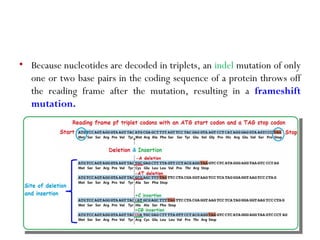
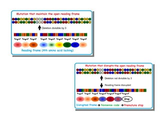
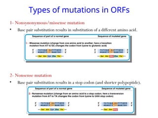
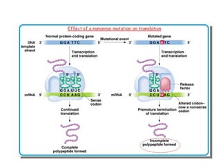
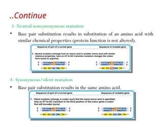
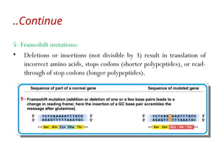
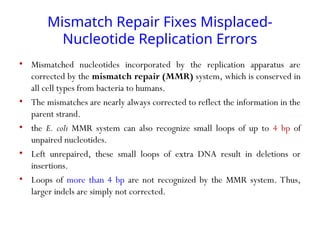
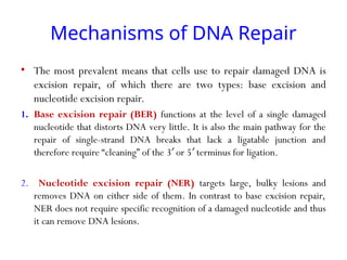
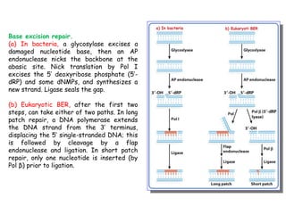
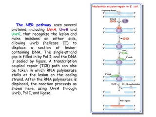
DNA Damage and Repair
An Overview
• Because each cell contains only one or two copies of its DNA, the
DNA sequence is highly protected from harm.
• DNA is a relatively stable molecule, but Earth’s natural environment
is quite toxic, and damage to DNA is inevitable.
• DNA can also be altered by mistakes made during its own replication
or recombination.
• Damage and sequence alterations to DNA are often quickly repaired,
but when they are not, the DNA becomes permanently altered and
harbors a mutation.
• Mutations are changes in DNA sequence, and when mutations occur
in germ-line cells, these changes are inheritable.
• A cancer cell has mutations that prevent cell death, resulting in loss of
cell cycle control and unregulated cell division, which leads to
malignant tumors that can end the life of the entire organism.
• The cell has a limited amount of time to fix the initial alteration and
restore the DNA to its normal sequence, before replication converts
the alteration into a mutation that will be passed on to the next
generation.
Continue
..
Continue
..
• Mutation
• Somatic mutations : occur in somatic cells and only affect the individual in
which the mutation arises.
• Germ-line mutations: alter gametes and passed to the next generation.
• Mutations are quantified in two ways:
1. Mutation rate = probability of a particular type of mutation per unit time
(or generation).
2. Mutation frequency = number of times a particular mutation occurs in a
population of cells or individuals.
Types of DNA mutations
• A mutation is a change in a DNA sequence that is propagated through
cellular generations. Mutations can be as small as a single base pair or
can range from a few base pairs to thousands.
• Mutations of one or a few base pairs usually result from errors in
replication or damaged nucleotides.
• Mutation can have different effects on gene function.
A mutation in a gene product can result in a loss of
function or a gain of function
e.g. Truncated protein,
disrupt the regulation of
gene expression
.
e.g. Increase the affinity of
an enzyme for its substrate,
up-regulate gene expression.
loss of
function
Gain of
function
Studies in Drosophila melanogaster suggest that mutations that alter
the protein sequence are most likely to be harmful; about 70% have a
negative effect, and the rest either are neutral or have a weak
beneficial effect.
A Point Mutation Can Alter One
Amino Acid
• A change in a single base pair is often
referred to as a point mutation.
• Point mutations fall into two categories:
i. A transition mutation is the exchange of
a purine-pyrimidine base pair for the other
purine-pyrimidine base pair: C G
≡ becomes
T=A, or T=A becomes C G.
≡
ii. A transversion mutation is the
replacement of a purine-pyrimidine base pair
with a pyrimidine-purine base pair, or vice
versa.
For example, C G
≡ becomes either G C
≡
or A=T.
• Transition mutations are nearly 10 times more frequent than
transversions.
• A point mutation in the protein-coding region of a gene can
result in an altered protein with partial or complete loss of
function.
• If the protein is central to cell viability, the cell could die.
• Point mutations are known to cause a wide variety of human diseases.
One example is sickle-cell anemia, resulting from a transversion that
produces an amino acid change in hemoglobin: a GAG encoding
glutamate (E) at residue 6 changes to GTG, encoding valine (V)
(E6V).
But: such type of mutation can considered as harmful
type of mutation?
• The most harmful mutations are those occurring in the genes involved in
DNA repair, because these often result in cancer.
• Many mutations in one cell can result in cancer because a mutation will
occur in a gene (or genes) that encodes a protein needed to control cell
division.
• In normal cells, oncogenes encode proteins that drive the cell division
cycle forward, and tumor suppressor genes encode proteins that
suppress cell division.
• Many tumor suppressors are transcription factors that regulate the
expression of genes that drive the cell cycle.
• The transcription factor p53 and the retinoblastoma protein are examples
of tumor suppressors that are mutated in many types of cancer.
Point mutations in a protein-coding region can
be classified by their effect on the protein
sequence
.
• The DNA sequence encoding a protein is read in codons. Each codon
corresponds to an amino acid .
• A silent mutation is a nucleotide change that produces a codon for
the same amino acid. For example, GAA and GAG both code for
glutamate.
• A missense mutation is a nucleotide change that results in a
different amino acid, such as a change from glutamate (GAA) to
glutamine (CAA).
• A nonsense mutation changes the nucleotide sequence so that
instead of encoding an amino acid, the triplet functions as a stop
codon, terminating the protein.
Small Insertion and Deletion
Mutations Change Protein Length
• Another type of mutation is the gain or loss of one or
more base pairs.
i. Insertion mutations occur when one or more base pairs are
added to the wild-type sequence.
i. Deletion mutations are due to the loss of one or more base
pairs.
• Insertion and deletion mutations are collectively referred
to as indels.
• The DNA sequence from the start codon to the stop
codon is referred to as a reading frame.
• Because nucleotides are decoded in triplets, an indel mutation of only
one or two base pairs in the coding sequence of a protein throws off
the reading frame after the mutation, resulting in a frameshift
mutation.
Types of mutations in ORFs
1- Nonsynonymous/missense mutation
• Base pair substitution results in substitution of a different amino acid.
2- Nonsense mutation
• Base pair substitution results in a stop codon (and shorter polypeptide).
Continue
..
3- Neutral nonsynonymous mutation
• Base pair substitution results in substitution of an amino acid with
similar chemical properties (protein function is not altered).
4- Synonymous/silent mutation
• Base pair substitution results in the same amino acid.
5- Frameshift mutations:
• Deletions or insertions (not divisible by 3) result in translation of
incorrect amino acids, stops codons (shorter polypeptides), or read-
through of stop codons (longer polypeptides).
Continue
..
Mechanisms of DNA Repair
Can you considered the
mistakes made during
replication and
recombination that do not
involve damaged bases as
mutations?
Mismatch Repair Fixes Misplaced-
Nucleotide Replication Errors
• Mismatched nucleotides incorporated by the replication apparatus are
corrected by the mismatch repair (MMR) system, which is conserved in
all cell types from bacteria to humans.
• The mismatches are nearly always corrected to reflect the information in the
parent strand.
• the E. coli MMR system can also recognize small loops of up to 4 bp of
unpaired nucleotides.
• Left unrepaired, these small loops of extra DNA result in deletions or
insertions.
• Loops of more than 4 bp are not recognized by the MMR system. Thus,
larger indels are simply not corrected.
Mechanisms of DNA Repair
• The most prevalent means that cells use to repair damaged DNA is
excision repair, of which there are two types: base excision and
nucleotide excision repair.
1. Base excision repair (BER) functions at the level of a single damaged
nucleotide that distorts DNA very little. It is also the main pathway for the
repair of single-strand DNA breaks that lack a ligatable junction and
therefore require “cleaning” of the 3 or 5 terminus for ligation.
′ ′
2. Nucleotide excision repair (NER) targets large, bulky lesions and
removes DNA on either side of them. In contrast to base excision repair,
NER does not require specific recognition of a damaged nucleotide and thus
it can remove DNA lesions.
Base excision repair.
(a) In bacteria, a glycosylase excises a
damaged nucleotide base, then an AP
endonuclease nicks the backbone at the
abasic site. Nick translation by Pol I
excises the 5′ deoxyribose phosphate (5′-
dRP) and some dNMPs, and synthesizes a
new strand. Ligase seals the gap.
(b) Eukaryotic BER, after the first two
steps, can take either of two paths. In long
patch repair, a DNA polymerase extends
the DNA strand from the 3′ terminus,
displacing the 5′ single-stranded DNA; this
is followed by cleavage by a flap
endonuclease and ligation. In short patch
repair, only one nucleotide is inserted (by
Pol β) prior to ligation.
The NER pathway uses several
proteins, including UvrA, UvrB and
UvrC, that recognize the lesion and
make incisions on either side,
allowing UvrD (helicase II) to
displace a section of lesion-
containing DNA. The single-strand
gap is filled in by Pol I, and the DNA
is sealed by ligase. A transcription
coupled repair (TCR) path can also
be taken in which RNA polymerase
stalls at the lesion on the coding
strand. After the RNA polymerase is
displaced, the reaction proceeds as
shown here, using UvrA through
UvrD, Pol I, and ligase.

DNA_Damage_and_Repair presentation for degree students

  • 1.
  • 2.
    An Overview • Becauseeach cell contains only one or two copies of its DNA, the DNA sequence is highly protected from harm. • DNA is a relatively stable molecule, but Earth’s natural environment is quite toxic, and damage to DNA is inevitable. • DNA can also be altered by mistakes made during its own replication or recombination. • Damage and sequence alterations to DNA are often quickly repaired, but when they are not, the DNA becomes permanently altered and harbors a mutation. • Mutations are changes in DNA sequence, and when mutations occur in germ-line cells, these changes are inheritable.
  • 3.
    • A cancercell has mutations that prevent cell death, resulting in loss of cell cycle control and unregulated cell division, which leads to malignant tumors that can end the life of the entire organism. • The cell has a limited amount of time to fix the initial alteration and restore the DNA to its normal sequence, before replication converts the alteration into a mutation that will be passed on to the next generation. Continue ..
  • 4.
    Continue .. • Mutation • Somaticmutations : occur in somatic cells and only affect the individual in which the mutation arises. • Germ-line mutations: alter gametes and passed to the next generation. • Mutations are quantified in two ways: 1. Mutation rate = probability of a particular type of mutation per unit time (or generation). 2. Mutation frequency = number of times a particular mutation occurs in a population of cells or individuals.
  • 5.
    Types of DNAmutations • A mutation is a change in a DNA sequence that is propagated through cellular generations. Mutations can be as small as a single base pair or can range from a few base pairs to thousands. • Mutations of one or a few base pairs usually result from errors in replication or damaged nucleotides. • Mutation can have different effects on gene function.
  • 6.
    A mutation ina gene product can result in a loss of function or a gain of function e.g. Truncated protein, disrupt the regulation of gene expression . e.g. Increase the affinity of an enzyme for its substrate, up-regulate gene expression. loss of function Gain of function Studies in Drosophila melanogaster suggest that mutations that alter the protein sequence are most likely to be harmful; about 70% have a negative effect, and the rest either are neutral or have a weak beneficial effect.
  • 7.
    A Point MutationCan Alter One Amino Acid • A change in a single base pair is often referred to as a point mutation. • Point mutations fall into two categories: i. A transition mutation is the exchange of a purine-pyrimidine base pair for the other purine-pyrimidine base pair: C G ≡ becomes T=A, or T=A becomes C G. ≡ ii. A transversion mutation is the replacement of a purine-pyrimidine base pair with a pyrimidine-purine base pair, or vice versa. For example, C G ≡ becomes either G C ≡ or A=T.
  • 8.
    • Transition mutationsare nearly 10 times more frequent than transversions. • A point mutation in the protein-coding region of a gene can result in an altered protein with partial or complete loss of function. • If the protein is central to cell viability, the cell could die.
  • 9.
    • Point mutationsare known to cause a wide variety of human diseases. One example is sickle-cell anemia, resulting from a transversion that produces an amino acid change in hemoglobin: a GAG encoding glutamate (E) at residue 6 changes to GTG, encoding valine (V) (E6V). But: such type of mutation can considered as harmful type of mutation?
  • 10.
    • The mostharmful mutations are those occurring in the genes involved in DNA repair, because these often result in cancer. • Many mutations in one cell can result in cancer because a mutation will occur in a gene (or genes) that encodes a protein needed to control cell division. • In normal cells, oncogenes encode proteins that drive the cell division cycle forward, and tumor suppressor genes encode proteins that suppress cell division. • Many tumor suppressors are transcription factors that regulate the expression of genes that drive the cell cycle. • The transcription factor p53 and the retinoblastoma protein are examples of tumor suppressors that are mutated in many types of cancer.
  • 11.
    Point mutations ina protein-coding region can be classified by their effect on the protein sequence . • The DNA sequence encoding a protein is read in codons. Each codon corresponds to an amino acid . • A silent mutation is a nucleotide change that produces a codon for the same amino acid. For example, GAA and GAG both code for glutamate. • A missense mutation is a nucleotide change that results in a different amino acid, such as a change from glutamate (GAA) to glutamine (CAA). • A nonsense mutation changes the nucleotide sequence so that instead of encoding an amino acid, the triplet functions as a stop codon, terminating the protein.
  • 12.
    Small Insertion andDeletion Mutations Change Protein Length • Another type of mutation is the gain or loss of one or more base pairs. i. Insertion mutations occur when one or more base pairs are added to the wild-type sequence. i. Deletion mutations are due to the loss of one or more base pairs. • Insertion and deletion mutations are collectively referred to as indels. • The DNA sequence from the start codon to the stop codon is referred to as a reading frame.
  • 13.
    • Because nucleotidesare decoded in triplets, an indel mutation of only one or two base pairs in the coding sequence of a protein throws off the reading frame after the mutation, resulting in a frameshift mutation.
  • 15.
    Types of mutationsin ORFs 1- Nonsynonymous/missense mutation • Base pair substitution results in substitution of a different amino acid. 2- Nonsense mutation • Base pair substitution results in a stop codon (and shorter polypeptide).
  • 17.
    Continue .. 3- Neutral nonsynonymousmutation • Base pair substitution results in substitution of an amino acid with similar chemical properties (protein function is not altered). 4- Synonymous/silent mutation • Base pair substitution results in the same amino acid.
  • 18.
    5- Frameshift mutations: •Deletions or insertions (not divisible by 3) result in translation of incorrect amino acids, stops codons (shorter polypeptides), or read- through of stop codons (longer polypeptides). Continue ..
  • 19.
    Mechanisms of DNARepair Can you considered the mistakes made during replication and recombination that do not involve damaged bases as mutations?
  • 20.
    Mismatch Repair FixesMisplaced- Nucleotide Replication Errors • Mismatched nucleotides incorporated by the replication apparatus are corrected by the mismatch repair (MMR) system, which is conserved in all cell types from bacteria to humans. • The mismatches are nearly always corrected to reflect the information in the parent strand. • the E. coli MMR system can also recognize small loops of up to 4 bp of unpaired nucleotides. • Left unrepaired, these small loops of extra DNA result in deletions or insertions. • Loops of more than 4 bp are not recognized by the MMR system. Thus, larger indels are simply not corrected.
  • 23.
    Mechanisms of DNARepair • The most prevalent means that cells use to repair damaged DNA is excision repair, of which there are two types: base excision and nucleotide excision repair. 1. Base excision repair (BER) functions at the level of a single damaged nucleotide that distorts DNA very little. It is also the main pathway for the repair of single-strand DNA breaks that lack a ligatable junction and therefore require “cleaning” of the 3 or 5 terminus for ligation. ′ ′ 2. Nucleotide excision repair (NER) targets large, bulky lesions and removes DNA on either side of them. In contrast to base excision repair, NER does not require specific recognition of a damaged nucleotide and thus it can remove DNA lesions.
  • 24.
    Base excision repair. (a)In bacteria, a glycosylase excises a damaged nucleotide base, then an AP endonuclease nicks the backbone at the abasic site. Nick translation by Pol I excises the 5′ deoxyribose phosphate (5′- dRP) and some dNMPs, and synthesizes a new strand. Ligase seals the gap. (b) Eukaryotic BER, after the first two steps, can take either of two paths. In long patch repair, a DNA polymerase extends the DNA strand from the 3′ terminus, displacing the 5′ single-stranded DNA; this is followed by cleavage by a flap endonuclease and ligation. In short patch repair, only one nucleotide is inserted (by Pol β) prior to ligation.
  • 25.
    The NER pathwayuses several proteins, including UvrA, UvrB and UvrC, that recognize the lesion and make incisions on either side, allowing UvrD (helicase II) to displace a section of lesion- containing DNA. The single-strand gap is filled in by Pol I, and the DNA is sealed by ligase. A transcription coupled repair (TCR) path can also be taken in which RNA polymerase stalls at the lesion on the coding strand. After the RNA polymerase is displaced, the reaction proceeds as shown here, using UvrA through UvrD, Pol I, and ligase.