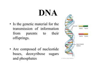
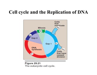
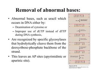
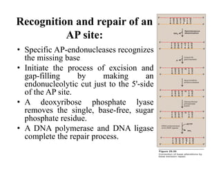
DNA
• Is the genetic material for the
transmission of information
from parents to their
offsprings.
• Are composed of nucleotide
bases, deoxyribose sugars
and phosphates
Cell cycle and the Replication of DNA
• The replication of DNA should be highly
regulated.
• If the damage is not repaired, a permanent
change (mutation) is introduced that can result
in any of a number of deleterious effects,
including loss of control over the proliferation
of the mutated cell, leading to cancer.
DNA Damage
• Errors like incorrect base-pairing or insertion of one - few
extra nucleotides can occur.
• DNA is also constantly subjected to environmental insults
that can cause the alteration or removal of nucleotide
bases.
• The damaging agents can be either
– Chemicals, for example, nitrous acid, or
– Radiation, for eg., UV light, which can fuse two pyrimidines
adjacent to each other in the DNA, and
– High energy ionizing radiation, which can cause double-
strand breaks.
• Bases are also altered or lost spontaneously from
mammalian DNA at a rate of many thousands per cell per
day.
• Cells are remarkably efficient at repairing
damage done to their DNA through DNA repair
systems.
• Most of the repair systems involves:
– Recognition of the damage (lesion) on the DNA,
– Removal or excision of the damage,
– Replacement or filling the gap left by excision using
the sister strand as a template for DNA synthesis,
and ligation.
• These repair systems thus perform excision
repair, with the removal of one to tens of
nucleotides.
Causes of DNA Damage
• Misincorporation of deoxynucleotides during
replication
• By spontaneous deamination of bases during normal
genetic functions
• From x-radiation that cause “nicks” in the DNA
• From UV irradiation that causes thymine dimer
formation
• From various chemicals that interact with DNA
The rapid repair of these damages to DNA are
necessary as they may be lethal to the cell or cause
mutations that may result in abnormal cell growth.
Repair Mechanisms
Various repair mechanisms includes:
A. Mismatch repair
B. Photoreactivation (Repair of damage
caused by UV light)
C. Base excision repair (Correction of base
alterations)
D. Recombinational repair (Repair of
double-strand breaks)
A. Methyl-directed mismatch repair
• Sometimes replication errors escape the
proofreading function during DNA
synthesis, causing a mismatch of one to
several bases.
• In E. coli, mismatch repair is mediated by a
group of proteins known as the Mut
proteins (Figure 29.27).
• Analogous proteins are present in humans.
Note: Mutation to the proteins involved in mismatch repair in
humans is associated with hereditary nonpolyposis colorectal
cancer (HNPCC), also known as Lynch syndrome. With HNPCC,
there is an increased risk for developing colon cancer (as well as
other cancers), but only about 5% of all colon cancer is the result
of mutations in mismatch repair.
Repair of damaged DNA:
When the strand containing the mismatch is
identified, an endonuclease nicks the strand and the
mismatched nucleotide(s) is/are removed by an
exonuclease.
• Additional nucleotides at the 5'- and 3'-ends of the
mismatch are also removed.
• The gap left by removal of the nucleotides is filled,
using the sister strand as a template, by a DNA
polymerase.
• The 3'-hydroxyl of the newly synthesized DNA is
joined to the 5'-phosphate of the remaining stretch
of the original DNA strand by DNA ligase.
B. Repair of damage caused by UV light
• Exposure of a cell to UV light can
result in the covalent joining of two
adjacent pyrimidines (usually
thymines), producing a dimer.
• These thymine dimers prevent DNA
polymerase from replicating the
DNA strand beyond the site of dimer
formation.
• Thymine dimers are excised in
bacteria by UVrABC proteins in a
process known as nucleotide excision
repair as illustrated in Figure 29.28.
• A related pathway involving XP
proteins is present in humans.
Recognition and excision of dimers by
UV-specific endonuclease:
• First, a UV-specific endonuclease
(called UVrABC excinuclease)
recognizes the dimer, and cleaves
the damaged strand on both the 5'-
side and 3'-side of the dimer.
• A short oligonucleotide containing
the dimer is released, leaving a gap
in the DNA strand that formerly
contained the dimer.
• This gap is filled in using the same
process described previously.
UV radiation and cancer:
• Pyrimidine dimers can be formed in the skin cells of
humans exposed to unfiltered sunlight.
• In the rare genetic disease xeroderma pigmentosum
(XP), the cells cannot repair the damaged DNA,
resulting in:
- Extensive accumulation of mutations and,
consequently,
- Early and numerous skin cancers
• XP can be caused by defects in any of the
several genes that code for the XP
proteins required for nucleotide excision
repair of UV damage in humans.
C. Base Excision Repair
(Correction of base alterations)
• The bases of DNA can be altered, either
spontaneously
– Such as Cytosine, which slowly undergoes
deamination to form uracil, or
• By the action of deaminating (Nitrous acid) or
alkylating compounds(S-adenosylmethionine)
– For example, nitrous acid, a potent compound that
deaminates cytosine, adenine (to hypoxanthine),
and guanine (to xanthine).
• Bases can also be lost spontaneously.
– For example, approximately 10,000 purine bases are
lost per cell per day.
• Lesions involving base alterations or loss can
be corrected by base excision repair.
Removal of abnormal bases:
• Abnormal bases, such as uracil which
occurs in DNA either by:
– Deamination of cytosine or
– Improper use of dUTP instead of dTTP
during DNA synthesis,
• Are recognized by specific glycosylases
that hydrolytically cleave them from the
deoxyribose-phosphate backbone of the
strand.
• This leaves an AP sites (apyrimidinic or
apurinic site).
Recognition and repair of an
AP site:
• Specific AP-endonucleases recognizes
the missing base
• Initiate the process of excision and
gap-filling by making an
endonucleolytic cut just to the 5'-side
of the AP site.
• A deoxyribose phosphate lyase
removes the single, base-free, sugar
phosphate residue.
• A DNA polymerase and DNA ligase
complete the repair process.
D. Recombinational Repair
• Also called as sister-strand exchange.
• In this process, the unmutated single stranded
segment from homologous DNA is excised
from the “Good” strand and inserted into the
“Gap” opposite the dimer.
• It occurs after the first round of DNA
replication.
Repair of double-strand breaks
• High-energy radiation or oxidative free radicals can
cause double- strand breaks in DNA, which are
potentially lethal to the cell.
• Such breaks also occur naturally during gene
rearrangements.
• dsDNA breaks cannot be corrected by the previously
described strategy of excising the damage on one strand
and using the remaining strand as a template for
replacing the missing nucleotide(s).
• They are repaired by one of two systems.
– Non-homologous end-joining repair system
– Homologous recombination repair system
• Nonhomologous end-joining repair:
– The break ends are directly ligated without the need for a
homologous template
– In which the ends of two DNA fragments are brought together by
a group of proteins that effect their religation.
– Some DNA is lost in the process, thus this mechanism of repair is
error prone and mutagenic.
– Defects in this repair system are associated with a predisposition
to cancer and immuno deficiency syndromes.
• Homologous recombination repair:
– It is most widely used by cells to accurately repair harmful breaks
that occur on both strands of DNA.
– Uses the enzymes that normally perform genetic recombination
between homologous chromosomes during meiosis.
– This system is much less error prone than nonhomologous end-
joining because any DNA that was lost is replaced using
homologous DNA as a template.
Mutation
• Alterations in DNA structure that produce permanent
changes in the genetic information encoded therein are
called mutations.
• There’s replacement of nitrogen base with another in
one or both the strands or addition or deletion of a base
pair in a DNA molecule.
• The changes in base sequence of genes can occur due
to:
– Accidental errors during replication
– External agents eg., UV light, ionizing radiation and
mutagen elements
• Repair mechanisms can correct most of these errors
and defects.
• Some errors escape correction and changed base
sequence becomes stably incorporated in genome.
• These stable changes in base sequence are k/a
Mutations.
Supression
mutation
Classification:
1. Point mutation
• It’s the replacement or changes in a single
base.
• Types:
– Transitions: changes of purine to purine or
pyrimidine to pyrimidine (T-C or A-G and vice
versa)
– Transversions: purine to pyrimidine or pyrimidine
to purine( T- A/G or C-A/G or A-T/C,G-T/C)
The consequences of either transitions or transversions when
translated into proteins may lead to changes as:
1. Silent mutation (No detectable effect)
2. Missense mutation (Missense effect)
3. Nonsense mutation(Nonsense effect)
Missense mutation
• Can have:
– Acceptable missence effect
– Partially acceptable missence effect or
– Unacceptable missence effect
HbA β chain 61Lys to
Hb(Hikari) β chain
61Aspn
Codon affected
AAA or AAG to Either AAU or
AAC ( By Transversion)
HbA β 61Gla to HbS β
chain 61Val
Codon affected
GAA or GAG (Glutamic acid) to
Either GUA or GUG ( Valine)
HbA α chain 58His to
HbM(Boston) α chain
58Tyr
Codon affected
CAU or CAC to Either UAU or
UAC ( By Transversion)
Substitution
• A substitution is a mutation that exchanges one base for
another (i.e., a change in a single "chemical letter" such as
switching an A to a G). Such a substitution could:
– change a codon to one that encodes a different amino acid and
cause a small change in the protein produced.
– Eg., sickle cell anemia is caused by a substitution in the beta-
hemoglobin gene, which alters a single amino acid in the protein
produced.
– change a codon to one that encodes the same amino acid and
causes no change in the protein produced. These are called silent
mutations.
– change an amino-acid-coding codon to a single "stop" codon and
cause an incomplete protein. This can have serious effects since
the incomplete protein probably won't function.
2. Frameshift mutation
• Since protein-coding DNA is divided into codons three
bases long, insertions and deletions can alter a gene so that
its message is no longer correctly parsed. These changes
are called frameshifts.
• For example, consider the sentence, "The fat cat sat." Each
word represents a codon. If we delete the first letter and
parse the sentence in the same way, it doesn't make sense.
• In frameshifts, a similar error occurs at the DNA level,
causing the codons to be parsed incorrectly. This usually
generates truncated proteins that are as useless as "hef atc
ats at" is uninformative.
• There are other types of mutations as well, but this short list
should give you an idea of the possibilities.
• Insertion
Insertions are mutations in which extra base
pairs are inserted into a new place in the DNA.
• Deletion
Deletions are mutations in which a section of
DNA is lost, or deleted.
3. Supression mutation
• Abnoramlly functionoing T-RNA molecules
formed as a result of alterations in their
“anticodon” regions.
• Some of these abnormal T-RNA are found to
suppress the effects of mutations in distant
structural genes.
DNA Repair and Mutation.pdf

DNA Repair and Mutation.pdf

  • 1.
    DNA • Is thegenetic material for the transmission of information from parents to their offsprings. • Are composed of nucleotide bases, deoxyribose sugars and phosphates
  • 2.
    Cell cycle andthe Replication of DNA
  • 3.
    • The replicationof DNA should be highly regulated. • If the damage is not repaired, a permanent change (mutation) is introduced that can result in any of a number of deleterious effects, including loss of control over the proliferation of the mutated cell, leading to cancer.
  • 4.
    DNA Damage • Errorslike incorrect base-pairing or insertion of one - few extra nucleotides can occur. • DNA is also constantly subjected to environmental insults that can cause the alteration or removal of nucleotide bases. • The damaging agents can be either – Chemicals, for example, nitrous acid, or – Radiation, for eg., UV light, which can fuse two pyrimidines adjacent to each other in the DNA, and – High energy ionizing radiation, which can cause double- strand breaks. • Bases are also altered or lost spontaneously from mammalian DNA at a rate of many thousands per cell per day.
  • 5.
    • Cells areremarkably efficient at repairing damage done to their DNA through DNA repair systems. • Most of the repair systems involves: – Recognition of the damage (lesion) on the DNA, – Removal or excision of the damage, – Replacement or filling the gap left by excision using the sister strand as a template for DNA synthesis, and ligation. • These repair systems thus perform excision repair, with the removal of one to tens of nucleotides.
  • 6.
    Causes of DNADamage • Misincorporation of deoxynucleotides during replication • By spontaneous deamination of bases during normal genetic functions • From x-radiation that cause “nicks” in the DNA • From UV irradiation that causes thymine dimer formation • From various chemicals that interact with DNA The rapid repair of these damages to DNA are necessary as they may be lethal to the cell or cause mutations that may result in abnormal cell growth.
  • 7.
    Repair Mechanisms Various repairmechanisms includes: A. Mismatch repair B. Photoreactivation (Repair of damage caused by UV light) C. Base excision repair (Correction of base alterations) D. Recombinational repair (Repair of double-strand breaks)
  • 8.
    A. Methyl-directed mismatchrepair • Sometimes replication errors escape the proofreading function during DNA synthesis, causing a mismatch of one to several bases. • In E. coli, mismatch repair is mediated by a group of proteins known as the Mut proteins (Figure 29.27). • Analogous proteins are present in humans. Note: Mutation to the proteins involved in mismatch repair in humans is associated with hereditary nonpolyposis colorectal cancer (HNPCC), also known as Lynch syndrome. With HNPCC, there is an increased risk for developing colon cancer (as well as other cancers), but only about 5% of all colon cancer is the result of mutations in mismatch repair.
  • 9.
    Repair of damagedDNA: When the strand containing the mismatch is identified, an endonuclease nicks the strand and the mismatched nucleotide(s) is/are removed by an exonuclease. • Additional nucleotides at the 5'- and 3'-ends of the mismatch are also removed. • The gap left by removal of the nucleotides is filled, using the sister strand as a template, by a DNA polymerase. • The 3'-hydroxyl of the newly synthesized DNA is joined to the 5'-phosphate of the remaining stretch of the original DNA strand by DNA ligase.
  • 10.
    B. Repair ofdamage caused by UV light • Exposure of a cell to UV light can result in the covalent joining of two adjacent pyrimidines (usually thymines), producing a dimer. • These thymine dimers prevent DNA polymerase from replicating the DNA strand beyond the site of dimer formation. • Thymine dimers are excised in bacteria by UVrABC proteins in a process known as nucleotide excision repair as illustrated in Figure 29.28. • A related pathway involving XP proteins is present in humans.
  • 11.
    Recognition and excisionof dimers by UV-specific endonuclease: • First, a UV-specific endonuclease (called UVrABC excinuclease) recognizes the dimer, and cleaves the damaged strand on both the 5'- side and 3'-side of the dimer. • A short oligonucleotide containing the dimer is released, leaving a gap in the DNA strand that formerly contained the dimer. • This gap is filled in using the same process described previously.
  • 12.
    UV radiation andcancer: • Pyrimidine dimers can be formed in the skin cells of humans exposed to unfiltered sunlight. • In the rare genetic disease xeroderma pigmentosum (XP), the cells cannot repair the damaged DNA, resulting in: - Extensive accumulation of mutations and, consequently, - Early and numerous skin cancers • XP can be caused by defects in any of the several genes that code for the XP proteins required for nucleotide excision repair of UV damage in humans.
  • 13.
    C. Base ExcisionRepair (Correction of base alterations) • The bases of DNA can be altered, either spontaneously – Such as Cytosine, which slowly undergoes deamination to form uracil, or • By the action of deaminating (Nitrous acid) or alkylating compounds(S-adenosylmethionine) – For example, nitrous acid, a potent compound that deaminates cytosine, adenine (to hypoxanthine), and guanine (to xanthine). • Bases can also be lost spontaneously. – For example, approximately 10,000 purine bases are lost per cell per day. • Lesions involving base alterations or loss can be corrected by base excision repair.
  • 14.
    Removal of abnormalbases: • Abnormal bases, such as uracil which occurs in DNA either by: – Deamination of cytosine or – Improper use of dUTP instead of dTTP during DNA synthesis, • Are recognized by specific glycosylases that hydrolytically cleave them from the deoxyribose-phosphate backbone of the strand. • This leaves an AP sites (apyrimidinic or apurinic site).
  • 15.
    Recognition and repairof an AP site: • Specific AP-endonucleases recognizes the missing base • Initiate the process of excision and gap-filling by making an endonucleolytic cut just to the 5'-side of the AP site. • A deoxyribose phosphate lyase removes the single, base-free, sugar phosphate residue. • A DNA polymerase and DNA ligase complete the repair process.
  • 16.
    D. Recombinational Repair •Also called as sister-strand exchange. • In this process, the unmutated single stranded segment from homologous DNA is excised from the “Good” strand and inserted into the “Gap” opposite the dimer. • It occurs after the first round of DNA replication.
  • 17.
    Repair of double-strandbreaks • High-energy radiation or oxidative free radicals can cause double- strand breaks in DNA, which are potentially lethal to the cell. • Such breaks also occur naturally during gene rearrangements. • dsDNA breaks cannot be corrected by the previously described strategy of excising the damage on one strand and using the remaining strand as a template for replacing the missing nucleotide(s). • They are repaired by one of two systems. – Non-homologous end-joining repair system – Homologous recombination repair system
  • 18.
    • Nonhomologous end-joiningrepair: – The break ends are directly ligated without the need for a homologous template – In which the ends of two DNA fragments are brought together by a group of proteins that effect their religation. – Some DNA is lost in the process, thus this mechanism of repair is error prone and mutagenic. – Defects in this repair system are associated with a predisposition to cancer and immuno deficiency syndromes. • Homologous recombination repair: – It is most widely used by cells to accurately repair harmful breaks that occur on both strands of DNA. – Uses the enzymes that normally perform genetic recombination between homologous chromosomes during meiosis. – This system is much less error prone than nonhomologous end- joining because any DNA that was lost is replaced using homologous DNA as a template.
  • 19.
    Mutation • Alterations inDNA structure that produce permanent changes in the genetic information encoded therein are called mutations. • There’s replacement of nitrogen base with another in one or both the strands or addition or deletion of a base pair in a DNA molecule.
  • 20.
    • The changesin base sequence of genes can occur due to: – Accidental errors during replication – External agents eg., UV light, ionizing radiation and mutagen elements • Repair mechanisms can correct most of these errors and defects. • Some errors escape correction and changed base sequence becomes stably incorporated in genome. • These stable changes in base sequence are k/a Mutations.
  • 21.
  • 22.
    1. Point mutation •It’s the replacement or changes in a single base. • Types: – Transitions: changes of purine to purine or pyrimidine to pyrimidine (T-C or A-G and vice versa) – Transversions: purine to pyrimidine or pyrimidine to purine( T- A/G or C-A/G or A-T/C,G-T/C)
  • 23.
    The consequences ofeither transitions or transversions when translated into proteins may lead to changes as: 1. Silent mutation (No detectable effect) 2. Missense mutation (Missense effect) 3. Nonsense mutation(Nonsense effect)
  • 24.
    Missense mutation • Canhave: – Acceptable missence effect – Partially acceptable missence effect or – Unacceptable missence effect HbA β chain 61Lys to Hb(Hikari) β chain 61Aspn Codon affected AAA or AAG to Either AAU or AAC ( By Transversion) HbA β 61Gla to HbS β chain 61Val Codon affected GAA or GAG (Glutamic acid) to Either GUA or GUG ( Valine) HbA α chain 58His to HbM(Boston) α chain 58Tyr Codon affected CAU or CAC to Either UAU or UAC ( By Transversion)
  • 25.
    Substitution • A substitutionis a mutation that exchanges one base for another (i.e., a change in a single "chemical letter" such as switching an A to a G). Such a substitution could: – change a codon to one that encodes a different amino acid and cause a small change in the protein produced. – Eg., sickle cell anemia is caused by a substitution in the beta- hemoglobin gene, which alters a single amino acid in the protein produced. – change a codon to one that encodes the same amino acid and causes no change in the protein produced. These are called silent mutations. – change an amino-acid-coding codon to a single "stop" codon and cause an incomplete protein. This can have serious effects since the incomplete protein probably won't function.
  • 26.
    2. Frameshift mutation •Since protein-coding DNA is divided into codons three bases long, insertions and deletions can alter a gene so that its message is no longer correctly parsed. These changes are called frameshifts. • For example, consider the sentence, "The fat cat sat." Each word represents a codon. If we delete the first letter and parse the sentence in the same way, it doesn't make sense. • In frameshifts, a similar error occurs at the DNA level, causing the codons to be parsed incorrectly. This usually generates truncated proteins that are as useless as "hef atc ats at" is uninformative. • There are other types of mutations as well, but this short list should give you an idea of the possibilities.
  • 27.
    • Insertion Insertions aremutations in which extra base pairs are inserted into a new place in the DNA. • Deletion Deletions are mutations in which a section of DNA is lost, or deleted.
  • 29.
    3. Supression mutation •Abnoramlly functionoing T-RNA molecules formed as a result of alterations in their “anticodon” regions. • Some of these abnormal T-RNA are found to suppress the effects of mutations in distant structural genes.