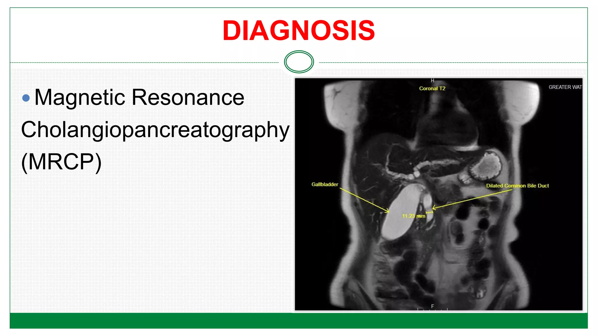
PANCREATITIS
PRESENTED BY
BADAPLIN RYNJAH
1St YEAR MSc
NURSING
OBJECTIVES
 Define acute pancreatitis
 Etiological factors of pancreatitis
 Pathophysiology of acute pancreatitis
 Enlist the clinical manifestations
 Identify the complication
 Diagnostic evaluation
 Discuss the treatment modalities of acute pancreatitis
 Surgical management of acute pancreatitis
 Chronic pancreatitis
 Clinical manifestations of chronic pancreatitis
 Diagnostic evaluation of chronic pancreatitis
 Management of chronic pancreatitis
 Preventive and health promotion measures for pancreatitis
REVIEW OF ANATOMY AND PHYSIOLOGY
FUNCTIONS
EXOCRINE : ENDOCRINE:
Pancreatic juice
Amylase
Lipase
Trypsin
Chymotripsin
Carboxypeptidase
Isets of langerhans
Alpha cells: Glucagon
(20%)
Beta cells: Insulin (75%)
Gamma cells :
Somatostatin
Polypeptide cells
MEANING
Acute pancreatitis is an acute inflammation of the
pancreas .
The degree of inflammation varies from mild edema
to severe haemorrhagic necrosis .
Acute pancreatitis is common in middle aged men
and women
CAUSES
Two main causes for pancreatitis are
 Gallstones (38%)
 Alcohol (36%)
Other, less common causes of acute pancreatitis
include
 Trauma (postsurgical, abdominal)
 Viral infections mumps
 Coxsackievirus B, HIV
 Penetrating duodenal ulcer cysts
CAUSES
 Metabolic disorders
 Hyperparathyroidism
 Hyperlipidemia
 Renal failure
 Vascular diseases.
 Pancreatitis may occur after surgical procedures on
the pancreas, stomach, duodenum or biliary tract.
CAUSES
Abscesses
Cystic fibrosis
 Kaposi sarcoma
Certain drugs (corticosteroids, thiazide , diuretics,
oral contraceptives, sulfonamides NSAIDs)
Pancreatitis can also occur after ERCP.
In some cases the cause is unknown (idiopathic)
PATHOPHYSIOLOGY
Etiological factors
Cause injury to pancreatic cells
Activation of the pancreatic enzymes
Reflux of bile acids into the pancreatic
ducts
Open distended sphincter of Oddi causes reflux
due to blockage
CLINICAL MANIFESTATION
FEVER
Rarely exceeds 102 degree F
ABDOMINAL FINDINGS
Rigidity, tenderness, guarding
Distension
Decreased or absent peristalsis
CLINICAL MANIFESTATION
ABDOMINAL PAIN
 Steady and severe excruciating
 Located in the left upper quadrant or in
the mid epigastrium may radiate to the
back
 Worsened by lying supine may be
lessened by flexed knee, curved back
positioning
CLINICAL MANIFESTATION
VOMITTING
 Varies in severity but is usually
protacted.
 Worsened by ingestion of food
or fluid.
 Does not relieve the pain.
 Usually accompanied by
nausea.
CLINICAL MANIFESTATION
Cullen’s sign Grey’s turner sign
Bluish discoloration around
the umbilicus
Bluish discoloration along
the flanks
COMPLICATIONS
 Hypotension or shock from hypovolemia or
hypoalbuminemia
 Leukocytosis, anemia , disseminated intravascular
coagulation from unknown causes
 Atelectasis, pneumonia, pleural effusion, acute
respiratory distress syndrome
 Gastrointestinal bleeding
COMPLICATIONS
Pancreatic pseudocysts, pancreatic necrosis,
pancreatic abscesses, pancreatic ascites
Oliguria and acute tubular necrosis
Hyperglycemia, hypocalcemia, hyperlipidemia
DIAGNOSIS
History and physical examination
Liver function tests: elevations commonly seen
Serum trigylycerides
• Serum amylase: Levels elevated within a few hours
of disease onset
• Serum lipase: Levels remain elevated up to 7 days
after disease onset
• Serum glucose: Hyperglycaemia of 500 to 900 mg/d
• Serum calcium: Hypocalcaemia from calcium
sequestering in abdomen; hypocalcemia is a poor
prognostic sign
DIAGNOSIS
 Abdominal ultrasound
Endoscopic ultrasound
DIAGNOSIS
DIAGNOSIS
 Magnetic Resonance
Cholangiopancreatography
(MRCP)
DIAGNOSIS
 Endoscopic Retrograde
Cholangiopancreatography
(ERCP)
COLLABORATIVE CARE
GOALS
1) Relief of pain
2) Prevention or alleviation of shock
3) Reduction of pancreatic secretions
4) Correction of fluid and electrolyte imbalances
5) Prevention and treatment of infections
6) Removal of the precipitating cause
CONSERVATIVE THERAPY
Focused on primary care:
1) Aggressive hydration
2) Management of metabolic complications
3) Minimization of pancreatic stimulation
CONSERVATIVE THERAPY
1) Management of pain
 IV morphine ( pain medication may be
combine with antispasmodic agents)
 Spasmolytics : Nitroglycerine or papaverine
2) Supplemental oxygen: To maintain oxygen
saturation > 95%
3) Serum glucose : Monitored for Hyperglycemia
CONSERVATIVE THERAPY
If shock present
1) Blood volume replacement
2) Plasma or plasma volume expanders : Dextran
or albumin may be given
3) Fluid and electrolyte: Ringer’s lactate solution
4) Increase vascular resistance with hypotension
: Vasoactive drug such as dopamine
CONSERVATIVE THERAPY
To Suppress the pancreatic enzymes
1)The patient to be kept in NPO
2) NG suction :
 To reduce vomiting and gastric distension
 To prevent gastric acid contents from entering into
the duodenum
3) Drugs such as Antacids, PPI, Acetazolamide
CONSERVATIVE THERAPY
4) Enteral nutrition : For patients who does not resume
oral intake
5) Antibiotic therapy: In patients with acute necrotizing
pancreatitis
6) Endoscopic or CT guided percutaneous aspiration
with gram stain and culture may be performed
SURGICAL THERAPY
1) Acute pancreatitis related to Gallstone:
ERCP together with endoscopic spinchterotomy
followed by laproscopic cholecystectomy to reduce
potential for recurrence
2) Severe acute pancreatitis:
 Drainage of necrotic fluid collections
DRUG THERAPY
DRUG MECHANISM OF ACTION
Morphine Relief of pain
Antispasmodic (e.g diclyclomine) Decrease vagal stimulation , motility,
pancreatic outflow
Carbonic anhydrase inhibitor
(acetazolamide)
Decrease volume and bicarbonate
concentration of pancreatic secretion
Proton pump inhibitors Decrease acid secretion (HCL acid
stimulate pancreatic activity )
Antacids Neutralization of gastric hydrochloride
acid secretion
NUTRITIONAL MANAGEMENT
 Initially the patient to kept on NPO to decrease the gastric
acid secretions
 Enteral feeding: Nasojejunal feeding tube
 IV lipids : Blood triglyceride levels are monitored
 When food is allowed, small, frequent feedings are given.
 Carbohydrate rich diet should be given
 Needs to abstain from alcohol
 Supplemental fat-soluble vitamins may be given
CHRONIC PANCREATITIS
DEFINITION
Chronic pancreatitis (CP) is characterised by
prolonged pancreatic inflammation and fibrosis
leading eventually to destruction of pancreatic
parenchyma and loss of exocrine and endocrine
function
ETIOLOGY
1) Alcohol abuse
2) Obstruction caused by cholelitiasis
3) Tumour
4) Pseudocyst
ETIOLOGY
5)Trauma
6)Systemic disease (Systemic lupus
erythematosus)
7)Auto immune pancreatitis
8)Cystic fibrosis
ETIOLOGY
OBSTRUCTIVE
PANCREATITIS
NON OBSTRUCTIVE
PANCREATITIS
Inflammation of
Sphincter of oddi
associated cholelitiasis
Cancer of ampulla of
vater duodenum,
pancreas
Inflammation and
sclerosis mainly in the
head of pancreas and
around the pancreatic
duct
CLINICAL MANIFESTATION
Abdominal pain :
oEpisode of acute pain and it remains almost
constant
oPain may be locate in the same area as acute
pancreatitis
oDescribe as heavy, gnawing feeling or
sometimes burning and cramplike
CLINICAL MANIFESTATION
Others include:
Malabsorption with weight loss constipation, mild
jaundice with dark urine, steatorrhea and diabetes
mellitus
Staetorrhea may be voluminous, foul smelling fatty
stools
 Urine and stool may be frothy
Some abdominal tenderness may be present
COMPLICATIONS
 Pseudocyst formation
 Bile duct or duodenal obstruction
 Pancreatic ascitis
 Pleural effusion
 Splenic vein thrombosis
 Pseudoaneurysm
 Pancreatic cancer
DIAGNOSIS
 Serum amylase
 Serum Lipase
 Serum bilirubin
  Alkaline phosphatase
  ESR, mild leucocytosis
 ERCP
 MRCP
DIAGNOSIS
 CT, MRI
 Abdominal ultrasound
 Stool sample : Fecal fat content
 Deficiencies of fat soluble vitamin and cobalamin,
glucose intolerance
 Secretin stimulation test
SURGICAL MANGEMENT
Pancreaticojejunostomy
Side-to-side anastomosis
of the pancreatic duct to the
jejunum, allows drainage of
the pancreatic secretions
into the jejunum.
SURGICAL MANGEMENT
Whipple resection
(pancreaticoduodenectomy )
Removal of the head of the pancreas, the
first part of the small intestine (duodenum),
the gallbladder and the bile duct.
SURGICAL MANGEMENT
Other surgical procedures:
Revision of the sphincter of the ampulla of Vater
Internal drainage of a pancreatic cyst into the
stomach
Insertion of a stent and wide resection or
removal of the pancreas.
SURGICAL MANGEMENT
Autotransplantation :
Implantation of the pancreatic islet cells
Moving the pancreas to another location within
the abdomen with revised vascular and enteric
anastomosis
SURGICAL MANGEMENT
 Gall bladder disease : The obstruction is treated by
surgery to explore the common duct and remove the
stones; the gallbladder is removed at the same time.
 Drainage : common bile duct and the pancreatic duct
 A T-tube usually is placed in the common bile duct,
requiring drainage system to collect the bile
NURSING MANAGEMENT
 Health History.
 Assess for
History of gallbladder disease
History of other GI diseases (e.g., peptic ulcer
disease, IBD)
History of alcohol use: amount and duration
Medications in use: prescription, over the counter,
and herbal preparation
NURSING MANAGEMENT
Onset and progression of symptoms such as:
Pain, which is often steady and severe; is located in
the epigastric or umbilical region or may radiate to the
back; worsens when patient is supine; is unrelieved
by vomiting
Nausea and vomiting
NURSING MANAGEMENT
Physical Examination.
 Assess for:
 Vital sign indications of hypovolemia: tachycardia,
tachypnea, normal to low blood pressure,
restlessness, and anxiety
 Abdominal rigidity, distention, guarding, and
tenderness to palpation
NURSING MANAGEMENT
Diminished or absent bowel sounds on
auscultation
Fever : > 102° F
Signs of third spacing: falling urinary output,
decreased skin turgor, dry or sticky mucous
membranes, increased abdominal girth
PRIORITIZED NURSING DIAGNOSIS
Acute pain related to inflammation, edema,
distension of pancreatic capsule and activation
of pancreatic enzyme
Ineffective breathing pattern related to severe
pain, pulmonary infiltrates , pleural effusion,
atelectasis and elevated diaphragm
PRIORITIZED NURSING DIAGNOSIS
 Risk for deficient fluid volume related to vomiting,
hyperglycemia, and increased capillary permeability
secondary to acute pancretitis
 Imbalanced nutrition less than body requirement related to
vomiting, NPO status and malabsorption secondary to
pancreatitis
 Impaired skin integrity related to poor nutritional status,
bed rest, multiple drains, and surgical wound
Does mortality occur early or late in acute
pancreatitis?
Abstract:
 Several prior studies have suggested that 80% of deaths in acute
pancreatitis occur late as a result of pancreatic infection. Others have
suggested that approximately half of deaths occur early as a result of
multisystem organ failure. The aim of the present study was to
determine the timing of mortality of acute pancreatitis at a large tertiary-
care hospital in the United States.
CONCLUSION
 Conclusion:
Approximately half of deaths in acute pancreatitis occur
within the first 14 days owing to organ failure and the
remainder of deaths occur later because of complications
associated with necrotizing pancreatitis. Improvement in
mortality in the future will require innovative approaches to
counteract early organ failure and late complications of
necrotizing pancreatitis.
Authors
 Muthoka Mutinga,Adam Rosenbluth,Scott M. Tenner,Robert R. Odze
Gregory T. Sica, Peter A. Bank
REFERENCES
BOOKS
 Lewis, Driksen, Heikemper, Bucher. Lewis Textbook of
medical surgical nursing- 2nd edition
 Urden D.L StacyM.K Laugh E.M Textbook For Critical
Care Nursing
 Myers/Gulanick, Nursing Care Plans. Nursing Diagnosis
And Interventions 6th edition
 Linda s. Williams, Paula D. Hopper. Textbook of medical
surgical nursing-4th edition
Pancreatitis topic for nursing students

Pancreatitis topic for nursing students

  • 1.
  • 2.
    OBJECTIVES  Define acutepancreatitis  Etiological factors of pancreatitis  Pathophysiology of acute pancreatitis  Enlist the clinical manifestations  Identify the complication  Diagnostic evaluation  Discuss the treatment modalities of acute pancreatitis
  • 3.
     Surgical managementof acute pancreatitis  Chronic pancreatitis  Clinical manifestations of chronic pancreatitis  Diagnostic evaluation of chronic pancreatitis  Management of chronic pancreatitis  Preventive and health promotion measures for pancreatitis
  • 4.
    REVIEW OF ANATOMYAND PHYSIOLOGY
  • 5.
    FUNCTIONS EXOCRINE : ENDOCRINE: Pancreaticjuice Amylase Lipase Trypsin Chymotripsin Carboxypeptidase Isets of langerhans Alpha cells: Glucagon (20%) Beta cells: Insulin (75%) Gamma cells : Somatostatin Polypeptide cells
  • 7.
    MEANING Acute pancreatitis isan acute inflammation of the pancreas . The degree of inflammation varies from mild edema to severe haemorrhagic necrosis . Acute pancreatitis is common in middle aged men and women
  • 8.
    CAUSES Two main causesfor pancreatitis are  Gallstones (38%)  Alcohol (36%) Other, less common causes of acute pancreatitis include  Trauma (postsurgical, abdominal)  Viral infections mumps  Coxsackievirus B, HIV  Penetrating duodenal ulcer cysts
  • 9.
    CAUSES  Metabolic disorders Hyperparathyroidism  Hyperlipidemia  Renal failure  Vascular diseases.  Pancreatitis may occur after surgical procedures on the pancreas, stomach, duodenum or biliary tract.
  • 10.
    CAUSES Abscesses Cystic fibrosis  Kaposisarcoma Certain drugs (corticosteroids, thiazide , diuretics, oral contraceptives, sulfonamides NSAIDs) Pancreatitis can also occur after ERCP. In some cases the cause is unknown (idiopathic)
  • 11.
    PATHOPHYSIOLOGY Etiological factors Cause injuryto pancreatic cells Activation of the pancreatic enzymes Reflux of bile acids into the pancreatic ducts Open distended sphincter of Oddi causes reflux due to blockage
  • 12.
    CLINICAL MANIFESTATION FEVER Rarely exceeds102 degree F ABDOMINAL FINDINGS Rigidity, tenderness, guarding Distension Decreased or absent peristalsis
  • 13.
    CLINICAL MANIFESTATION ABDOMINAL PAIN Steady and severe excruciating  Located in the left upper quadrant or in the mid epigastrium may radiate to the back  Worsened by lying supine may be lessened by flexed knee, curved back positioning
  • 14.
    CLINICAL MANIFESTATION VOMITTING  Variesin severity but is usually protacted.  Worsened by ingestion of food or fluid.  Does not relieve the pain.  Usually accompanied by nausea.
  • 15.
    CLINICAL MANIFESTATION Cullen’s signGrey’s turner sign Bluish discoloration around the umbilicus Bluish discoloration along the flanks
  • 16.
    COMPLICATIONS  Hypotension orshock from hypovolemia or hypoalbuminemia  Leukocytosis, anemia , disseminated intravascular coagulation from unknown causes  Atelectasis, pneumonia, pleural effusion, acute respiratory distress syndrome  Gastrointestinal bleeding
  • 17.
    COMPLICATIONS Pancreatic pseudocysts, pancreaticnecrosis, pancreatic abscesses, pancreatic ascites Oliguria and acute tubular necrosis Hyperglycemia, hypocalcemia, hyperlipidemia
  • 18.
    DIAGNOSIS History and physicalexamination Liver function tests: elevations commonly seen Serum trigylycerides
  • 19.
    • Serum amylase:Levels elevated within a few hours of disease onset • Serum lipase: Levels remain elevated up to 7 days after disease onset • Serum glucose: Hyperglycaemia of 500 to 900 mg/d • Serum calcium: Hypocalcaemia from calcium sequestering in abdomen; hypocalcemia is a poor prognostic sign DIAGNOSIS
  • 20.
  • 21.
  • 22.
  • 23.
    COLLABORATIVE CARE GOALS 1) Reliefof pain 2) Prevention or alleviation of shock 3) Reduction of pancreatic secretions 4) Correction of fluid and electrolyte imbalances 5) Prevention and treatment of infections 6) Removal of the precipitating cause
  • 24.
    CONSERVATIVE THERAPY Focused onprimary care: 1) Aggressive hydration 2) Management of metabolic complications 3) Minimization of pancreatic stimulation
  • 25.
    CONSERVATIVE THERAPY 1) Managementof pain  IV morphine ( pain medication may be combine with antispasmodic agents)  Spasmolytics : Nitroglycerine or papaverine 2) Supplemental oxygen: To maintain oxygen saturation > 95% 3) Serum glucose : Monitored for Hyperglycemia
  • 26.
    CONSERVATIVE THERAPY If shockpresent 1) Blood volume replacement 2) Plasma or plasma volume expanders : Dextran or albumin may be given 3) Fluid and electrolyte: Ringer’s lactate solution 4) Increase vascular resistance with hypotension : Vasoactive drug such as dopamine
  • 27.
    CONSERVATIVE THERAPY To Suppressthe pancreatic enzymes 1)The patient to be kept in NPO 2) NG suction :  To reduce vomiting and gastric distension  To prevent gastric acid contents from entering into the duodenum 3) Drugs such as Antacids, PPI, Acetazolamide
  • 28.
    CONSERVATIVE THERAPY 4) Enteralnutrition : For patients who does not resume oral intake 5) Antibiotic therapy: In patients with acute necrotizing pancreatitis 6) Endoscopic or CT guided percutaneous aspiration with gram stain and culture may be performed
  • 29.
    SURGICAL THERAPY 1) Acutepancreatitis related to Gallstone: ERCP together with endoscopic spinchterotomy followed by laproscopic cholecystectomy to reduce potential for recurrence 2) Severe acute pancreatitis:  Drainage of necrotic fluid collections
  • 30.
    DRUG THERAPY DRUG MECHANISMOF ACTION Morphine Relief of pain Antispasmodic (e.g diclyclomine) Decrease vagal stimulation , motility, pancreatic outflow Carbonic anhydrase inhibitor (acetazolamide) Decrease volume and bicarbonate concentration of pancreatic secretion Proton pump inhibitors Decrease acid secretion (HCL acid stimulate pancreatic activity ) Antacids Neutralization of gastric hydrochloride acid secretion
  • 31.
    NUTRITIONAL MANAGEMENT  Initiallythe patient to kept on NPO to decrease the gastric acid secretions  Enteral feeding: Nasojejunal feeding tube  IV lipids : Blood triglyceride levels are monitored  When food is allowed, small, frequent feedings are given.  Carbohydrate rich diet should be given  Needs to abstain from alcohol  Supplemental fat-soluble vitamins may be given
  • 32.
    CHRONIC PANCREATITIS DEFINITION Chronic pancreatitis(CP) is characterised by prolonged pancreatic inflammation and fibrosis leading eventually to destruction of pancreatic parenchyma and loss of exocrine and endocrine function
  • 33.
    ETIOLOGY 1) Alcohol abuse 2)Obstruction caused by cholelitiasis 3) Tumour 4) Pseudocyst
  • 34.
    ETIOLOGY 5)Trauma 6)Systemic disease (Systemiclupus erythematosus) 7)Auto immune pancreatitis 8)Cystic fibrosis
  • 35.
    ETIOLOGY OBSTRUCTIVE PANCREATITIS NON OBSTRUCTIVE PANCREATITIS Inflammation of Sphincterof oddi associated cholelitiasis Cancer of ampulla of vater duodenum, pancreas Inflammation and sclerosis mainly in the head of pancreas and around the pancreatic duct
  • 36.
    CLINICAL MANIFESTATION Abdominal pain: oEpisode of acute pain and it remains almost constant oPain may be locate in the same area as acute pancreatitis oDescribe as heavy, gnawing feeling or sometimes burning and cramplike
  • 37.
    CLINICAL MANIFESTATION Others include: Malabsorptionwith weight loss constipation, mild jaundice with dark urine, steatorrhea and diabetes mellitus Staetorrhea may be voluminous, foul smelling fatty stools  Urine and stool may be frothy Some abdominal tenderness may be present
  • 38.
    COMPLICATIONS  Pseudocyst formation Bile duct or duodenal obstruction  Pancreatic ascitis  Pleural effusion  Splenic vein thrombosis  Pseudoaneurysm  Pancreatic cancer
  • 39.
    DIAGNOSIS  Serum amylase Serum Lipase  Serum bilirubin   Alkaline phosphatase   ESR, mild leucocytosis  ERCP  MRCP
  • 40.
    DIAGNOSIS  CT, MRI Abdominal ultrasound  Stool sample : Fecal fat content  Deficiencies of fat soluble vitamin and cobalamin, glucose intolerance  Secretin stimulation test
  • 41.
    SURGICAL MANGEMENT Pancreaticojejunostomy Side-to-side anastomosis ofthe pancreatic duct to the jejunum, allows drainage of the pancreatic secretions into the jejunum.
  • 42.
    SURGICAL MANGEMENT Whipple resection (pancreaticoduodenectomy) Removal of the head of the pancreas, the first part of the small intestine (duodenum), the gallbladder and the bile duct.
  • 44.
    SURGICAL MANGEMENT Other surgicalprocedures: Revision of the sphincter of the ampulla of Vater Internal drainage of a pancreatic cyst into the stomach Insertion of a stent and wide resection or removal of the pancreas.
  • 45.
    SURGICAL MANGEMENT Autotransplantation : Implantationof the pancreatic islet cells Moving the pancreas to another location within the abdomen with revised vascular and enteric anastomosis
  • 46.
    SURGICAL MANGEMENT  Gallbladder disease : The obstruction is treated by surgery to explore the common duct and remove the stones; the gallbladder is removed at the same time.  Drainage : common bile duct and the pancreatic duct  A T-tube usually is placed in the common bile duct, requiring drainage system to collect the bile
  • 47.
    NURSING MANAGEMENT  HealthHistory.  Assess for History of gallbladder disease History of other GI diseases (e.g., peptic ulcer disease, IBD) History of alcohol use: amount and duration Medications in use: prescription, over the counter, and herbal preparation
  • 48.
    NURSING MANAGEMENT Onset andprogression of symptoms such as: Pain, which is often steady and severe; is located in the epigastric or umbilical region or may radiate to the back; worsens when patient is supine; is unrelieved by vomiting Nausea and vomiting
  • 49.
    NURSING MANAGEMENT Physical Examination. Assess for:  Vital sign indications of hypovolemia: tachycardia, tachypnea, normal to low blood pressure, restlessness, and anxiety  Abdominal rigidity, distention, guarding, and tenderness to palpation
  • 50.
    NURSING MANAGEMENT Diminished orabsent bowel sounds on auscultation Fever : > 102° F Signs of third spacing: falling urinary output, decreased skin turgor, dry or sticky mucous membranes, increased abdominal girth
  • 51.
    PRIORITIZED NURSING DIAGNOSIS Acutepain related to inflammation, edema, distension of pancreatic capsule and activation of pancreatic enzyme Ineffective breathing pattern related to severe pain, pulmonary infiltrates , pleural effusion, atelectasis and elevated diaphragm
  • 52.
    PRIORITIZED NURSING DIAGNOSIS Risk for deficient fluid volume related to vomiting, hyperglycemia, and increased capillary permeability secondary to acute pancretitis  Imbalanced nutrition less than body requirement related to vomiting, NPO status and malabsorption secondary to pancreatitis  Impaired skin integrity related to poor nutritional status, bed rest, multiple drains, and surgical wound
  • 53.
    Does mortality occurearly or late in acute pancreatitis? Abstract:  Several prior studies have suggested that 80% of deaths in acute pancreatitis occur late as a result of pancreatic infection. Others have suggested that approximately half of deaths occur early as a result of multisystem organ failure. The aim of the present study was to determine the timing of mortality of acute pancreatitis at a large tertiary- care hospital in the United States.
  • 54.
    CONCLUSION  Conclusion: Approximately halfof deaths in acute pancreatitis occur within the first 14 days owing to organ failure and the remainder of deaths occur later because of complications associated with necrotizing pancreatitis. Improvement in mortality in the future will require innovative approaches to counteract early organ failure and late complications of necrotizing pancreatitis. Authors  Muthoka Mutinga,Adam Rosenbluth,Scott M. Tenner,Robert R. Odze Gregory T. Sica, Peter A. Bank
  • 55.
    REFERENCES BOOKS  Lewis, Driksen,Heikemper, Bucher. Lewis Textbook of medical surgical nursing- 2nd edition  Urden D.L StacyM.K Laugh E.M Textbook For Critical Care Nursing  Myers/Gulanick, Nursing Care Plans. Nursing Diagnosis And Interventions 6th edition  Linda s. Williams, Paula D. Hopper. Textbook of medical surgical nursing-4th edition