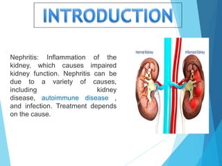
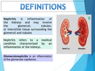
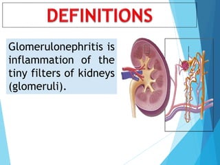
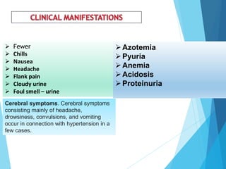
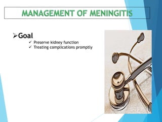
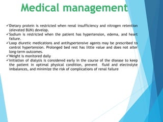
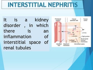
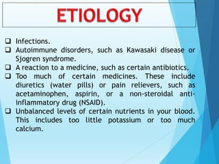
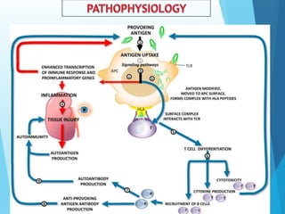
Nephritis: Inflammation of the
kidney, which causes impaired
kidney function. Nephritis can be
due to a variety of causes,
including kidney
disease, autoimmune disease ,
and infection. Treatment depends
on the cause.
Nephritis is inflammation of
the kidneys and may involve
the glomeruli, tubules,
or interstitial tissue surrounding the
glomeruli and tubules
Nephritis refers to a medical
condition characterized by an
inflammation of the kidneys.
Glomerulonephritis is an inflammation
of the glomerular capillaries.
 overall prevalence was 4.4 per
100,000 population
 7.1 per 100,000 in women,
 1.4 per 100,000 in men.
There are different types of nephritis. Each type is based
on the part of the kidney that has been affected with the
condition. The three main areas commonly affected are the
glomeruli, tubule, and interstitial renal tissue.
Glomerulonephritis
Pyelonephritis
Interstitial Nephritis
This condition refers to the
inflammation of the tiny
capillaries in the kidneys called
glomeruli, which task is to filter
blood. When they become
inflamed, they are unable to
effectively filter the blood
Glomerulonephritis is
inflammation of the
tiny filters of kidneys
(glomeruli).
 Infections
 Post-streptococcal glomerulonephritis.
Glomerulonephritis may develop a week or two after recovery
from a strep throat infection or, rarely, a skin infection (impetigo). To fight
the infection, your body produces extra antibodies that can eventually
settle in the glomeruli, causing inflammation.
Children are more likely to develop post-streptococcal
glomerulonephritis than are adults, and they're also more likely to recover
quickly.
 Bacterial endocarditis.
Bacteria occasionally can spread through your bloodstream
and lodge in your heart, causing an infection of one or more of your
heart valves. You're at greater risk of this condition if you have a
heart defect, such as a damaged or artificial heart valve. Bacterial
endocarditis is associated with glomerular disease, but the
connection between the two is unclear.
 Viral infections.
Viral infections, such as the human immunodeficiency virus
(HIV), hepatitis B and hepatitis C, can trigger glomerulonephritis
 Vasculitis
Polyarteritis.
This form of vasculitis affects small and medium blood vessels in many
parts of your body, such as your heart, kidneys and intestines.
Granulomatosis with polyangiitis.
This form of vasculitis, formerly known as Wegener's granulomatosis,
affects small and medium blood vessels in your lungs, upper airways and kidneys.
 Immune Response
Lupus
A chronic inflammatory disease, lupus can affect many parts of your body, including
your skin, joints, kidneys, blood cells, heart and lungs.
Goodpasture's syndrome
A rare immunological lung disorder that can mimic pneumonia, Goodpasture's
syndrome causes bleeding in your lungs as well as glomerulonephritis.
IgA nephropathy
Characterized by recurrent episodes of blood in the urine, this primary glomerular
disease results from deposits of immunoglobulin A (IgA) in the
glomeruli. IgA nephropathy can progress for years with no noticeable symptoms
 Risk factors :
HTN-Diabetic Nephropathy - Focal segmental Glomerulo sclerosis
Acute glomerulonephritis
Primarily a disease of children older than 2 years of age, but it
can occur at nearly any age
Chronic glomerulonephritis:
One of a group of kidney diseases characterized by long-
term inflammation and scarring of
the glomeruli (microscopic structures in the kidney that
filter blood and produce urine). This form of kidney
disease usually develops slowly (over years) and may not
produce symptoms at the outset. When symptoms and
signs do appear, they typically include blood in the urine
(hematuria), swelling (edema), high blood pressure, foamy
urine (due to protein content), and frequent nighttime
urination.
 The primary presenting feature
:Hematuria, which may be
microscopic identifiable
 through microscopic: examination or
(visible to the eye)
 cola- colored Urine because of RBCs
and protein plugs or casts. (RBC casts
indicate glomerular injury.)
 Oliguria : Patient has acute renal
failure
 Elevated BUN and serum creatinine levels
 feet are slightly swollen at night
 Loss of weight and strength
 increasing irritability
 Increased need to urinate at night
(nocturia).
 Headaches,
 Dizziness
 Digestive disturbances
 Blood pressure may be normal or severely
elevated.
 Retinalfindings include hemorrhage,
exudate, narrowed tortuous arterioles, and
papilledema.
 Mucous membranes are pale because of
anemia.
 Cardiomegaly, a gallop rhythm, distended
neck veins,
 Crackles can be heard in the lungs
 Fewer
 Chills
 Nausea
 Headache
 Flank pain
 Cloudy urine
 Foul smell – urine
Azotemia
Pyuria
Anemia
Acidosis
Proteinuria
Cerebral symptoms. Cerebral symptoms
consisting mainly of headache,
drowsiness, convulsions, and vomiting
occur in connection with hypertension in a
few cases.
 HISTORY TAKING
 PHYSICAL EXAMINATION
 BLOOD TEST :
 creatinine Elevated
 blood urea nitrogen Elevated
 Hyperkalemia due to decreased potassium
excretion, acidosis, catabolism, and excessive
potassium intake from food and medications
 ABG :Metabolic acidosis from decreased acid
secretion by the kidney and inability to
regenerate bicarbonate
 Anemia secondary to decreased erythropoiesis
(production of RBCs)
 Hypoalbuminemia with edema secondary to
protein loss through the damaged glomerular
membrane
 Increased serum phosphorus & magnesium
level due to decreased renal excretion of
phosphorus & Mg
 Decreased serum calcium level (calcium binds
to phosphorus to compensate for elevated
serum phosphorus levels)
 Impaired nerve conduction due to electrolyte
abnormalities and uremia
 KIDNEY BIOPSY PROCEDURE
 URINE TEST.
 might show red
blood cells and red
cell casts in your
urine, an indicator of
possible damage to
the glomeruli.
 white blood cells, a
common indicator of
infection or
inflammation,
 Increased protein,
which can indicate
nephron damage.
 Other indicators,
such as increased
blood levels of
creatinine or urea,
are red flags.
 Imaging tests.
kidney X-ray, an
ultrasound exam or a CT
scan
Goal
 Preserve kidney function
 Treating complications promptly
Dietary protein is restricted when renal insufficiency and nitrogen retention
(elevated BUN) develop.
Sodium is restricted when the patient has hypertension, edema, and heart
failure.
Loop diuretic medications and antihypertensive agents may be prescribed to
control hypertension. Prolonged bed rest has little value and does not alter
long-term outcomes.
Weight is monitored daily
Initiation of dialysis is considered early in the course of the disease to keep
the patient in optimal physical condition, prevent fluid and electrolyte
imbalances, and minimize the risk of complications of renal failure
Pharmacological management
 Antibiotic therapy to prevent streptococcal
infection ( Prophylaxis ).
 Steroids to suppress immunity.
 Corticosteroids. These strong anti-inflammatory drugs can
decrease inflammation
 Immunosuppressive drugs. These drugs, which are related to the
ones used to treat cancer or prevent the rejection of transplanted
organs, work by suppressing immune system activity that
damages the kidneys. They
include cyclophosphamide (Cytoxan), azathioprine (Imuran) and
mycophenolate (Cellcept)
 Medications to prevent blood clots or lower blood pressure if
needed
Dialysis
Pyelonephritis is inflammation
of renal pelvis and parenchyma
caused by a bacterial infection
 Renal calculi
 Malignancy
 Hydro nephrosis
 Trauma
 Urinary tract infection
 E. Coli
 Acute
 Chronic
 Fewer
 Chills
 Nausea
 Headache
 Flank pain
 Cloudy urine
 Foul smell – urine
Azotemia
Pyuria
Anemia
Acidosis
Proteinuria
 HISTORY TAKING
 PHYSICAL EXAMINATION
 BLOOD TEST :
 creatinine Elevated
 blood urea nitrogen Elevated
 Hyperkalemia due to decreased potassium
excretion, acidosis, catabolism, and excessive
potassium intake from food and medications
 ABG :Metabolic acidosis from decreased acid
secretion by the kidney and inability to
regenerate bicarbonate
 Anemia secondary to decreased erythropoiesis
(production of RBCs)
 Hypoalbuminemia with edema secondary to
protein loss through the damaged glomerular
membrane
 Increased serum phosphorus & magnesium
level due to decreased renal excretion of
phosphorus & Mg
 Decreased serum calcium level (calcium binds
to phosphorus to compensate for elevated
serum phosphorus levels)
 Impaired nerve conduction due to electrolyte
abnormalities and uremia
 KIDNEY BIOPSY PROCEDURE
 URINE TEST.
 might show red
blood cells and red
cell casts in your
urine, an indicator of
possible damage to
the glomeruli.
 white blood cells, a
common indicator of
infection or
inflammation,
 Increased protein,
which can indicate
nephron damage.
 Other indicators,
such as increased
blood levels of
creatinine or urea,
are red flags.
 Imaging tests.
kidney X-ray, an
ultrasound exam or a CT
scan
Goal
 Preserve kidney function
 Treating complications promptly
Dietary protein is restricted when renal insufficiency and nitrogen retention
(elevated BUN) develop.
Sodium is restricted when the patient has hypertension, edema, and heart
failure.
Loop diuretic medications and antihypertensive agents may be prescribed to
control hypertension. Prolonged bed rest has little value and does not alter
long-term outcomes.
Weight is monitored daily
Initiation of dialysis is considered early in the course of the disease to keep
the patient in optimal physical condition, prevent fluid and electrolyte
imbalances, and minimize the risk of complications of renal failure
Pharmacological management
 Antibiotic therapy : sulphonamide
 Steroids to suppress immunity.
 Analgesics
 Corticosteroids. These strong anti-inflammatory drugs can
decrease inflammation
 Immunosuppressive drugs. These drugs, which are related to the
ones used to treat cancer or prevent the rejection of transplanted
organs, work by suppressing immune system activity that
damages the kidneys. They
include cyclophosphamide (Cytoxan), azathioprine (Imuran) and
mycophenolate (Cellcept)
 Medications to prevent blood clots or lower blood pressure if
needed
Dialysis
It is a kidney
disorder , in which
there is an
inflammation of
interstitial space of
renal tubules
 Infections.
 Autoimmune disorders, such as Kawasaki disease or
Sjogren syndrome.
 A reaction to a medicine, such as certain antibiotics.
 Too much of certain medicines. These include
diuretics (water pills) or pain relievers, such as
acetaminophen, aspirin, or a non-steroidal anti-
inflammatory drug (NSAID).
 Unbalanced levels of certain nutrients in your blood.
This includes too little potassium or too much
calcium.
 The primary presenting feature
:Hematuria, which may be
microscopic identifiable
 through microscopic: examination or
(visible to the eye)
 cola- colored Urine because of RBCs
and protein plugs or casts. (RBC casts
indicate glomerular injury.)
 Oliguria : Patient has acute renal
failure
 Elevated BUN and serum creatinine levels
 feet are slightly swollen at night
 Loss of weight and strength
 increasing irritability
 Increased need to urinate at night
(nocturia).
 Headaches,
 Dizziness
 Digestive disturbances
 Blood pressure may be normal or severely
elevated.
 Retinalfindings include hemorrhage,
exudate, narrowed tortuous arterioles, and
papilledema.
 Mucous membranes are pale because of
anemia.
 Cardiomegaly, a gallop rhythm, distended
neck veins,
 Crackles can be heard in the lungs
 Fewer
 Chills
 Nausea
 Headache
 Flank pain
 Cloudy urine
 Foul smell – urine

Azotemia
Pyuria
Anemia
Acidosis
Proteinuria
 HISTORY TAKING
 PHYSICAL EXAMINATION
 BLOOD TEST :
 creatinine Elevated
 blood urea nitrogen Elevated
 Hyperkalemia due to decreased potassium
excretion, acidosis, catabolism, and excessive
potassium intake from food and medications
 ABG :Metabolic acidosis from decreased acid
secretion by the kidney and inability to
regenerate bicarbonate
 Anemia secondary to decreased erythropoiesis
(production of RBCs)
 Hypoalbuminemia with edema secondary to
protein loss through the damaged glomerular
membrane
 Increased serum phosphorus & magnesium
level due to decreased renal excretion of
phosphorus & Mg
 Decreased serum calcium level (calcium binds
to phosphorus to compensate for elevated
serum phosphorus levels)
 Impaired nerve conduction due to electrolyte
abnormalities and uremia
 KIDNEY BIOPSY PROCEDURE
 URINE TEST.
 might show red
blood cells and red
cell casts in your
urine, an indicator of
possible damage to
the glomeruli.
 white blood cells, a
common indicator of
infection or
inflammation,
 Increased protein,
which can indicate
nephron damage.
 Other indicators,
such as increased
blood levels of
creatinine or urea,
are red flags.
 Imaging tests.
kidney X-ray, an
ultrasound exam or a CT
scan
Goal
 Preserve kidney function
 Treating complications promptly
Dietary protein is restricted when renal insufficiency and nitrogen retention
(elevated BUN) develop.
Sodium is restricted when the patient has hypertension, edema, and heart
failure.
Loop diuretic medications and antihypertensive agents may be prescribed to
control hypertension. Prolonged bed rest has little value and does not alter
long-term outcomes.
Weight is monitored daily
Initiation of dialysis is considered early in the course of the disease to keep
the patient in optimal physical condition, prevent fluid and electrolyte
imbalances, and minimize the risk of complications of renal failure
Pharmacological management
 Antibiotic therapy : sulphonamide
 Steroids to suppress immunity.
 Analgesics
 Corticosteroids. These strong anti-inflammatory drugs can
decrease inflammation- CONTRA INDICATED IN DRUG
INDUCED
 Immunosuppressive drugs. These drugs, which are related to the
ones used to treat cancer or prevent the rejection of transplanted
organs, work by suppressing immune system activity that
damages the kidneys. They
include cyclophosphamide (Cytoxan), azathioprine (Imuran) and
mycophenolate (Cellcept)
 Medications to prevent blood clots or lower blood pressure if
needed
Dialysis
 Ineffective breathing pattern related to the inflammatory process.
 Altered urinary elimination related to decreased bladder capacity
or irritation secondary to infection.
 Excess fluid volume related to a decrease in regulatory
mechanisms (renal failure) with the potential of water.
 Risk for infection related to a decrease in the immunological
defense.
 Imbalanced nutrition less than body requirements related
to anorexia, nausea, vomiting.
 Risk for impaired skin integrity related to edema and pruritus.
 Hyperthermia related to the ineffectiveness of thermoregulation
secondary to infection.
The prognosis is poor. At least 80% of people
who are not treated develop end-stage kidney
failure within 6 months. The prognosis is
better for people younger than 60 years
 Renal failure
 Encephalopathy,
 Heart failure
 Pulmonary
edema
BOOK REFERENCES
1. BASVANTHAPPA, MEDICAL SURGICAL NURSING, 2NDEDITION, JAYPEE
PUBLISHERS,NEW DELHI
2. BRUNNER AND SUDDARTHS, TEXT BOOK OF MEDICAL SURGICAL NURSING,
11NTHEDITION, LIPPINCOTT WILLIAMS AND WILKINS, WOLTER KLUWER (INDIA)
PVT LTD,2008
3. DANIIEL RICK et-al, CONTEMPARARY MEDICAL SURGICAL NURSING, 2NDEDITION
2007, SWAT PRINTERS,
4. DONNA D Et-al, MEDICAL SURGICAL NURSING, 2ndEDITION WB SAUNDERS COMPANY
5. ELIZEBATH A MARTIN Et-al
MINI DICTIONARY FOR NURSES, OXFORD UNIVERSITY PRESS.
6. JAYA KURUVILA, ESSENTIALS OF CRITICAL CARE NURSING, JAYPEE BROTHERS
MEDICAL PUBLISHERS PVT LTD, NEWDELHI , 2007.
7. JOYCE M BLACK, Et-all, MEDICAL SURGICAL NURSING,CLINICAL MANAGEMENT
FOR POSITIVE OUTCOMES, 8THEDITION,ELSAVIER INDIA PVT LTD, 2010.
8. MOSBY, 2006 DRUG CONSULT FOR NURSES, ELSAVIER PUBLICATIONS 2006.
9. NANCY HOLMES Et-al, MASTERING MEDICAL SURGICAL NURSING DISORDERS &
TREATMENT & NURSING TIPS ANDGUIDELINES PATIENT TEACHING AND OUT COME,
SPRINGHOUSE .
10. SANDRA N NETTINA, THE LIPPINCOTT MANUAL OF NURSING PRACTICE, 7NTH
EDITION, LIPPINCOTT PUBLISHERS, PHILADELPHIA, 2003.
Nephritis

Nephritis

  • 2.
    Nephritis: Inflammation ofthe kidney, which causes impaired kidney function. Nephritis can be due to a variety of causes, including kidney disease, autoimmune disease , and infection. Treatment depends on the cause.
  • 3.
    Nephritis is inflammationof the kidneys and may involve the glomeruli, tubules, or interstitial tissue surrounding the glomeruli and tubules Nephritis refers to a medical condition characterized by an inflammation of the kidneys. Glomerulonephritis is an inflammation of the glomerular capillaries.
  • 4.
     overall prevalencewas 4.4 per 100,000 population  7.1 per 100,000 in women,  1.4 per 100,000 in men.
  • 5.
    There are differenttypes of nephritis. Each type is based on the part of the kidney that has been affected with the condition. The three main areas commonly affected are the glomeruli, tubule, and interstitial renal tissue. Glomerulonephritis Pyelonephritis Interstitial Nephritis
  • 6.
    This condition refersto the inflammation of the tiny capillaries in the kidneys called glomeruli, which task is to filter blood. When they become inflamed, they are unable to effectively filter the blood
  • 7.
    Glomerulonephritis is inflammation ofthe tiny filters of kidneys (glomeruli).
  • 8.
     Infections  Post-streptococcalglomerulonephritis. Glomerulonephritis may develop a week or two after recovery from a strep throat infection or, rarely, a skin infection (impetigo). To fight the infection, your body produces extra antibodies that can eventually settle in the glomeruli, causing inflammation. Children are more likely to develop post-streptococcal glomerulonephritis than are adults, and they're also more likely to recover quickly.  Bacterial endocarditis. Bacteria occasionally can spread through your bloodstream and lodge in your heart, causing an infection of one or more of your heart valves. You're at greater risk of this condition if you have a heart defect, such as a damaged or artificial heart valve. Bacterial endocarditis is associated with glomerular disease, but the connection between the two is unclear.  Viral infections. Viral infections, such as the human immunodeficiency virus (HIV), hepatitis B and hepatitis C, can trigger glomerulonephritis
  • 9.
     Vasculitis Polyarteritis. This formof vasculitis affects small and medium blood vessels in many parts of your body, such as your heart, kidneys and intestines. Granulomatosis with polyangiitis. This form of vasculitis, formerly known as Wegener's granulomatosis, affects small and medium blood vessels in your lungs, upper airways and kidneys.  Immune Response Lupus A chronic inflammatory disease, lupus can affect many parts of your body, including your skin, joints, kidneys, blood cells, heart and lungs. Goodpasture's syndrome A rare immunological lung disorder that can mimic pneumonia, Goodpasture's syndrome causes bleeding in your lungs as well as glomerulonephritis. IgA nephropathy Characterized by recurrent episodes of blood in the urine, this primary glomerular disease results from deposits of immunoglobulin A (IgA) in the glomeruli. IgA nephropathy can progress for years with no noticeable symptoms  Risk factors : HTN-Diabetic Nephropathy - Focal segmental Glomerulo sclerosis
  • 10.
    Acute glomerulonephritis Primarily adisease of children older than 2 years of age, but it can occur at nearly any age Chronic glomerulonephritis: One of a group of kidney diseases characterized by long- term inflammation and scarring of the glomeruli (microscopic structures in the kidney that filter blood and produce urine). This form of kidney disease usually develops slowly (over years) and may not produce symptoms at the outset. When symptoms and signs do appear, they typically include blood in the urine (hematuria), swelling (edema), high blood pressure, foamy urine (due to protein content), and frequent nighttime urination.
  • 12.
     The primarypresenting feature :Hematuria, which may be microscopic identifiable  through microscopic: examination or (visible to the eye)  cola- colored Urine because of RBCs and protein plugs or casts. (RBC casts indicate glomerular injury.)  Oliguria : Patient has acute renal failure  Elevated BUN and serum creatinine levels  feet are slightly swollen at night  Loss of weight and strength  increasing irritability  Increased need to urinate at night (nocturia).  Headaches,  Dizziness  Digestive disturbances  Blood pressure may be normal or severely elevated.  Retinalfindings include hemorrhage, exudate, narrowed tortuous arterioles, and papilledema.  Mucous membranes are pale because of anemia.  Cardiomegaly, a gallop rhythm, distended neck veins,  Crackles can be heard in the lungs
  • 13.
     Fewer  Chills Nausea  Headache  Flank pain  Cloudy urine  Foul smell – urine Azotemia Pyuria Anemia Acidosis Proteinuria Cerebral symptoms. Cerebral symptoms consisting mainly of headache, drowsiness, convulsions, and vomiting occur in connection with hypertension in a few cases.
  • 14.
     HISTORY TAKING PHYSICAL EXAMINATION  BLOOD TEST :  creatinine Elevated  blood urea nitrogen Elevated  Hyperkalemia due to decreased potassium excretion, acidosis, catabolism, and excessive potassium intake from food and medications  ABG :Metabolic acidosis from decreased acid secretion by the kidney and inability to regenerate bicarbonate  Anemia secondary to decreased erythropoiesis (production of RBCs)  Hypoalbuminemia with edema secondary to protein loss through the damaged glomerular membrane  Increased serum phosphorus & magnesium level due to decreased renal excretion of phosphorus & Mg  Decreased serum calcium level (calcium binds to phosphorus to compensate for elevated serum phosphorus levels)  Impaired nerve conduction due to electrolyte abnormalities and uremia  KIDNEY BIOPSY PROCEDURE  URINE TEST.  might show red blood cells and red cell casts in your urine, an indicator of possible damage to the glomeruli.  white blood cells, a common indicator of infection or inflammation,  Increased protein, which can indicate nephron damage.  Other indicators, such as increased blood levels of creatinine or urea, are red flags.  Imaging tests. kidney X-ray, an ultrasound exam or a CT scan
  • 15.
    Goal  Preserve kidneyfunction  Treating complications promptly
  • 16.
    Dietary protein isrestricted when renal insufficiency and nitrogen retention (elevated BUN) develop. Sodium is restricted when the patient has hypertension, edema, and heart failure. Loop diuretic medications and antihypertensive agents may be prescribed to control hypertension. Prolonged bed rest has little value and does not alter long-term outcomes. Weight is monitored daily Initiation of dialysis is considered early in the course of the disease to keep the patient in optimal physical condition, prevent fluid and electrolyte imbalances, and minimize the risk of complications of renal failure
  • 17.
    Pharmacological management  Antibiotictherapy to prevent streptococcal infection ( Prophylaxis ).  Steroids to suppress immunity.  Corticosteroids. These strong anti-inflammatory drugs can decrease inflammation  Immunosuppressive drugs. These drugs, which are related to the ones used to treat cancer or prevent the rejection of transplanted organs, work by suppressing immune system activity that damages the kidneys. They include cyclophosphamide (Cytoxan), azathioprine (Imuran) and mycophenolate (Cellcept)  Medications to prevent blood clots or lower blood pressure if needed Dialysis
  • 18.
    Pyelonephritis is inflammation ofrenal pelvis and parenchyma caused by a bacterial infection
  • 19.
     Renal calculi Malignancy  Hydro nephrosis  Trauma  Urinary tract infection  E. Coli
  • 20.
  • 22.
     Fewer  Chills Nausea  Headache  Flank pain  Cloudy urine  Foul smell – urine Azotemia Pyuria Anemia Acidosis Proteinuria
  • 23.
     HISTORY TAKING PHYSICAL EXAMINATION  BLOOD TEST :  creatinine Elevated  blood urea nitrogen Elevated  Hyperkalemia due to decreased potassium excretion, acidosis, catabolism, and excessive potassium intake from food and medications  ABG :Metabolic acidosis from decreased acid secretion by the kidney and inability to regenerate bicarbonate  Anemia secondary to decreased erythropoiesis (production of RBCs)  Hypoalbuminemia with edema secondary to protein loss through the damaged glomerular membrane  Increased serum phosphorus & magnesium level due to decreased renal excretion of phosphorus & Mg  Decreased serum calcium level (calcium binds to phosphorus to compensate for elevated serum phosphorus levels)  Impaired nerve conduction due to electrolyte abnormalities and uremia  KIDNEY BIOPSY PROCEDURE  URINE TEST.  might show red blood cells and red cell casts in your urine, an indicator of possible damage to the glomeruli.  white blood cells, a common indicator of infection or inflammation,  Increased protein, which can indicate nephron damage.  Other indicators, such as increased blood levels of creatinine or urea, are red flags.  Imaging tests. kidney X-ray, an ultrasound exam or a CT scan
  • 24.
    Goal  Preserve kidneyfunction  Treating complications promptly
  • 25.
    Dietary protein isrestricted when renal insufficiency and nitrogen retention (elevated BUN) develop. Sodium is restricted when the patient has hypertension, edema, and heart failure. Loop diuretic medications and antihypertensive agents may be prescribed to control hypertension. Prolonged bed rest has little value and does not alter long-term outcomes. Weight is monitored daily Initiation of dialysis is considered early in the course of the disease to keep the patient in optimal physical condition, prevent fluid and electrolyte imbalances, and minimize the risk of complications of renal failure
  • 26.
    Pharmacological management  Antibiotictherapy : sulphonamide  Steroids to suppress immunity.  Analgesics  Corticosteroids. These strong anti-inflammatory drugs can decrease inflammation  Immunosuppressive drugs. These drugs, which are related to the ones used to treat cancer or prevent the rejection of transplanted organs, work by suppressing immune system activity that damages the kidneys. They include cyclophosphamide (Cytoxan), azathioprine (Imuran) and mycophenolate (Cellcept)  Medications to prevent blood clots or lower blood pressure if needed Dialysis
  • 28.
    It is akidney disorder , in which there is an inflammation of interstitial space of renal tubules
  • 29.
     Infections.  Autoimmunedisorders, such as Kawasaki disease or Sjogren syndrome.  A reaction to a medicine, such as certain antibiotics.  Too much of certain medicines. These include diuretics (water pills) or pain relievers, such as acetaminophen, aspirin, or a non-steroidal anti- inflammatory drug (NSAID).  Unbalanced levels of certain nutrients in your blood. This includes too little potassium or too much calcium.
  • 32.
     The primarypresenting feature :Hematuria, which may be microscopic identifiable  through microscopic: examination or (visible to the eye)  cola- colored Urine because of RBCs and protein plugs or casts. (RBC casts indicate glomerular injury.)  Oliguria : Patient has acute renal failure  Elevated BUN and serum creatinine levels  feet are slightly swollen at night  Loss of weight and strength  increasing irritability  Increased need to urinate at night (nocturia).  Headaches,  Dizziness  Digestive disturbances  Blood pressure may be normal or severely elevated.  Retinalfindings include hemorrhage, exudate, narrowed tortuous arterioles, and papilledema.  Mucous membranes are pale because of anemia.  Cardiomegaly, a gallop rhythm, distended neck veins,  Crackles can be heard in the lungs
  • 33.
     Fewer  Chills Nausea  Headache  Flank pain  Cloudy urine  Foul smell – urine  Azotemia Pyuria Anemia Acidosis Proteinuria
  • 34.
     HISTORY TAKING PHYSICAL EXAMINATION  BLOOD TEST :  creatinine Elevated  blood urea nitrogen Elevated  Hyperkalemia due to decreased potassium excretion, acidosis, catabolism, and excessive potassium intake from food and medications  ABG :Metabolic acidosis from decreased acid secretion by the kidney and inability to regenerate bicarbonate  Anemia secondary to decreased erythropoiesis (production of RBCs)  Hypoalbuminemia with edema secondary to protein loss through the damaged glomerular membrane  Increased serum phosphorus & magnesium level due to decreased renal excretion of phosphorus & Mg  Decreased serum calcium level (calcium binds to phosphorus to compensate for elevated serum phosphorus levels)  Impaired nerve conduction due to electrolyte abnormalities and uremia  KIDNEY BIOPSY PROCEDURE  URINE TEST.  might show red blood cells and red cell casts in your urine, an indicator of possible damage to the glomeruli.  white blood cells, a common indicator of infection or inflammation,  Increased protein, which can indicate nephron damage.  Other indicators, such as increased blood levels of creatinine or urea, are red flags.  Imaging tests. kidney X-ray, an ultrasound exam or a CT scan
  • 35.
    Goal  Preserve kidneyfunction  Treating complications promptly
  • 36.
    Dietary protein isrestricted when renal insufficiency and nitrogen retention (elevated BUN) develop. Sodium is restricted when the patient has hypertension, edema, and heart failure. Loop diuretic medications and antihypertensive agents may be prescribed to control hypertension. Prolonged bed rest has little value and does not alter long-term outcomes. Weight is monitored daily Initiation of dialysis is considered early in the course of the disease to keep the patient in optimal physical condition, prevent fluid and electrolyte imbalances, and minimize the risk of complications of renal failure
  • 37.
    Pharmacological management  Antibiotictherapy : sulphonamide  Steroids to suppress immunity.  Analgesics  Corticosteroids. These strong anti-inflammatory drugs can decrease inflammation- CONTRA INDICATED IN DRUG INDUCED  Immunosuppressive drugs. These drugs, which are related to the ones used to treat cancer or prevent the rejection of transplanted organs, work by suppressing immune system activity that damages the kidneys. They include cyclophosphamide (Cytoxan), azathioprine (Imuran) and mycophenolate (Cellcept)  Medications to prevent blood clots or lower blood pressure if needed Dialysis
  • 39.
     Ineffective breathingpattern related to the inflammatory process.  Altered urinary elimination related to decreased bladder capacity or irritation secondary to infection.  Excess fluid volume related to a decrease in regulatory mechanisms (renal failure) with the potential of water.  Risk for infection related to a decrease in the immunological defense.  Imbalanced nutrition less than body requirements related to anorexia, nausea, vomiting.  Risk for impaired skin integrity related to edema and pruritus.  Hyperthermia related to the ineffectiveness of thermoregulation secondary to infection.
  • 41.
    The prognosis ispoor. At least 80% of people who are not treated develop end-stage kidney failure within 6 months. The prognosis is better for people younger than 60 years
  • 42.
     Renal failure Encephalopathy,  Heart failure  Pulmonary edema
  • 44.
    BOOK REFERENCES 1. BASVANTHAPPA,MEDICAL SURGICAL NURSING, 2NDEDITION, JAYPEE PUBLISHERS,NEW DELHI 2. BRUNNER AND SUDDARTHS, TEXT BOOK OF MEDICAL SURGICAL NURSING, 11NTHEDITION, LIPPINCOTT WILLIAMS AND WILKINS, WOLTER KLUWER (INDIA) PVT LTD,2008 3. DANIIEL RICK et-al, CONTEMPARARY MEDICAL SURGICAL NURSING, 2NDEDITION 2007, SWAT PRINTERS, 4. DONNA D Et-al, MEDICAL SURGICAL NURSING, 2ndEDITION WB SAUNDERS COMPANY
  • 45.
    5. ELIZEBATH AMARTIN Et-al MINI DICTIONARY FOR NURSES, OXFORD UNIVERSITY PRESS. 6. JAYA KURUVILA, ESSENTIALS OF CRITICAL CARE NURSING, JAYPEE BROTHERS MEDICAL PUBLISHERS PVT LTD, NEWDELHI , 2007. 7. JOYCE M BLACK, Et-all, MEDICAL SURGICAL NURSING,CLINICAL MANAGEMENT FOR POSITIVE OUTCOMES, 8THEDITION,ELSAVIER INDIA PVT LTD, 2010. 8. MOSBY, 2006 DRUG CONSULT FOR NURSES, ELSAVIER PUBLICATIONS 2006. 9. NANCY HOLMES Et-al, MASTERING MEDICAL SURGICAL NURSING DISORDERS & TREATMENT & NURSING TIPS ANDGUIDELINES PATIENT TEACHING AND OUT COME, SPRINGHOUSE . 10. SANDRA N NETTINA, THE LIPPINCOTT MANUAL OF NURSING PRACTICE, 7NTH EDITION, LIPPINCOTT PUBLISHERS, PHILADELPHIA, 2003.