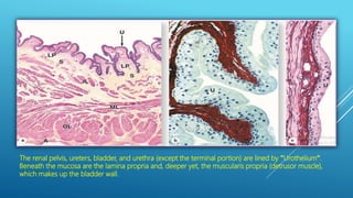
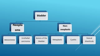
This document discusses various pathologies of the ureter, bladder, and urethra. It begins by describing common congenital anomalies of the ureter including double and bifid ureters as well as ureteropelvic junction obstruction. It then discusses non-neoplastic conditions of the bladder such as vesical diverticula, cystitis, and various metaplastic lesions. Neoplastic conditions covered include urothelial carcinoma and adenocarcinomas of the bladder. Finally, it briefly mentions primary carcinoma of the urethra.
Urological symptoms can involve pain, changes to urination, changes in urine appearance, or abnormalities of the male genitalia. A complete history divides into chief complaint, history of present illness, past medical history, family history, and review of systems. Physical exam includes inspection and palpation of the abdomen with specific examination of the kidneys, bladder, and genitalia. Common urological complaints involve sites of pain, irritative urinary symptoms, obstructive symptoms, incontinence, hematuria, and sexual or genital abnormalities.
Membranous glomerulopathy is a slowly progressive disease that most commonly affects people between 30-50 years old. It is the second most common cause of nephrotic syndrome in adults. There are primary and secondary types, with 85% being primary/idiopathic and the remainder being secondary to conditions like autoimmune diseases, infections, drugs, or tumors. It is caused by immune complex formation in the glomerulus resulting from antibodies binding to antigens in the glomerular basement membrane. On pathology, it shows diffuse thickening of the glomerular basement membrane. Treatment involves immunosuppressive drugs and anti-proteinuria measures, with prognosis being one-third spontaneous remission, one-third progression
Chronic pyelonephritis is a chronic inflammation of the renal tubules and interstitium that occurs due to recurrent urinary tract infections and scarring. It most commonly affects children with congenital anomalies or spinal cord injuries and is the leading cause of end-stage renal disease. It is typically caused by either chronic obstructive pyelonephritis due to obstruction of urine outflow, or reflux nephropathy caused by vesicoureteral reflux allowing urine to flow back into the kidneys. Symptoms may include fever, flank pain, and symptoms of chronic renal failure like hypertension. The condition can lead to complications like proteinuria, focal glomerulosclerosis, and papillary necrosis.
The document discusses tumors of the small and large intestines. It classifies intestinal tumors and provides details on various benign and malignant tumor types. The most common tumors are epithelial tumors, with colorectal cancer representing 70% of all gastrointestinal malignancies. Adenomas are precursors to most colorectal cancers. Risk factors include inflammatory bowel disease, familial polyposis, and diet. Prognosis and treatment depend on tumor stage and characteristics.
(1) Gastric polyps are nodules that project above the surrounding mucosa and can develop due to hyperplasia, inflammation, ectopia, or neoplasia. The most common types are inflammatory or hyperplastic polyps.
(2) Gastric adenomas are uncommon neoplasms that are often antral and can have dysplastic epithelium. Larger or villous adenomas have a higher malignant potential.
(3) Gastric adenocarcinoma is one of the most common cancers worldwide. Risk factors include H. pylori infection and atrophic gastritis. Prognosis depends on depth of invasion and extent of metastasis.
The document discusses the pathology of the renal system. It provides an overview of the goals and topics that will be covered in a lecture series on renal disorders. These include acute and chronic kidney injury, glomerular diseases, urinary tract infections, tubulointerstitial diseases, vascular diseases, obstructive uropathy, renal neoplasms, congenital and cystic kidney diseases, and electrolyte and acid-base disorders. The series will describe the causes, mechanisms, and effects of various renal conditions.
This document discusses tubulo-interstitial pathology and chronic glomerulonephritis. It defines acute pyelonephritis, its causes and morphology. It describes chronic pyelonephritis and reflux nephropathy, including forms of chronic pyelonephritis and their gross and microscopic morphology. It also discusses drug-induced tubulo-interstitial nephritis, analgesic nephropathy, causes of chronic glomerulonephritis, and interpreting morphology of chronic glomerulonephritis.
The document contains multiple choice questions about the kidney and renal pathology. Some key points addressed include:
- The correct location of electron dense deposits in Heymann glomerulonephritis.
- The most specific finding on renal biopsy for diabetic glomerulosclerosis.
- Rapidly progressive glomerulonephritis presenting as crescent forming glomerulonephritis.
- Clinical vignettes and the most likely diagnoses for the patients described.
1. The document discusses various types of ovarian tumours, including benign, borderline, and malignant surface epithelial tumours as well as germ cell tumours and sex cord-stromal tumours.
2. Etiological factors for ovarian tumours include nulliparity, heredity such as BRCA gene mutations, and genetic syndromes like Lynch syndrome.
3. Common epithelial tumours include serous and mucinous tumours. Germ cell tumours include mature teratomas, dysgerminomas, and choriocarcinomas. Sex cord-stromal tumours include granulosa cell tumours and thecomas.
This document provides an overview of the hepatobiliary system and various liver diseases. It discusses chronic venous congestion of the liver, fatty change, alcoholic liver disease, cirrhosis, cholelithiasis, chronic cholecystitis, and tumors of the liver. For each topic, it describes the gross and microscopic pathology findings. The goal is to educate about the introduction, causes, presentations and histopathological features of common hepatobiliary conditions.
Hematuria is defined as the presence of blood or red blood cells in the urine. It can be macroscopic (visible to the naked eye) or microscopic. Common causes include urinary tract infections, kidney diseases, cancers, and stones. A urinalysis and urine culture can help identify infection, while imaging tests like ultrasound, CT, and cystoscopy can locate any abnormalities in the kidneys, ureters, bladder, or urethra. Further blood tests may also indicate underlying conditions. Hematuria is investigated through examination of urine and imaging of the urinary tract to diagnose the cause.
The document discusses various diseases of the kidney. It begins by describing the normal structure and functions of the kidney. It then discusses various glomerular and non-glomerular diseases in detail, including their classification, clinical manifestations, and microscopic pathology. Some of the diseases covered include glomerulonephritis, pyelonephritis, diabetic nephropathy, benign and malignant nephrosclerosis, and hydronephrosis.
This document provides an overview of inflammatory bowel disease (IBD), including ulcerative colitis and Crohn's disease. Some key points:
- IBD results from a combination of genetic and immunological factors. Several genes have been identified that increase risk of developing IBD, including NOD2 for Crohn's.
- Ulcerative colitis is limited to the colon and rectum, while Crohn's can affect any part of the GI tract and is typically transmural.
- Histologically, ulcerative colitis shows continuous mucosal inflammation while Crohn's is discontinuous with features like granulomas and cobblestoning.
- The role of the microbiome
This document summarizes renal pathology and glomerular diseases. It discusses how diseases can affect the glomeruli, tubules, interstitium and vasculature. The glomeruli are described as a network of capillaries lined by endothelial cells, the glomerular basement membrane, and podocytes. Immunological and toxic mechanisms can cause glomerular injury. Membranous glomerulopathy is described as the most common cause of nephrotic syndrome in adults, characterized by thickening of the glomerular capillary wall.
Cystic kidney diseases can be genetic or acquired. Autosomal dominant polycystic kidney disease (ADPKD) is a common genetic condition characterized by numerous fluid-filled cysts in the kidneys that worsen over time, potentially leading to kidney failure. Autosomal recessive polycystic kidney disease (ARPKD) presents in infancy with enlarged cystic kidneys and often liver disease, and can be fatal. Other cystic conditions include medullary sponge kidney cysts in the kidney papillae and nephronophthiasis-medullary cystic complex cysts at the corticomedullary junction. Simple cortical cysts are very common incidental findings. Ren
Chronic pancreatitis is a progressive inflammatory condition of the pancreas characterized by irreversible morphological changes and loss of function. It is most commonly caused by long term heavy alcohol use. Symptoms include recurrent abdominal pain, steatorrhea due to exocrine insufficiency, and diabetes mellitus due to endocrine insufficiency. Diagnosis involves functional tests like fecal elastase and imaging modalities like CT, MRI, ERCP and EUS which demonstrate findings of pancreatic duct abnormalities, parenchymal changes and calcifications.
This 68-year-old male carpenter presents with urinary symptoms including frequency, urgency, terminal dribbling, poor stream, and occasional incontinence. His differential diagnosis includes benign prostatic hyperplasia, prostatitis, prostate cancer, stones, and other causes. Further workup is needed to determine the underlying pathology and guide treatment.
This document discusses tumours of the kidney. It begins by defining benign and malignant tumours in general. It then describes several types of benign kidney tumours, including cortical adenomas, oncocytomas, angiomyolipomas, and metanephric adenomas. Malignant kidney tumours are also briefly mentioned. The document focuses on Wilms' tumour, providing details on its epidemiology, etiology, pathogenesis, clinical presentation, morphological features, diagnosis and treatment. Wilms' tumour is described as the most common malignant kidney tumour in children.
The document summarizes tumors of the kidney and urinary tract. The most common malignant kidney tumor is renal cell carcinoma, followed by nephroblastoma (Wilms tumor). Tumors of the lower urinary tract are more common than renal cancers. Renal cell carcinoma originates from renal tubular epithelium and accounts for 80-85% of primary malignant kidney tumors. Clear cell carcinoma is the most common type of renal cell carcinoma.
This document summarizes renal cell carcinoma (RCC), including its incidence, risk factors, pathology, subtypes, clinical presentation, and prognosis. RCC accounts for 2-3% of adult cancers and is the most lethal urologic cancer. Clear cell RCC is the most common subtype, accounting for 70-80% of cases. Other subtypes include papillary RCC, chromophobe RCC, collecting duct carcinoma, and unclassified RCC. Tumor size, grade, stage, and histologic subtype are important prognostic factors. Over 60% of RCCs are now detected incidentally with improved imaging techniques.
The document discusses two causes of obstructive uropathy: nephrolithiasis and hydronephrosis. Nephrolithiasis is the formation of urinary calculi (stones) in the kidneys or urinary tract. Calculi most commonly occur in middle-aged men and can cause pain. Hydronephrosis is the dilation of the renal pelvis and calyces due to partial or intermittent blockage of urine flow. It is usually caused by obstruction in the ureter but can also be congenital or due to conditions affecting the bladder. Advanced hydronephrosis leads to compression of the renal cortex. Both conditions are demonstrated in photographs showing enlarged kidneys with dilated pelvis and
This document provides an overview of pathology related to the female genital tract, including the vulva, vagina, cervix, and endometrium. It discusses infections, pre-malignant and malignant neoplasms of these structures. Some key points include that HPV is associated with most cervical cancers, endometrial carcinoma is most common in post-menopausal women due to unopposed estrogen, and endometrial hyperplasia is a risk factor for the development of endometrial cancer if not treated.
This document discusses the anatomy, functions, diseases, and surgical procedures related to the spleen. It begins by describing the embryological development and functions of the spleen. It then discusses various spleen diseases including anomalies, infections, tumors, and those related to blood disorders. The indications for splenectomy and techniques for both open and laparoscopic splenectomy are explained. Finally, it covers postoperative complications such as hemorrhage, infection, and recommendations for immunization and antibiotic prophylaxis.
The document describes a case of a 37-year-old woman with type 1 diabetes presenting with various symptoms including dysuria, urinary frequency, fatigue, and edema. Laboratory tests showed elevated blood sugar, proteinuria, and urine positive for nitrates, ketones, glucose, protein, and red blood cells. A renal biopsy showed glomerulonephritis.
There are many different types of kidney cysts that can be classified in various ways. An ideal classification system would take into account morphological features, pathogenesis, and therapeutic potential. Autosomal dominant polycystic kidney disease (ADPKD) is the most common cystic kidney disease and is characterized by multiple bilateral cysts of varying sizes in the kidneys and liver. It has a prevalence of 1 in 400 to 1,000 people and is caused by mutations in the PKD1 or PKD2 genes.
The document discusses various types of glomerular and tubulointerstitial diseases:
1. Glomerular diseases are caused by damage to the glomerulus and can manifest as nephritic syndrome, nephrotic syndrome, or hematuria. Common glomerular diseases include minimal change disease, membranous nephropathy, and focal segmental glomerulosclerosis.
2. Tubulointerstitial diseases involve damage to the renal tubules and interstitium. Acute interstitial nephritis is often caused by drug reactions while chronic interstitial nephritis can result from various insults like drugs, infections, toxins, and inherited conditions.
3
This document summarizes renal cell carcinoma (RCC), the most common type of kidney cancer. Key points include:
- RCC originates in the renal cortex and arises mostly from the upper pole of the kidney.
- Common subtypes include clear cell, papillary, and chromophobe carcinomas.
- Risk factors include male sex, older age, smoking, obesity, and genetic conditions like von Hippel-Lindau syndrome.
- Symptoms may include hematuria, flank pain, and palpable flank mass. Imaging like CT and MRI are used to diagnose and stage disease.
- Treatment depends on stage but typically involves surgical removal (radical or partial nephrectomy
1. The document provides tips for using a PowerPoint presentation on benign lung tumors. It recommends asking students questions about blank slides to promote active learning and revisiting topics through multiple revisions.
2. The presentation covers learning objectives on the introduction, anatomy, aetiology, pathophysiology, classification, clinical features, investigations, management, prevention and other topics related to benign lung tumors.
3. A solitary pulmonary nodule is defined as a rounded opacity less than 3 cm that is surrounded by lung parenchyma. Common benign tumors include hamartomas, adenomas, and papillomas.
This document discusses pathology of the lower urinary tract, including congenital anomalies, inflammation, obstructive lesions, and tumors of the urinary bladder. Some key points include:
- Congenital anomalies of the lower urinary tract include bifid ureters, ureteropelvic junction obstruction, and bladder diverticula.
- Inflammation of the lower urinary tract can cause ureteritis and cystitis.
- Obstructive lesions are a major cause of hydronephrosis and hydroureter and can be caused by various factors including calculi, strictures, and tumors.
- Tumors of the bladder include both noninvasive papillary tumors and carcinoma in situ.
The patient, a 63-year-old man, has noted increasing back pain for 7 months and has had recurrent respiratory infections. Laboratory tests show decreased kidney function with proteinuria. A renal biopsy shows deposits of pink amorphous material in the glomeruli, interstitium, and arteries. These findings are most consistent with a diagnosis of multiple myeloma.
The document discusses the anatomy and histology of the bladder and various benign and malignant tumors that can occur. It provides details on:
1. The anatomy of the bladder including its shape, surfaces and location in the pelvis.
2. The normal histology of the bladder wall and urothelial lining.
3. Various benign tumors of the bladder including epithelial metaplasia, leukoplakia, inverted papilloma, nephrogenic adenoma, leiomyoma, cystitis cystica and glandularis.
4. Risk factors, pathology, clinical features and staging of urothelial cancer of the bladder, which represents the majority of bladder cancers.
This document discusses various congenital anomalies, inflammations, and tumors of the lower urinary tract. It begins by describing common congenital anomalies like vesicoureteric reflux and double ureters. It then covers inflammations of the ureter, bladder, and urethra. The majority of the document discusses tumors of the bladder, including classification of urothelial/transitional cell tumors and their histological features. Non-epithelial bladder tumors and tumors of the renal pelvis and ureters are also briefly covered.
This document provides information on bladder anatomy, risk factors, classification, grading, staging, and tumor biology of bladder cancer. It discusses the histologic layers of the bladder wall and lists several genetic and environmental risk factors. Papillary and flat urothelial lesions are classified according to the WHO/ISUP system. Grading is described for non-invasive and invasive papillary urothelial carcinomas. Staging is outlined using the TNM system. Immunohistochemistry markers and their roles in diagnosis are reviewed. The molecular pathways and progression of bladder cancer are summarized in a figure.
The peritoneum is a membrane that lines the abdominal cavity. In women, the peritoneum is an "open system" interrupted by the fallopian tubes, allowing transmission between the genital tract and peritoneal cavity. Many inflammatory conditions can involve the peritoneum. Endometriosis is a common condition where endometrial tissue grows outside the uterus, often on the ovaries or peritoneum. Symptoms include pelvic pain and infertility. Mesothelial tumors of the peritoneum range from benign to aggressive malignancies. Serous tumors are the most common type of peritoneal tumor and can be borderline or adenocarcinomas.
This document discusses various rectal diseases including prolapse, proctitis, polyps, benign lesions, and carcinoma. Rectal prolapse is classified as mucosal or full-thickness and can be caused by straining, weak pelvic floor muscles, or trauma from childbirth. Treatment depends on the type but may include injections, banding, or surgery. Proctitis is inflammation that can be caused by infection, radiation, or inflammatory bowel disease. Polyps are growths that can be removed endoscopically if small or via surgery if large. Benign lesions include endometriosis, hemangiomas, and neuroendocrine tumors. Rectal carcinoma is often treated with surgery such as anterior resection or
The document discusses the anatomy and pathology of the male urethra and genital system. It describes the anatomy of the urethra in males, noting it is longer than in females and has three regions. It also discusses inflammation of the urethra, tumors and tumor-like conditions, congenital anomalies of the penis like hypospadias, and infections. For the testis, it outlines the anatomy, congenital anomalies like cryptorchidism, atrophy, vascular disorders like torsion, and inflammation.
This document discusses the diagnosis and management of adnexal masses. It provides information on the normal size and variation of ovaries and lifetime risk of ovarian neoplasms. Differential diagnoses of adnexal masses are outlined based on patient age. Diagnostic evaluation includes physical exam, ultrasound, CT or MRI if needed. Common ovarian tumors at different life stages are described. Functional cysts, endometriomas, benign and borderline tumors are discussed. Complications, clinical presentation, imaging and tumor markers are summarized. Guidelines for management of asymptomatic cysts and indications for surgery are provided.
1) The document discusses various precancerous lesions of the colon and rectum, including adenomas, hyperplastic polyps, sessile serrated lesions, and traditional serrated adenomas.
2) It describes the histological features and progression of these lesions, noting that sessile serrated lesions and traditional serrated adenomas have a significant malignant potential, whereas hyperplastic polyps have a very low malignant potential.
3) Two pathways of colorectal carcinogenesis are discussed: the classic adenoma-carcinoma sequence and the serrated neoplastic pathway, which involves certain serrated polyps.
This document summarizes various pathologies that can present as cystic or solid masses in the peritoneum based on CT imaging findings. It describes mucinous carcinomatosis, pseudomyxoma peritonei, lymphangioma, enteric duplication cyst, tuberculosis, echinococcal cyst, peritoneal metastases, lymphoma, carcinoid tumors, gastrointestinal stromal tumors, inflammatory pseudotumor, desmoid tumors, sclerosing mesenteritis, malignant mesothelioma, primary peritoneal serous carcinoma and desmoplastic small round cell tumor. Key distinguishing imaging features for each condition are provided.
This document discusses pathology of the male reproductive system, specifically focusing on the prostate. It provides information on normal prostate structure and histology. It then discusses three main pathologic processes that can occur in the prostate - prostatitis, nodular hyperplasia, and carcinoma. For prostatitis and nodular hyperplasia, it describes the etiology, gross and microscopic morphology, and clinical features. It provides detailed information on the different types of prostatitis and the morphological features of nodular hyperplasia. The document is intended as an educational guide on prostate pathology.
Cancer of the uterus is a common cancer that affects the female reproductive system. Abnormal vaginal bleeding is the most common symptom. Diseases that can affect the reproductive system include cancers of the prostate, breast, ovaries, penis, uterus, and testicles. The uterus is a major female reproductive organ located in the pelvis. Diseases of the uterus include prolapse, cancers of the cervix and uterus, fibroids, adenomyosis, infections, and uterine malformations. Benign lesions of the uterus include endometrial polyps and Asherman's syndrome. Uterine fibroids are a common benign tumor of the uterus that can cause heavy bleeding, pain, and infertility. Treatment options depend on symptoms
The document discusses surgical anatomy and neoplasms of the peritoneum. It describes the peritoneum as the largest serous membrane in the body, which can be divided into parietal and visceral portions. It covers various primary and secondary neoplasms that can affect the peritoneum, including malignant mesothelioma, peritoneal carcinomatosis, and others. Malignant peritoneal mesothelioma is described as the most common primary malignant peritoneal neoplasm, with diffuse forms being highly aggressive and incurable in most cases.
Cryptorchidism is the failure of one or both testes to descend into the scrotum. It occurs in about 1% of males and can lead to testicular atrophy and increased risk of testicular cancer. Undescended testes may be due to hormonal abnormalities, testicular abnormalities, or mechanical problems. Orchiopexy, or surgical placement of undescended testes into the scrotum, may help prevent atrophy but not necessarily decrease cancer risk. Testicular atrophy is a regressive change seen as the end stage of various conditions like inflammation, irradiation, malnutrition, and cryptorchidism. It is characterized microscopically by tubular hyalinization and interstitial fibrosis
This document discusses urinary bladder tumors. It begins by covering the anatomy and histology of the bladder. It then describes the WHO classification of bladder tumors, which includes urothelial, squamous, glandular, urachal, mullerian, and mesenchymal tumors, among others. Non-invasive urothelial lesions like carcinoma in situ, papillomas, and non-invasive papillary carcinomas are summarized. Invasive urothelial carcinoma is also covered, noting the range of architectural patterns and cell types seen microscopically. Variants with divergent differentiation like squamous or glandular are also common. The document provides an overview of bladder tumor pathology.
The gallbladder is a pear-shaped organ that stores and concentrates bile produced by the liver. It can be affected by various congenital anomalies and gallstones. Gallstones are usually cholesterol stones but some may be pigment stones related to hemolytic conditions. Cholecystitis, or inflammation of the gallbladder, can be acute or chronic and is usually caused by gallstones. Gallbladder cancer is rare but associated with conditions like gallstones and chronic infection. Cancers can also occur in the bile ducts and ampulla of Vater.
1. The document discusses diseases of the female reproductive system including the endometrium, fallopian tubes, and ovaries. It covers conditions such as endometritis, adenomyosis, endometrial hyperplasia and cancer, salpingitis, and ovarian cysts and cancers.
2. Various tumors are described such as endometrial polyps, endometrial cancer, and the different subtypes of ovarian tumors including serous and mucinous cystadenomas and cystadenocarcinomas.
3. The causes, histological features, clinical presentations and diagnoses of these diseases are provided, with examples of microscopic images.
This document discusses benign liver tumors. It describes the three most common types: hemangioma, focal nodular hyperplasia (FNH), and hepatocellular adenoma. For each type, it covers characteristics, clinical presentation, diagnosis, and treatment. Hemangiomas are the most frequent solid benign liver tumor. FNH predominantly affects young women and usually presents as an asymptomatic mass. Hepatocellular adenomas occur in reproductive-aged women and have been linked to oral contraceptive use.
This document discusses Covid-19 in children. It notes that while fewer Covid cases have been diagnosed in children, most pediatric cases are mild. Hospitalization rates are significantly lower in children. The pathophysiology and risk factors for children are also discussed. Common symptoms in children are cough and fever. Diagnostic methods, treatment options, and prevention strategies are summarized.
Facial trauma can cause injuries to soft tissues, bones, or both from accidents like car crashes or assaults. Common signs are pain, swelling, bleeding, and changes to facial structure or function. Treatment involves airway management, bleeding control, wound care, and treating underlying bone fractures. Fractures of the maxilla and mandible are classified by location and require techniques like interdental wiring, plates or screws for fixation.
This document defines hyphaema as blood in the anterior chamber of the eye. It can result from compressive trauma that injures blood vessels in the iris, ciliary body, or trabecular meshwork. Hyphaema is classified based on etiology (traumatic, medical conditions, etc.) and severity (mild, moderate, severe). Presentation includes blurry vision, pain, and blood visible in the anterior chamber. Examination involves assessing injury, vision, pressure, and ruling out other issues. Treatment focuses on preventing increased pressure through medications, limiting activity, and sometimes surgery to remove blood if pressure remains high.
Oliguria is a low urine output defined as less than 1 mL/kg/hr in infants, less than 0.5 mL/kg/hr in children, and less than 300 mL daily in adults. It indicates an underlying disorder and can lead to acute renal failure if left untreated. Anuria is even less urine output at less than 50 mL/day. Causes of oliguria and anuria include pre-renal (low blood volume), renal (kidney damage), and post-renal (urinary tract obstruction). Evaluation and management depends on determining the cause through history, physical exam, urinalysis, and blood tests to guide volume replacement or other interventions to prevent further kidney injury.
Hepatitis viruses A, B, C, D, E and G can cause infection of the liver. Hepatitis A and E are transmitted via the fecal-oral route while hepatitis B and C are bloodborne. Hepatitis A is common in children in areas with poor sanitation and causes jaundice and abdominal symptoms for 2-6 weeks before recovery without treatment. Hepatitis B infects over 350 million carriers worldwide and can be transmitted sexually or via contaminated needles. It may cause acute or chronic infection. Hepatitis C is a major cause of post-transfusion hepatitis in developed countries and can lead to liver cancer. Vaccines exist to prevent hepatitis A and B but treatment relies on supportive care or antiviral drugs for
Memory is the ability of the brain to store and recall information over time. There are four main types of memory: sensory memory (up to 0.5 seconds), short-term memory (minutes to hours), long-term memory (hours to years), and permanent memory. Information enters the brain and is either selected and stored in memory traces through processes like long-term potentiation, or neglected and forgotten. The hippocampus plays a key role in consolidating memories from short-term to long-term storage. Disorders like amnesia and Alzheimer's disease can impair memory formation and recall.
The document discusses abnormal hemoglobin metabolism and hemoglobinopathies. It provides information on hemoglobin structure, definitions of thalassemias and hemoglobinopathies, specifics on sickle cell anemia and thalassemia, Bart's hydrops fetalis, and diagnosis of hemoglobinopathies. Sickle cell anemia results from a single amino acid substitution causing hemoglobin S, which polymerizes and makes red blood cells rigid and sickle-shaped. Thalassemias involve absent or reduced alpha or beta globin chains. Diagnosis involves family history, symptoms, lab tests, and analyzing hemoglobin structure.
This document discusses hemoglobin metabolism and heme synthesis. It notes that hemoglobin is made up of heme and globin, with heme containing an iron atom bound to a porphyrin ring. Heme synthesis occurs in multiple steps, starting with the formation of alpha-aminolevulinate from succinyl-CoA and glycine in the mitochondria. Several other reactions then occur to ultimately form protoporphyrin, into which iron is inserted by ferrochelatase to form heme. Heme is broken down to bilirubin, which is excreted in bile. Key enzymes involved in heme synthesis and the regulation of the process are also outlined.
This document provides information about performing a complete blood count (CBC) test. It discusses the objectives and aims of the practical, which include counting red blood cells, white blood cells, determining hemoglobin concentration, and calculating red blood cell indices. It describes manual and automated methods for obtaining CBC measurements using a hemocytometer or Coulter counter. Key steps in the procedures and normal reference ranges for CBC components are also outlined. The document concludes by discussing some clinical applications and implications of abnormal CBC results.
The erythrocyte sedimentation rate (ESR) is a nonspecific screening test used to detect inflammation. It measures how fast red blood cells sediment in a tube in one hour. Faster sedimentation indicates more inflammation in the body from conditions like infections, cancers, and autoimmune diseases. The ESR is directly related to red cell mass and fibrinogen levels, and inversely related to plasma viscosity. It is an inexpensive and widely available initial screening test to monitor disease progression or remission.
Multiple myeloma is a cancer of plasma cells that produce abnormal proteins called monoclonal proteins (M proteins). The most common M protein is IgG, followed by IgA. In some cases, only free light chains are produced. Free light chains can cause kidney damage and are detected in urine as Bence Jones proteins. Myeloma cells secrete cytokines like IL-6 that support their growth and cause bone damage. Patients experience symptoms like bone pain, fractures, anemia, and kidney problems. Diagnosis involves tests to detect M proteins in blood and urine and check for organ damage and immune suppression.
The lungs have several metabolic functions beyond gas exchange, including host defence, water balance maintenance, temperature and acid-base regulation, and metabolizing substances. The lungs are protected by physical, mucociliary, and cellular defences like macrophages. The lungs also play roles in water loss, heat loss, acid-base balance maintenance through CO2 exchange, fibrinolysis, renin-angiotensin system effects, and synthesizing/releasing/metabolizing various substances that enter the bloodstream.
This document summarizes several tissues and organs examined in histology labs, including the tongue, salivary glands, esophagus, liver, gallbladder, and pancreas. Key features are highlighted such as the papillae and taste buds on the tongue, the serous and mucous acini in salivary glands, the stratified squamous epithelium and esophageal glands in the esophagus, the liver lobule structure around the central vein and portal areas, the simple columnar epithelium of the gallbladder, and the islets of Langerhans surrounded by pancreatic acini. Diagrams are included illustrating histological structures.
Mammography uses low-dose x-rays to examine the breast for early cancer detection. It has advanced from film to digital mammography and tomosynthesis, which creates 3D breast images. Computer-aided detection highlights abnormal areas. Screening mammograms aim to detect cancer in asymptomatic women, while diagnostic mammograms investigate symptoms. Benefits include early detection, but limitations include false positives and negatives due to breast density. Yearly mammograms after age 40 are recommended for breast cancer screening. Ultrasound provides localized breast images without radiation but cannot screen whole breasts. MRI is superior for dense breasts but has no radiation risk.
This document summarizes several pathology slides including fatty degenerative liver, cirrhosis liver, primary carcinoma of the liver, ulcerative colitis of the rectum, adenocarcinoma of the pancreas, acute and chronic peptic ulcers, mixed tumor of the salivary gland, acute suppurative appendicitis, gastric carcinoma, and colon adenocarcinoma. For each slide, it describes the histological features such as presence of malignant cells, inflammatory cells, fibrosis, hemorrhage, and other abnormalities. The document aims to provide a histological overview of various diseases and tissues examined under the microscope on pathology slides.
Osteoporosis is a disease where bones become weak and fragile from loss of tissue. There are drugs that can help prevent and treat osteoporosis by slowing bone loss, making bone stronger, or replacing hormones. The main drug types used for osteoporosis are bisphosphonates, selective estrogen receptor modulators (SERMs), parathyroid hormone (PTH) therapy, and RANK ligand inhibitors.
Vasculitis refers to inflammation of blood vessels. There are many types classified by the size of vessels involved and pathogenic mechanisms. Noninfectious vasculitis can be immune complex-mediated, associated with ANCAs, anti-endothelial cell antibodies, or autoreactive T cells. Infectious vasculitis involves direct invasion of vessels by pathogens. Common noninfectious vasculitides include giant cell arteritis, Takayasu arteritis, polyarteritis nodosa, Kawasaki disease, microscopic polyangiitis, Wegener's granulomatosis, Churg-Strauss syndrome, and Buerger's disease. Clinical manifestations depend on the organs involved. Treatment involves immunos
This document discusses the causes, types, diagnosis, and treatment of jaundice. It begins by describing the normal breakdown process of red blood cells and hemoglobin. It then covers various types of congenital and acquired jaundice, including physiological jaundice in newborns, breast milk jaundice, hemolytic diseases, hepatic/hepatocellular jaundice, and obstructive jaundice. Diagnosis involves urine, liver, and imaging tests. Treatment depends on the underlying cause but may include discontinuing drugs, antibiotics, blood transfusions, surgery, or phototherapy for newborns.
Malabsorption results from defects in intraluminal digestion, terminal digestion, transepithelial transport, and lymphatic transport. It is characterized by weight loss, abdominal distention, diarrhea, and steatorrhea. Causes of malabsorption include cystic fibrosis, celiac disease, lactase deficiency, environmental enteropathy, abetalipoproteinemia, microscopic colitis, graft-versus-host disease, and Whipple's disease. Histological examination often reveals villous atrophy, increased intraepithelial lymphocytes, and lipid-laden macrophages in the lamina propria.
Integrating Ayurveda into Parkinson’s Management: A Holistic ApproachAyurveda ForAll
Explore the benefits of combining Ayurveda with conventional Parkinson's treatments. Learn how a holistic approach can manage symptoms, enhance well-being, and balance body energies. Discover the steps to safely integrate Ayurvedic practices into your Parkinson’s care plan, including expert guidance on diet, herbal remedies, and lifestyle modifications.
Promoting Wellbeing - Applied Social Psychology - Psychology SuperNotesPsychoTech Services
A proprietary approach developed by bringing together the best of learning theories from Psychology, design principles from the world of visualization, and pedagogical methods from over a decade of training experience, that enables you to: Learn better, faster!
- Video recording of this lecture in English language: https://youtu.be/kqbnxVAZs-0
- Video recording of this lecture in Arabic language: https://youtu.be/SINlygW1Mpc
- Link to download the book free: https://nephrotube.blogspot.com/p/nephrotube-nephrology-books.html
- Link to NephroTube website: www.NephroTube.com
- Link to NephroTube social media accounts: https://nephrotube.blogspot.com/p/join-nephrotube-on-social-media.html
Basavarajeeyam is an important text for ayurvedic physician belonging to andhra pradehs. It is a popular compendium in various parts of our country as well as in andhra pradesh. The content of the text was presented in sanskrit and telugu language (Bilingual). One of the most famous book in ayurvedic pharmaceutics and therapeutics. This book contains 25 chapters called as prakaranas. Many rasaoushadis were explained, pioneer of dhatu druti, nadi pareeksha, mutra pareeksha etc. Belongs to the period of 15-16 century. New diseases like upadamsha, phiranga rogas are explained.
NVBDCP.pptx Nation vector borne disease control programSapna Thakur
NVBDCP was launched in 2003-2004 . Vector-Borne Disease: Disease that results from an infection transmitted to humans and other animals by blood-feeding arthropods, such as mosquitoes, ticks, and fleas. Examples of vector-borne diseases include Dengue fever, West Nile Virus, Lyme disease, and malaria.
Adhd Medication Shortage Uk - trinexpharmacy.comreignlana06
The UK is currently facing a Adhd Medication Shortage Uk, which has left many patients and their families grappling with uncertainty and frustration. ADHD, or Attention Deficit Hyperactivity Disorder, is a chronic condition that requires consistent medication to manage effectively. This shortage has highlighted the critical role these medications play in the daily lives of those affected by ADHD. Contact : +1 (747) 209 – 3649 E-mail : sales@trinexpharmacy.com
Does Over-Masturbation Contribute to Chronic Prostatitis.pptxwalterHu5
In some case, your chronic prostatitis may be related to over-masturbation. Generally, natural medicine Diuretic and Anti-inflammatory Pill can help mee get a cure.
ABDOMINAL TRAUMA in pediatrics part one.drhasanrajab
Abdominal trauma in pediatrics refers to injuries or damage to the abdominal organs in children. It can occur due to various causes such as falls, motor vehicle accidents, sports-related injuries, and physical abuse. Children are more vulnerable to abdominal trauma due to their unique anatomical and physiological characteristics. Signs and symptoms include abdominal pain, tenderness, distension, vomiting, and signs of shock. Diagnosis involves physical examination, imaging studies, and laboratory tests. Management depends on the severity and may involve conservative treatment or surgical intervention. Prevention is crucial in reducing the incidence of abdominal trauma in children.
2. OBJECTIVES:
Congenital Anomalies of the ureter :
* Double and bifid ureters
* Ureteropelvic junction (UPJ) obstruction
non-neoplastic conditions of the bladder : Vesical diverticula, Cystitis, and Metaplastic
lesions of the bladder
Neoplastic conditions of bladder: Urothelial carcinoma and Adenocarcinomas of the
bladder.
Urethra: primary carcinoma
3. The renal pelvis, ureters, bladder, and urethra (except the terminal portion) are lined by "Urothelium".
Beneath the mucosa are the lamina propria and, deeper yet, the muscularis propria (detrusor muscle),
which makes up the bladder wall.
4. URETER
The main pathological lesions involving the ureter include:
Congenital Anomalies:
* Double and bifid ureters
* Ureteropelvic junction (UPJ) obstruction
Obstructive Lesions: as a result of calculi, tumours, blood clots
Primary malignant tumours:
Primary malignant tumours of the ureter follow patterns similar to those arising in the
renal pelvis, calyces, and bladder, and a majority are urothelial carcinomas. Primary
tumors of the ureter are rare.
5. DOUBLE AND BIFID URETERS
Double ureters are almost invariably
associated with totally distinct double renal
pelves or with the anomalous development
of a large kidney having a partially bifid
pelvis terminating in separate ureters.
Double ureters may pursue separate
courses to the bladder but commonly are
joined within the bladder wall and drain
through a single ureteral orifice.
Most are unilateral and of no clinical
significance.
6. URETEROPELVIC JUNCTION (UPJ)
OBSTRUCTION
a congenital disorder, results in
hydronephrosis.
It usually manifests in infancy or childhood,
much more commonly in boys.
It is the most frequent cause of
hydronephrosis in infants and children.
Grossly: the renal pelvis is markedly
dilated, but the ureter is not, indicating
that the point of obstruction is at the
ureteropelvic junction.
8. RETROPERITONEAL FIBROSIS
It is an uncommon cause of ureteral narrowing or obstruction,
characterized by a fibrous proliferative inflammatory process encasing the retroperitoneal
structures and causing hydronephrosis.
The disorder occurs in middle to old age.
The affected sites include: the pancreas, retroperitoneum, and salivary glands, to mention a
few.
Causes:
1. The majority of cases have no obvious cause and are considered primary, or idiopathic (Ormond
disease).
2. At least a proportion of these cases are related to the newly described entity in which elevations of
serum IgG4 are associated with fibroinflammatory lesions that are rich in IgG4-secreting plasma cells.
3. Other cases are associated with drug exposures (ergot derivatives, adrenergic blockers), or malignant
disease (lymphomas, urinary tract carcinomas).
11. BLADDER OR VESICAL DIVERTICULUM
Consists of a pouchlike evagination of the
bladder wall.
Diverticula may be congenital but more
commonly are acquired lesions that arise
as a consequence of persistent urethral
obstruction caused, for example, by
benign prostatic hyperplasia.
Although most diverticula are small and
asymptomatic, they sometimes lead to
urinary stasis and predispose to infection.
12. CYSTITIS
Cystitis takes many forms.
Most cases stem from nonspecific acute or chronic
inflammation of the bladder.
Etiology:
1. The common etiologic agents of bacterial cystitis are
coliform bacteria.
2. Patients receiving cytotoxic antitumor drugs, such as
cyclophosphamide, sometimes develop hemorrhagic
cystitis.
3. Adenovirus infection also causes a hemorrhagic cystitis.
Several distinct variants of cystitis are defined by
either morphologic appearance or causation
(Interstitial cystitis, Malakoplakia, Polypoid cystitis).
13. INTERSTITIAL CYSTITIS (I.E., CHRONIC
PELVIC PAIN SYNDROME)
is a persistent, painful form of chronic cystitis
occurring most frequently in women.
It is characterized by intermittent, often severe
suprapubic pain, urinary frequency, urgency,
hematuria and dysuria without evidence of
bacterial infection.
There are cystoscopic findings of fissures and
punctate hemorrhages (glomerulations) in the
bladder mucosa.
The histologic findings are nonspecific.
Late in the course, transmural fibrosis may
ensue, leading to a contracted bladder.
14. No epithelium and
plenty of ulceration. Not high powered,
therefore can't
see mast cells.
Difficult to treat
due to
unknown
etiology.
15. Ulcerating, no
PMN, mast
cells, chronic
inflammation.
Difficult to know
how to treat
these patients.
Sometimes
steroids are
given.
16. MALAKOPLAKIA
Malakoplakia: from Greek Malako "soft" + Plako "plaque"
Most commonly occurs in the bladder.
Results from defects in phagocytic or degradative function of macrophages, such that
phagosomes become overloaded with undigested bacterial products. The macrophages have
abundant granular cytoplasm filled with phagosomes stuffed with particulate and
membranous bacterial debris.
In addition, laminated mineralized concretions resulting from deposition of calcium in
enlarged lysosomes, known as Michaelis-Gutmann bodies, typically are present within the
macrophages.
17. POLYPOID CYSTITIS
Is an inflammatory condition resulting from irritation to the bladder mucosa in which the urothelium is thrown
into broad bulbous polypoid projections as a result of marked submucosal edema.
Polypoid cystitis may be confused with papillary urothelial carcinoma both clinically and histologically.
18. METAPLASIA
Various metaplastic lesions may occur in the bladder:
1. Cystitis glandularis: Nests of urothelium (Brunn nests) may grow downward into the
lamina propria, and their central epithelial cells may variously differentiate into a
cuboidal or columnar epithelium lining.
2. Cystitis cystica: cystic spaces filled with clear fluid lined by flattened urothelium.
Maybe there are goblet cells resembling intestinal mucosa (intestinal or
colonic metaplasia).
As a response to injury, the urothelium often undergoes squamous
metaplasia, which must be differentiated from normal glycogenated
squamous epithelium, commonly found at the trigone in women.
19. METAPLASIA (BRUNN NESTS)
Solid nests of benign urothelial cells often with
regular contour.
Cells have normal cytology and orderly
arrangement.
21. METAPLASIA (CYSTITIS CYSTICA)
May appear grossly as pearly or
luminescent cysts with intact surface
urothelium.
Well-defined nests of urothelium with a centrally dilated
lumen (like von Brunn's nests but with a hole in the
middle).
25. NEOPLASMS
Bladder cancer accounts for approximately 7% of cancers and 3% of cancer deaths in the
United States.
The vast majority of bladder cancers (90%) are urothelial carcinomas.
Carcinoma of the bladder is more common in men than in women, in industrialized than
in developing nations, and in urban than in rural dwellers.
About 80% of patients are between the ages of 50 and 80 years.
PATHOGENESIS OF BLADDER CANCER
Bladder cancer, with rare exceptions, is not familial.
Some of the most common factors implicated in the causation of urothelial carcinoma
include:
1. cigarette smoking
2. various occupational carcinogens
3. Schistosoma haematobium infections in areas where it is endemic, such as Egypt.
26. PATHOGENESIS OF BLADDER CANCERS
Genetic Models for bladder carcinogenesis include:
1. A model for bladder carcinogenesis has been proposed in which the tumor is initiated by
deletions of tumor-suppressor genes on 9p and 9q, leading to formation of superficial
papillary tumors, a few of which may then acquire TP53 mutations and progress to
invasion.
2. A second pathway, possibly initiated by TP53 mutations, leads first to carcinoma in situ
and then, with loss of chromosome 9, progresses to invasion.
3. The underlying genetic alterations in superficial tumors include fibroblast growth factor
receptor 3 (FGFR3) mutations and activation of the Ras pathway.
27. MORPHOLOGY
Two distinct precursor lesions to invasive urothelial
carcinoma are recognized:
1. Noninvasive papillary neoplasms (maybe low or high
grade)
2. Flat noninvasive carcinoma in situ (uniformly high
grade).
In about half of the patients with invasive bladder
cancer, no precursor lesion is found; in such cases, it
is presumed that the precursor lesion was
overgrown by the high-grade invasive component.
28. NON INVASIVE PAPILLARY UROTHELIAL
NEOPLASMS
The most common precursor lesion to invasive urothelial carcinoma.
Demonstrate range of atypia and are graded to reflect their biologic behavior
The most common grading system classifies tumors as follows:
1. Papilloma.
2. Papillary urothelial neoplasm of low malignant potential (PUNLMP).
3. Low grade papillary urothelial carcinoma.
4. High grade papillary urothelial carcinoma
These exophytic papillary neoplasms are to be distinguished from inverted urothelial
papilloma, which is entirely benign and not associated with an increased risk for subsequent
carcinoma.
30. CARCINOMA IN SITU (CIS)
CIS is defined by the presence of cytologically
malignant cells within a flat urothelium (Fig. 17–18).
Like high-grade papillary urothelial carcinoma, CIS
tumor cells lack cohesiveness. This leads to the
shedding of malignant cells into the urine, where
they can be detected by cytology.
CIS commonly is multifocal and sometimes involves
most of the bladder surface or extends into the
ureters and urethra.
On cystoscopic examination it may appear only as a
flat area of erythema or granularity. It is often
multifocal
CIS is often asymptomatic.
Without treatment, 50% to 75% of CIS cases
progress to muscle-invasive cancer
31. A urothelial CIS is shown. The atypical cells form a
disorganized epithelial layer that occupies the full
thickness of the urothelium but does not invade
through the basement membrane .
For the urothelium, any malignant cells above the
basement membrane qualify as CIS.
CARCINOMA IN SITU (CIS)
32. INVASIVE UROTHELIAL CANCER
Invasive urothelial cancer associated with papillary urothelial cancer (usually of high grade) or
CIS may superficially invade the lamina propria or extend more deeply into underlying muscle.
Underestimation of the extent of invasion in biopsy specimens is a significant problem.
The extent of invasion and spread (staging) at the time of initial diagnosis is the most
important prognostic factor.
Almost all infiltrating urothelial carcinomas are of high grade.
35. OTHER EPITHELIAL BLADDER TUMORS
Squamous cell carcinomas:
resembling squamous cancers occurring at other sites
Make up about 3% to 7% of bladder cancers in the United
States but are much more common in countries where urinary
schistosomiasis is endemic.
Pure squamous cell carcinomas are nearly always associated
with chronic bladder irritation and infection.
Mixed urothelial carcinomas with areas of squamous
carcinoma are more frequent than pure squamous cell
carcinomas.
Most are invasive, fungating tumors or are infiltrative and
ulcerative
The level of cellular differentiation varies widely, from well
differentiated lesions producing abundant keratin to
anaplastic tumors with only focal evidence of squamous
differentiation.
Squamous cell carcinoma : showing area of keratinization
(sample taken from Al kindy college of medicine pathology
lab)
37. OTHER EPITHELIAL BLADDER TUMORS
Adenocarcinomas of the bladder are rare and are histologically identical to adenocarcinomas
seen in the gastrointestinal tract.
Some arise from urachal remnants in the dome of the bladder or in association with extensive
intestinal metaplasia.
38. STAGING OF BLADDER CANCERS
Grading: tumor grade is the description of a tumor based on how abnormal the tumor cells
and the tumor tissue look under a microscope.
Staging: cancer stage refers to the size and/or extent (reach) of the original (primary) tumor
and whether or not cancer cells have spread in the body.
According to the TNM staging system (Tumor, Lymph node, Metastasis),The majority of
bladder cancers fall into one of the following categories:
40. CLINICAL FEATURES
Bladder tumors most commonly present with painless hematuria.
Patients with urothelial tumors, whatever their grade, have a tendency to develop new tumors after
excision, and recurrences may exhibit a higher grade.
The risk of recurrence is related to several factors, including tumor size, stage, grade, multifocality,
mitotic index, and associated dysplasia and/or CIS in the surrounding mucosa.
Most recurrent tumors arise at sites different than that of the original lesion, yet share the same
clonal abnormalities as those of the initial tumor, thus, these are true recurrences that stem from
shedding and implantation of the original tumor cells at new sites.
Whereas high-grade papillary urothelial carcinomas frequently are associated with either concurrent
or subsequent invasive urothelial carcinoma.
lower-grade papillary urothelial neoplasms often recur but infrequently invade
41. TREATMENT
The treatment for bladder cancer depends on tumor grade and stage and on whether the lesion is
flat or papillary.
For small localized papillary tumors that are not high grade, the initial transurethral resection is
both diagnostic and therapeutically sufficient.
Patients with tumors that are at high risk for recurrence or progression typically receive topical
immunotherapy consisting of intravesical instillation of an attenuated strain of the tuberculosis
bacillus called Bacille Calmette-Guérin (BCG).
BCG elicits a typical granulomatous reaction, and in doing so also triggers an effective local
antitumor immune response.
Patients are closely monitored for tumor recurrence with periodic cystoscopy and urine cytologic
studies for the rest of their lives.
Radical cystectomy typically is reserved for (1) tumor invading the muscularis propria; (2) CIS or
high-grade papillary cancer refractory to BCG; and (3) CIS extending into the prostatic urethra and
down the prostatic ducts, where BCG cannot contact the neoplastic cells.
Advanced bladder cancer is treated using chemotherapy, which can palliate but is not curatives
42. TUMORS OF THE URETHRA
Primary carcinoma of the urethra is an uncommon
lesion
Tumors arising within the proximal urethra tend to
show urothelial differentiation and are analogous to
those occurring within the bladder,
whereas lesions found within the distal urethra are
more often squamous cell carcinomas.
Adeno carcinomas are infrequent in the urethra and
generally occur in women.
Some neoplastic lesions of the urethra are similar to
those described in the bladder, arising through
metaplasia or, less commonly, from periurethral
glands.
Cancers arising within the prostatic urethra are dealt
with in the section on the prostate.
43. REFERENCES
Kumar, V., & Robbins, S. L. 1. (2013). Robbins basic pathology 9th ed.).
Philadelphia, PA: Saunders/Elsevier, 668-671.
Klatt, Edward C., 1951- author. (2015). Robbins and Cotran atlas of pathology.
Philadelphia, PA :Elsevier/Saunders, 343-349
Husain A. Sattar., (2011). Fundamentals of pathology. 1st ed.). 135
Kumar, V., Abbas, A. K., & Aster, J. C. (2015). Robbins and Cotran pathologic
basis of disease (Ninth edition.). Philadelphia, PA: Elsevier/Saunders. 959-969
Harsh Mohan, (2015). Textbook of PATHOLOGY 7th ed.). Philadelphia, PA 19106,
USA. 685
Editor's Notes
U = urothelium
S = submucosa
LP = lamina propria
Inner, middle, outer layers of smooth muscle (IL , ML, OL)
A = adventitia
Congenital anomalies of the ureters are found in about 2% or 3% of all autopsies. Although most have little clinical significance, certain anomalies may contribute to obstruction of the flow of urine and thus cause clinical disease.
Double ureters, gross
Complete ureteral duplication is shown, with two ureters (◀ ) exiting from each kidney and extending to the bladder, opened anteriorly. A segment of aorta lies between the normal kidneys. A partial or complete duplication of one or both ureters occurs in 1 in 150 people. There is a potential for urinary obstruction because of abnormal flow of urine and the entrance of two ureters into the bladder in close proximity, but most of the time this condition is an incidental finding.
Ureteropelvic junction stenosis, gross
There is irregular scarring over the cortical surface of this kidney as a consequence of chronic obstruction and development of acute and chronic pyelonephritis. The renal pelvis (*) is markedly dilated, but the ureter ( ♦) is not, indicating that the point of obstruction is at the ureteropelvic junction (▲ ). This condition usually manifests in childhood and most often affects boys. This is the most common cause of hydronephrosis in infants and children.
Hydronephrosis is the swelling of a kidney due to a build-up of urine. It happens when urine cannot drain out from the kidney to the bladder from a blockage or obstruction. Hydronephrosis can occur in one or both kidneys. Causes of hydronephrosis include, risk factors :Kidney stone , Congenital blockage (a defect that is present at birth) , Blood clot , Scarring of tissue (from injury or previous surgery)Tumor or cancer (examples include bladder, cervical, colon, or prostate) , Enlarged prostate (noncancerous) , Pregnancy , Urinary tract infection (or other diseases that cause inflammation of the urinary tract). How is Hydronephrosis Diagnosed ? An ultrasound is typically used to confirm a diagnosis.
Ergot is a fungus that grows on rye and less commonly on other grasses such as wheat. Ergot contains chemicals that can help reduce bleeding by causing a narrowing of the blood vessels.
There are two diverticula ( ) in this urinary bladder, opened anteriorly at autopsy. The urethral outlet is on the left, and the dome of the bladder is on the right
Cystitis, gross
This bladder has been opened anteriorly to reveal extensive mucosal hyperemia with an acute cystitis.
Cystitis, microscopic
Increased numbers of inflammatory cells can be seen within the submucosa. Urinary tract infections tend to be recurrent, and so episodes of acute cystitis become chronic cystitis with acute and chronic inflammatory components along with fibrous thickening of the muscularis. The typical clinical findings include increased urinary frequency, suprapubic pain, and dysuria marked by burning or pain on urination. More extensive cases may be marked by fever and malaise. Urinary tract infections are common, particularly in women, in whom the urethra is shorter than in men. Urinary tract obstruction increases the risk for infection.
Urgency : حاجة للتبول
Note the rounded Michaelis-Gutmann bodies ( ▶), which are calcium-containing concretions, within macrophages, shown with H&E stain in the left panel and with PAS stain in the right panel. Malacoplakia produces grossly visible mucosal plaques on cystoscopy, which must be distinguished from carcinoma on biopsy. Malacoplakia is a peculiar inflammatory response to chronic infection, usually with Escherichia coli or Proteus species. The increased numbers of macrophages suggest phagocytic defects with accumulation of bacterial products.
Cystoscopy shows friable edematous irregular mucosa with multiple small polypoid (<5 mm) nodules (image A).
Early lesion consists of broad based edematous papillae with tapered end lined by normal urothelium (polypoid cystitis) (image B)
(indeed, bladder cancer was one of the first human neoplasms found to have activating mutations in the Ras oncogene),
The TP53 gene provides instructions for making a protein called tumor protein p53 (or p53). This protein acts as a tumor suppressor, which means that it regulates cell division by keeping cells from growing and dividing (proliferating) too fast or in an uncontrolled way.
Ras is a family of related proteins. When Ras is 'switched on' by incoming signals, it subsequently switches on other proteins, which ultimately turn on genes involved in cell growth, differentiation and survival. Mutations in ras genes can lead to the production of permanently activated Ras proteins. As a result, this can cause unintended and overactive signaling inside the cell, even in the absence of incoming signals.Because these signals result in cell growth and division, overactive Ras signaling can ultimately lead to cancer.[1]
*Rare cases of progression have occurred in immunocompromised patients.
Erythema = منطقة احمرار
Cohesiveness = التماسك
High-grade cancer cells tend to grow and spread more quickly than low-grade cancer cells
Pure = نقي
Squamous cell carcinoma may occur at multiple areas in the bladder, but the lateral wall and trigone are the most common sites. [30, 31] On cystoscopy, the tumor appears nodular and has a plaquelike, irregular surface. There is deep invasion into the muscularis and often involvement of the extravesical organs (see image below). Most of the tumors are large, exophytic, and necrotic and bulge into the bladder cavity.
Microscopically, the tumors arise in epithelium and infiltrate in sheets, nests, and islands (see images below); they resemble epidermal tumors, with some combination of individual cell keratinization, keratin pearls, and intercellular bridges. Transurethral resection of bladder tumor (TURBT) biopsies may contain only keratinous debris. Keratinization of cells at the stromal interface is a sign of invasion
The urachus is a fibrous remnant of the allantois, a canal that drains the urinary bladder of the fetus that joins and runs within the umbilical cord.
Several morphologic patterns such as enteric (looks like colorectal adenocarcinoma!) (image B), (image C), & (image D), adenocarcinoma not otherwise specified, mucinous, signet ring cell, hepatoid or mixed (2 or >patterns).
Urachal: More often the tumor has the appearance of a mucinous ("colloid") carcinoma (tumor cells floating in a sea of mucin)
May also have enteric morphology (looks like colorectal adenocarcinoma).
Other morphologies include signet ring cell, which can diffusely spread into the bladder, and adenocarcinoma, not otherwise classifiable, or a mixed of these different patterns.
Figure : Carcinoma of urethra with typical fungating growth.