Downloaded 34 times


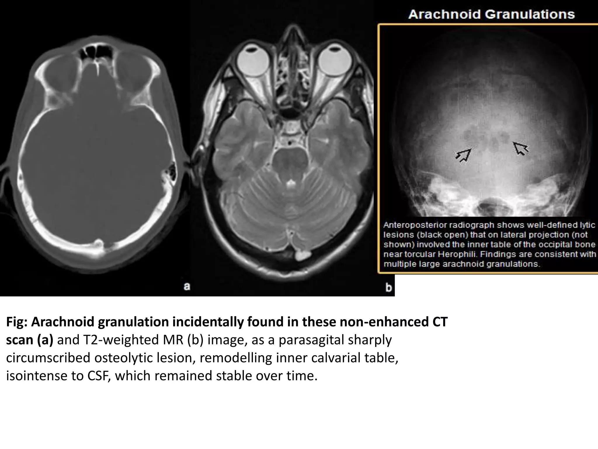

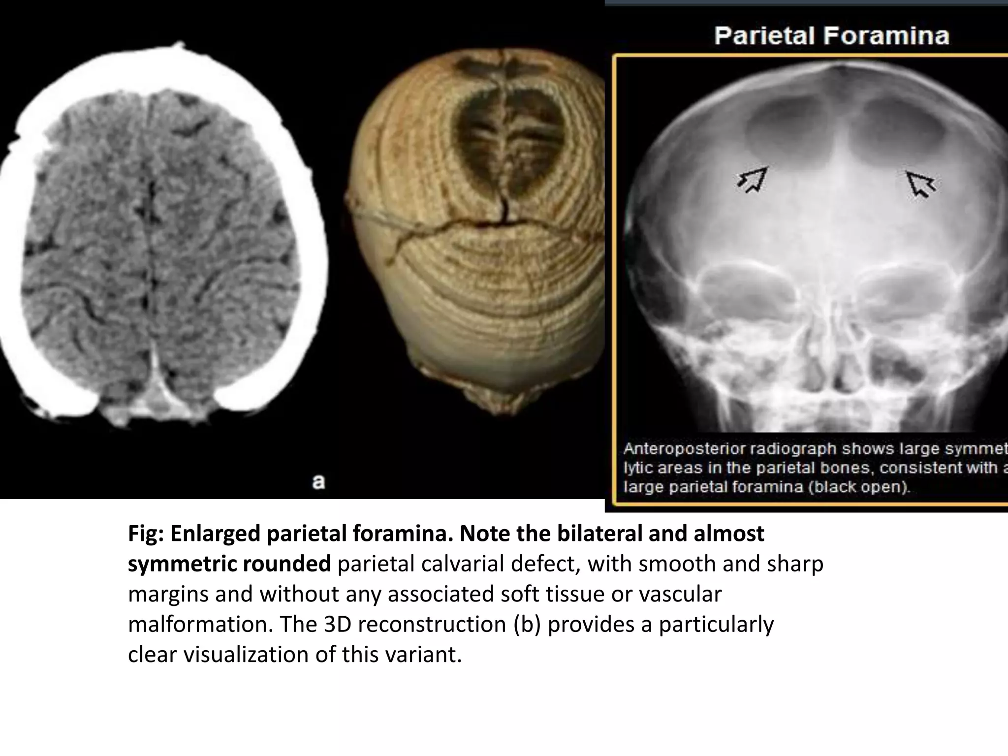

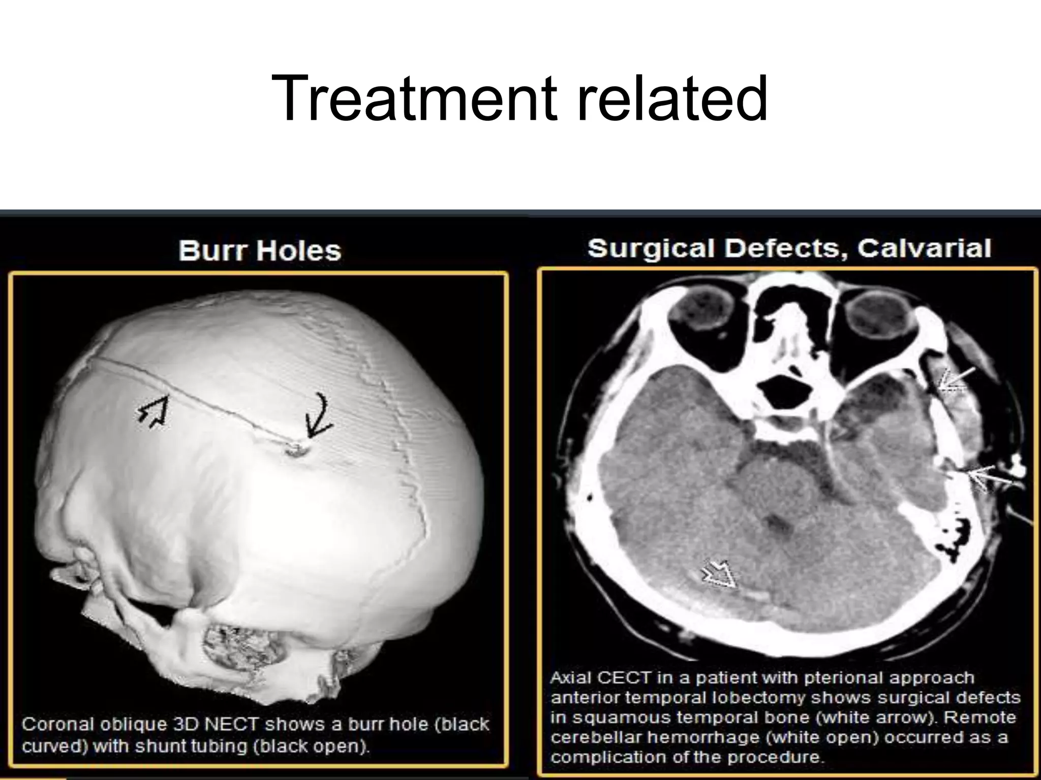


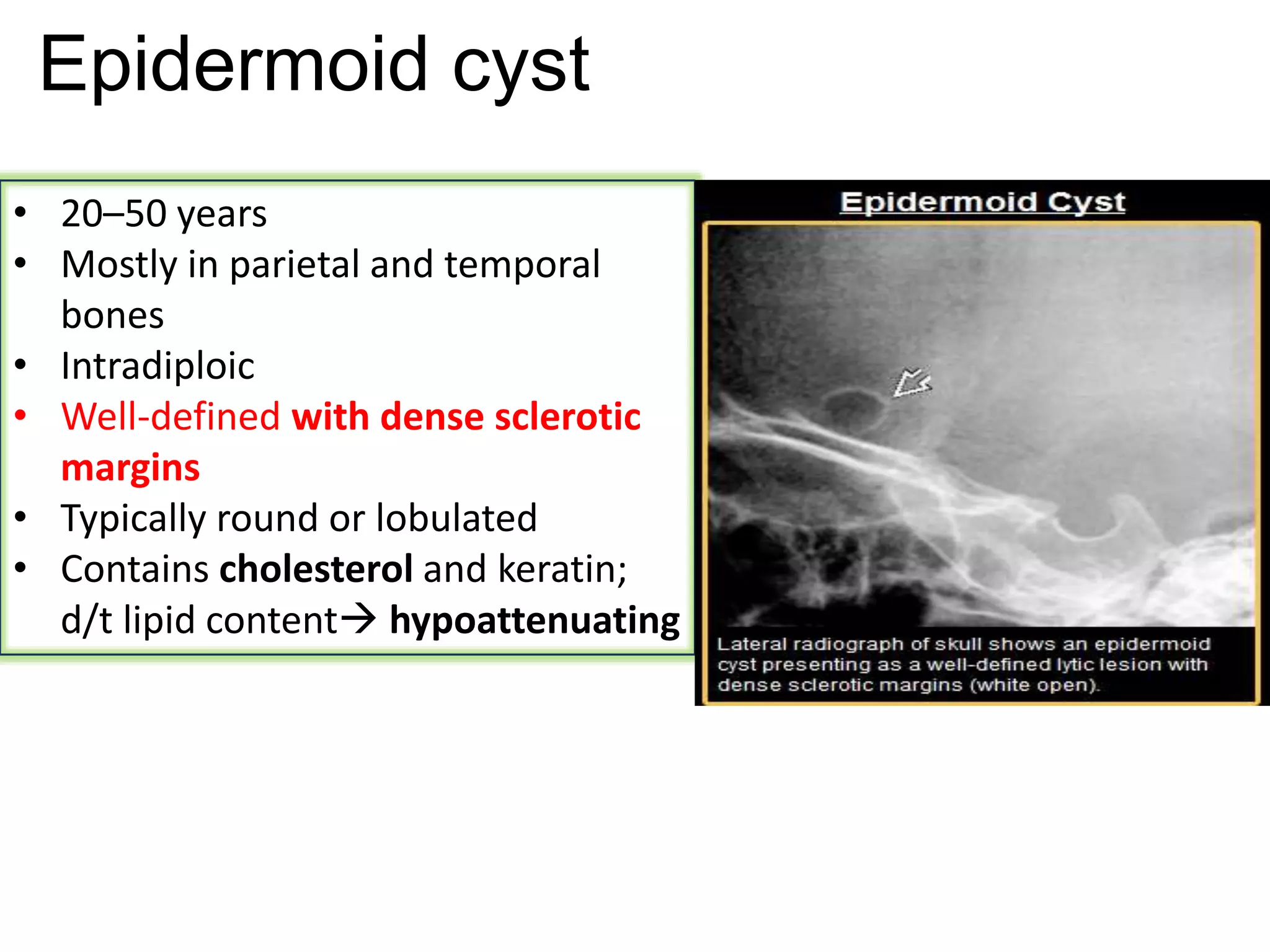


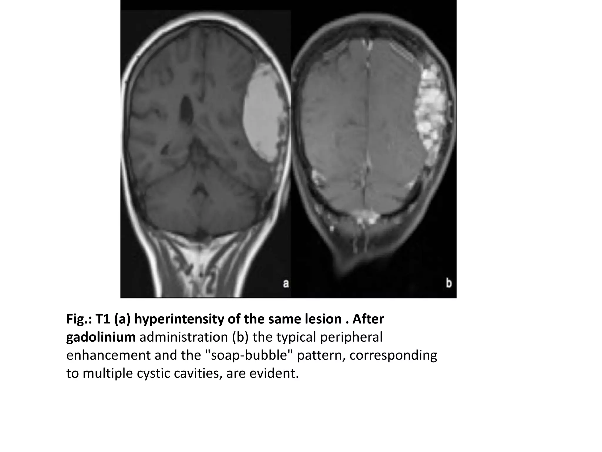


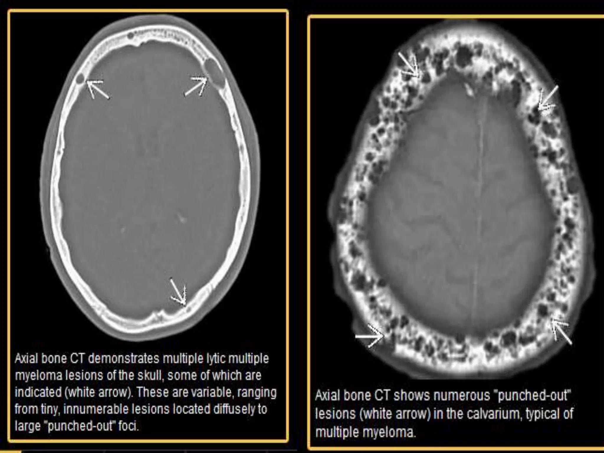


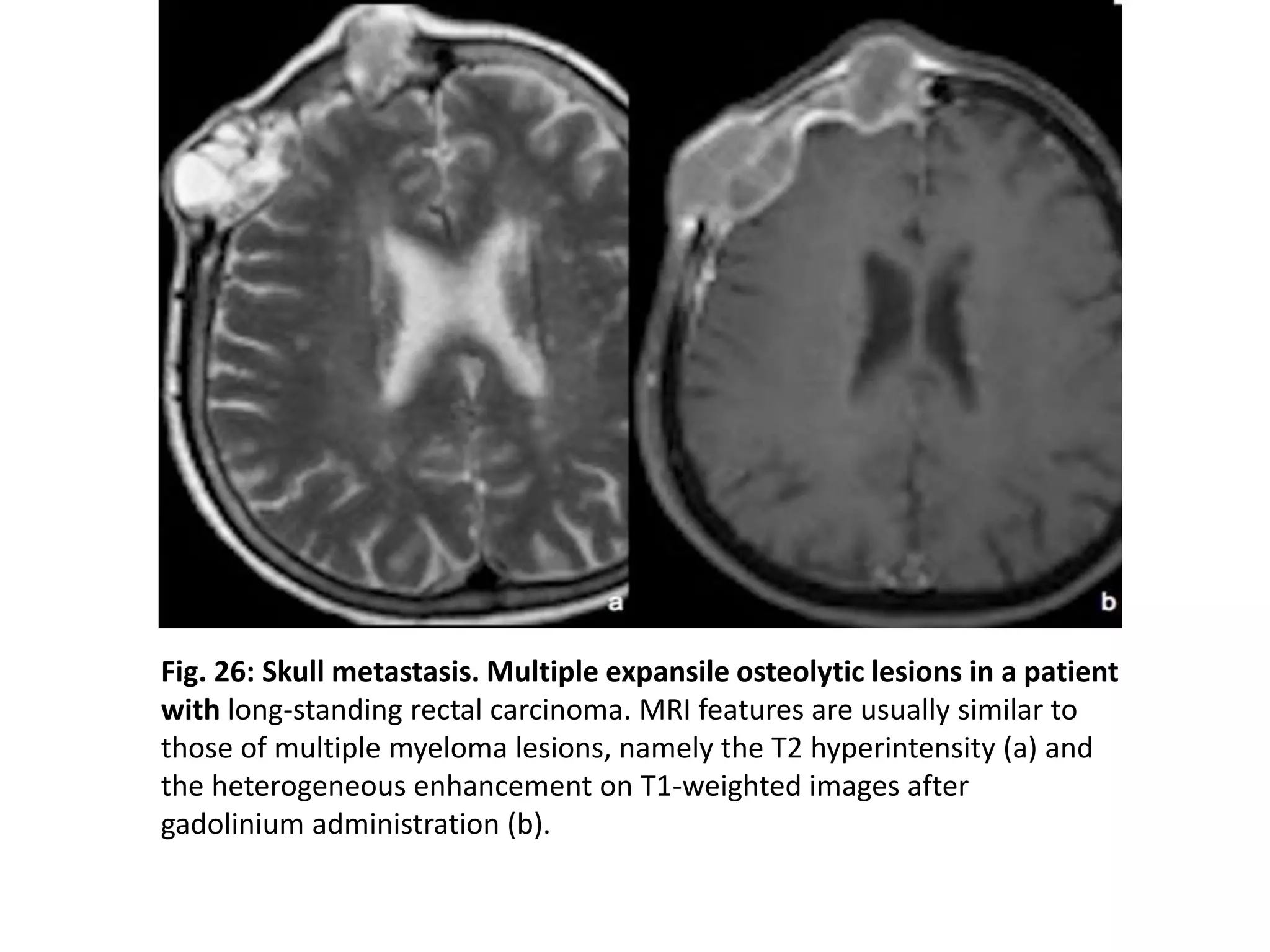
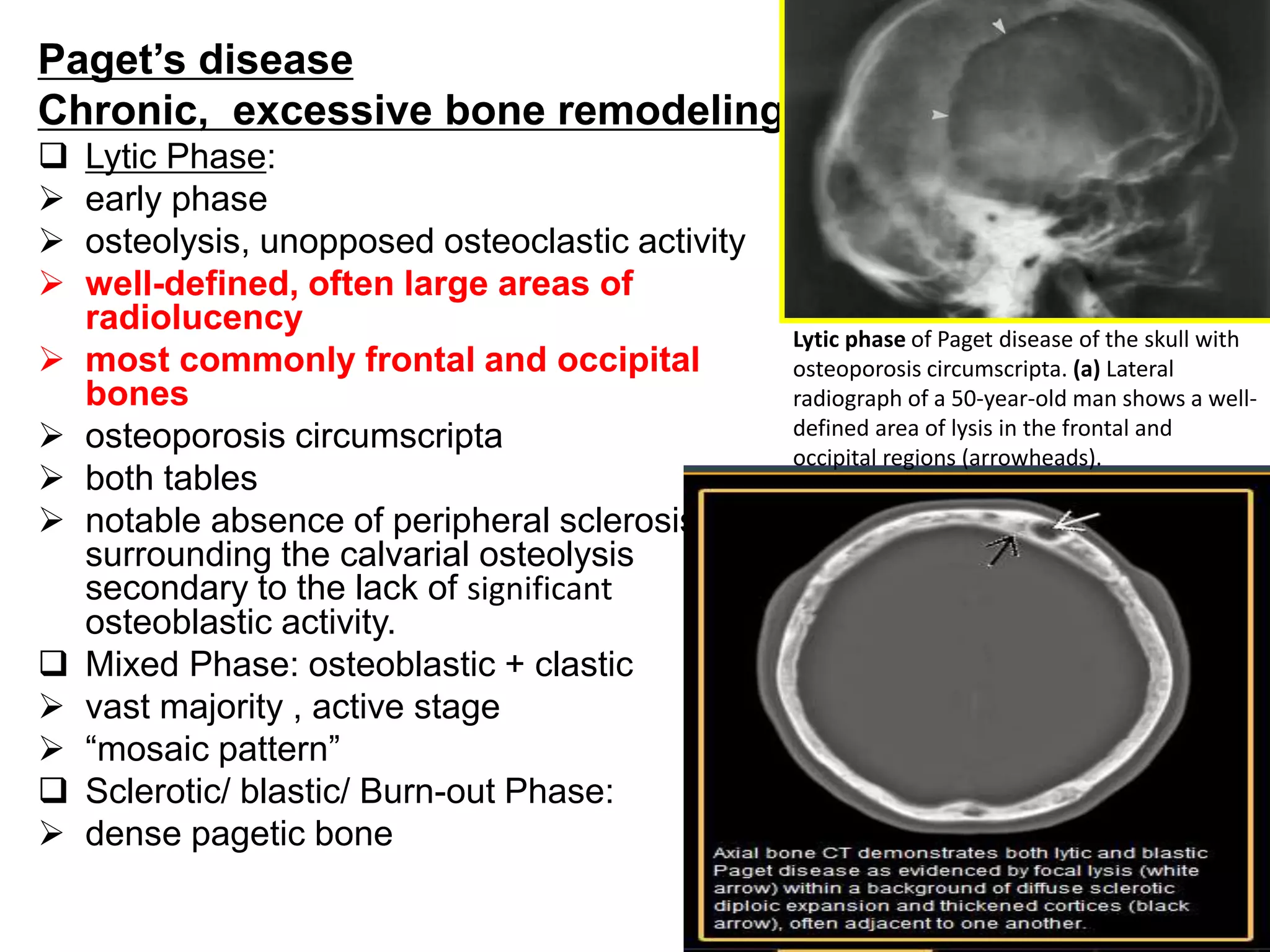


Lytic lesions of the skull can have many potential etiologies ranging from normal variants to neoplastic lesions. Imaging plays an important role in the evaluation and diagnosis of lytic skull lesions. CT and MRI are often used to characterize the lesions and assess bone and soft tissue involvement. The differential diagnosis depends on factors like the patient's age, lesion characteristics such as appearance, location and whether it is solitary or multiple. Common etiologies include metastases, multiple myeloma, epidermoid cysts, hemangioma and fibrous dysplasia among others. A thorough clinical history and imaging findings are needed to establish the correct diagnosis.


