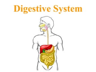
The digestive system includes the organs of the alimentary canal and accessory structures. The alimentary canal forms a continuous tube that is open to the outside environment at both ends. The organs of the alimentary canal are the mouth, pharynx, esophagus, stomach, small intestine, and large intestine.
Human digestive system structure and function
overview
Major organs
Mouth
Esophagus
Stomach
small intestine
large intestine
Acessory organs:
Liver
gall bladder
Pancreas.
Human digestive system
Major organs
Mouth
Esophagus
Stomach
small intestine
large intestine.
Acessory organs:
Liver
Gall bladder
Pancreas.
MAJOR ORGANS The Mouth
pH: 7
The first part of the digestive system
the entry point of food.
Structures in the mouth that aids digestion
Teeth – cut, tear, crush and grind food.
Salivary glands – produce and secrete saliva into the oral cavity.
saliva
moistens the food
contains enzymes (ptyalin or salivary amylase)
begins digestion of starch into smaller polysaccharides.
Function:
Mechanical digestion.
increasing surface area for faster chemical digestion.
The Esophagus
a tube connecting the mouth to the stomach
running through the Thoracic cavity.
Location:
lies behind windpipe (Trachea).
The trachea has as an epiglottis
preventing food from entering the windpipe,
moving the food to the esophagus while swallowing.
Food travels down the esophagus, through a series of involuntary rhythmic contractions (wave-like) called peristalsis.
Function:
The lining of the esophagus secretes mucus
lubricating
to support the movement of food.
Esophageal sphincter:
bolus reaches the stomach
must pass through a muscular ringed valve called the esophageal sphincter (Cardiac Sphincter).
Function:
prevent stomach acids from back flowing into the esophagus.
Stomach
J-shaped muscular sac
Has inner folds (rugae)
Increasing surface area of the stomach.
Function:
Stomach performs mechanical digestion
HOW By churning the bolus and mixing it with the gastric juices
secreted by the lining of the stomach.
GASTRIC JUICES HCl, salts, enzymes, water and mucus)
HCL helps break down of food and kills bacteria that came along with the food.
The bolus is now called Chyme.
Enzymes in stomach:
Acidic environment
HCl secreation
kill any microbes that are found in the bolus,
creating a pH of 2.
Mucus prevents the stomach from digesting itself.
Pepsin secreation
responsible for initiating the breakdown of proteins (in )food.
hydrolyzes proteins to yield polypeptides.
pH is 2, the enzyme from the salivary glands stops breaking down carbohydrates.
Pyloric sphincter:
chyme moves from the stomach to the small intestine.
It passes through a muscular ringed sphincter called the pyloric sphincter.
stomach does not digest itself Why ?
Protective Mechanism:
three protective mechanisms.
First the stomach only secretes small amounts of gastric juices until food is present.
Second the secretion of mucus coats the lining of the stomach protecting it from the gastric juices.
The third mechanism is the digestive enzyme pepsin is secreted in an inactive protein c
This PowerPoint presentation details out the anatomy of the human digestive system. Their are general terminologies that involves the topic but over-all this work focuses on how digestion takes place in the human body. The details coming from this presentation are combined from four different and liable sources/references including Biology (Thomson Asian Edition). I can say that this presentation is brief and well-organized so I hope this could help you in your class or seminars. Thanks.
Anatomy and Physiology of Digestive system.
Different Digestive process for absorption of food in GIT.
Different parts GIT Tract where food move from Mouth to the anus.
With this presentation we will learn to develop an understanding of the relationships between the structures and functions of the human digestive system and digestive processes which include the processes of digestion include six activities: ingestion, propulsion, mechanical or physical digestion, chemical digestion, absorption, and defecation
Peritoneum, abdomen, quadrant and regions.
a) Alimentary digestive organs: Oral cavity, pharynx, esophagus,
stomach, location and parts of stomach, parts of small and large
intestine, villi.
b) Accessory digestive organs: Salivary gland, liver, gall bladder,
pancreas.
Cardiovascular System: Mediastinum, divisions of mediastinum,
anatomy of heart, chambers of heart, opening and valves of the heart,
circulatory system
Knee anatomy and clinical tests 2024.pdfvimalpl1234
This includes all relevant anatomy and clinical tests compiled from standard textbooks, Campbell,netter etc..It is comprehensive and best suited for orthopaedicians and orthopaedic residents.
These lecture slides, by Dr Sidra Arshad, offer a quick overview of physiological basis of a normal electrocardiogram.
Learning objectives:
1. Define an electrocardiogram (ECG) and electrocardiography
2. Describe how dipoles generated by the heart produce the waveforms of the ECG
3. Describe the components of a normal electrocardiogram of a typical bipolar leads (limb II)
4. Differentiate between intervals and segments
5. Enlist some common indications for obtaining an ECG
Study Resources:
1. Chapter 11, Guyton and Hall Textbook of Medical Physiology, 14th edition
2. Chapter 9, Human Physiology - From Cells to Systems, Lauralee Sherwood, 9th edition
3. Chapter 29, Ganong’s Review of Medical Physiology, 26th edition
4. Electrocardiogram, StatPearls - https://www.ncbi.nlm.nih.gov/books/NBK549803/
5. ECG in Medical Practice by ABM Abdullah, 4th edition
6. ECG Basics, http://www.nataliescasebook.com/tag/e-c-g-basics
micro teaching on communication m.sc nursing.pdfAnurag Sharma
Microteaching is a unique model of practice teaching. It is a viable instrument for the. desired change in the teaching behavior or the behavior potential which, in specified types of real. classroom situations, tends to facilitate the achievement of specified types of objectives.
Acute scrotum is a general term referring to an emergency condition affecting the contents or the wall of the scrotum.
There are a number of conditions that present acutely, predominantly with pain and/or swelling
A careful and detailed history and examination, and in some cases, investigations allow differentiation between these diagnoses. A prompt diagnosis is essential as the patient may require urgent surgical intervention
Testicular torsion refers to twisting of the spermatic cord, causing ischaemia of the testicle.
Testicular torsion results from inadequate fixation of the testis to the tunica vaginalis producing ischemia from reduced arterial inflow and venous outflow obstruction.
The prevalence of testicular torsion in adult patients hospitalized with acute scrotal pain is approximately 25 to 50 percent
New Drug Discovery and Development .....NEHA GUPTA
The "New Drug Discovery and Development" process involves the identification, design, testing, and manufacturing of novel pharmaceutical compounds with the aim of introducing new and improved treatments for various medical conditions. This comprehensive endeavor encompasses various stages, including target identification, preclinical studies, clinical trials, regulatory approval, and post-market surveillance. It involves multidisciplinary collaboration among scientists, researchers, clinicians, regulatory experts, and pharmaceutical companies to bring innovative therapies to market and address unmet medical needs.
These simplified slides by Dr. Sidra Arshad present an overview of the non-respiratory functions of the respiratory tract.
Learning objectives:
1. Enlist the non-respiratory functions of the respiratory tract
2. Briefly explain how these functions are carried out
3. Discuss the significance of dead space
4. Differentiate between minute ventilation and alveolar ventilation
5. Describe the cough and sneeze reflexes
Study Resources:
1. Chapter 39, Guyton and Hall Textbook of Medical Physiology, 14th edition
2. Chapter 34, Ganong’s Review of Medical Physiology, 26th edition
3. Chapter 17, Human Physiology by Lauralee Sherwood, 9th edition
4. Non-respiratory functions of the lungs https://academic.oup.com/bjaed/article/13/3/98/278874
NVBDCP.pptx Nation vector borne disease control programSapna Thakur
NVBDCP was launched in 2003-2004 . Vector-Borne Disease: Disease that results from an infection transmitted to humans and other animals by blood-feeding arthropods, such as mosquitoes, ticks, and fleas. Examples of vector-borne diseases include Dengue fever, West Nile Virus, Lyme disease, and malaria.
Recomendações da OMS sobre cuidados maternos e neonatais para uma experiência pós-natal positiva.
Em consonância com os ODS – Objetivos do Desenvolvimento Sustentável e a Estratégia Global para a Saúde das Mulheres, Crianças e Adolescentes, e aplicando uma abordagem baseada nos direitos humanos, os esforços de cuidados pós-natais devem expandir-se para além da cobertura e da simples sobrevivência, de modo a incluir cuidados de qualidade.
Estas diretrizes visam melhorar a qualidade dos cuidados pós-natais essenciais e de rotina prestados às mulheres e aos recém-nascidos, com o objetivo final de melhorar a saúde e o bem-estar materno e neonatal.
Uma “experiência pós-natal positiva” é um resultado importante para todas as mulheres que dão à luz e para os seus recém-nascidos, estabelecendo as bases para a melhoria da saúde e do bem-estar a curto e longo prazo. Uma experiência pós-natal positiva é definida como aquela em que as mulheres, pessoas que gestam, os recém-nascidos, os casais, os pais, os cuidadores e as famílias recebem informação consistente, garantia e apoio de profissionais de saúde motivados; e onde um sistema de saúde flexível e com recursos reconheça as necessidades das mulheres e dos bebês e respeite o seu contexto cultural.
Estas diretrizes consolidadas apresentam algumas recomendações novas e já bem fundamentadas sobre cuidados pós-natais de rotina para mulheres e neonatos que recebem cuidados no pós-parto em unidades de saúde ou na comunidade, independentemente dos recursos disponíveis.
É fornecido um conjunto abrangente de recomendações para cuidados durante o período puerperal, com ênfase nos cuidados essenciais que todas as mulheres e recém-nascidos devem receber, e com a devida atenção à qualidade dos cuidados; isto é, a entrega e a experiência do cuidado recebido. Estas diretrizes atualizam e ampliam as recomendações da OMS de 2014 sobre cuidados pós-natais da mãe e do recém-nascido e complementam as atuais diretrizes da OMS sobre a gestão de complicações pós-natais.
O estabelecimento da amamentação e o manejo das principais intercorrências é contemplada.
Recomendamos muito.
Vamos discutir essas recomendações no nosso curso de pós-graduação em Aleitamento no Instituto Ciclos.
Esta publicação só está disponível em inglês até o momento.
Prof. Marcus Renato de Carvalho
www.agostodourado.com
Title: Sense of Taste
Presenter: Dr. Faiza, Assistant Professor of Physiology
Qualifications:
MBBS (Best Graduate, AIMC Lahore)
FCPS Physiology
ICMT, CHPE, DHPE (STMU)
MPH (GC University, Faisalabad)
MBA (Virtual University of Pakistan)
Learning Objectives:
Describe the structure and function of taste buds.
Describe the relationship between the taste threshold and taste index of common substances.
Explain the chemical basis and signal transduction of taste perception for each type of primary taste sensation.
Recognize different abnormalities of taste perception and their causes.
Key Topics:
Significance of Taste Sensation:
Differentiation between pleasant and harmful food
Influence on behavior
Selection of food based on metabolic needs
Receptors of Taste:
Taste buds on the tongue
Influence of sense of smell, texture of food, and pain stimulation (e.g., by pepper)
Primary and Secondary Taste Sensations:
Primary taste sensations: Sweet, Sour, Salty, Bitter, Umami
Chemical basis and signal transduction mechanisms for each taste
Taste Threshold and Index:
Taste threshold values for Sweet (sucrose), Salty (NaCl), Sour (HCl), and Bitter (Quinine)
Taste index relationship: Inversely proportional to taste threshold
Taste Blindness:
Inability to taste certain substances, particularly thiourea compounds
Example: Phenylthiocarbamide
Structure and Function of Taste Buds:
Composition: Epithelial cells, Sustentacular/Supporting cells, Taste cells, Basal cells
Features: Taste pores, Taste hairs/microvilli, and Taste nerve fibers
Location of Taste Buds:
Found in papillae of the tongue (Fungiform, Circumvallate, Foliate)
Also present on the palate, tonsillar pillars, epiglottis, and proximal esophagus
Mechanism of Taste Stimulation:
Interaction of taste substances with receptors on microvilli
Signal transduction pathways for Umami, Sweet, Bitter, Sour, and Salty tastes
Taste Sensitivity and Adaptation:
Decrease in sensitivity with age
Rapid adaptation of taste sensation
Role of Saliva in Taste:
Dissolution of tastants to reach receptors
Washing away the stimulus
Taste Preferences and Aversions:
Mechanisms behind taste preference and aversion
Influence of receptors and neural pathways
Impact of Sensory Nerve Damage:
Degeneration of taste buds if the sensory nerve fiber is cut
Abnormalities of Taste Detection:
Conditions: Ageusia, Hypogeusia, Dysgeusia (parageusia)
Causes: Nerve damage, neurological disorders, infections, poor oral hygiene, adverse drug effects, deficiencies, aging, tobacco use, altered neurotransmitter levels
Neurotransmitters and Taste Threshold:
Effects of serotonin (5-HT) and norepinephrine (NE) on taste sensitivity
Supertasters:
25% of the population with heightened sensitivity to taste, especially bitterness
Increased number of fungiform papillae
2. The digestive system is a collection of organs that can be divided into two parts: 1. The alimentary or gastrointestinal tract . 2. The accessory organs Pharynx Rectum Large Intestine Anus Small Intestine Stomach Esophagus Mouth
3. The first group of organs constitute the alimentary or gastrointestinal tract . The organs of the gastrointestinal tract are: 1. oral cavity (mouth) Pharynx Rectum Large Intestine Anus Small Intestine Stomach Esophagus Mouth 2. pharynx 3. esophagus 4. stomach 5. small intestine 6. large intestine 7. rectum 8. anus
4. The alimentary tract is a continuous, hollow tube about 30 ft. long that runs from the oral cavity to the anus . Food taken into the tube remains within it and is broken down (digested) into usable, simpler components. These substances are absorbed into the body (outside the tract) while the unusable part of the food passes on and out of the tube.
5.
6. The second group of organs are the accessory organs . These contribute to the break down of food into usable components (digestion) but are not part of the alimentary tract. The accessory organs are: 1. Tongue 2. Teeth
9. The oral cavity (mouth) – This is the first area of the alimentary canal. It is also where both the mechanical and chemical digestive processes begin. The oral cavity extends from the lips to the oropharynx.
10. The oral cavity is equipped with three different accessory organs. These are the tongue , teeth , and salivary glands and these each help the digestive processes begin. These organs cut, grind, lubricate, taste, manipulate, and move food, and have secretions that chemically break down food.
11. 1. Mechanical – Action of the digestive organs serve to move food materials along, moisten and liquefy, and to pulverize them to increase the surface area of the food. This makes things more susceptible to the actions of chemicals. The two ways food is “digested” and to be further explored later: 2. Chemical – These actions convert the large, complicated food molecules into smaller, simpler units (like glucose, amino acids, lipids) that can pass through cell membranes and be used by cells.
12.
13.
14. Esophagus A straight, collapsible muscular tube which lies behind (posterior to) the trachea and is about 1 inch in diameter and 10 inches long.
15.
16. The esophagus is composed mostly of smooth muscle arranged in two layers, one circular and one longitudinal, lined internally with a layer of mucus secreting cells. lumen Longitudinal Smooth muscle Circular Smooth muscle Mucous membrane
17. In fact, the entire alimentary tract is composed this way and secretions range from mucus to acid or digestive enzymes.
18. At the gastroesophageal junction (“bottom” of the esophagus where it connects to the stomach), the circular layer of smooth muscle fibers contract and relax creating a valve like action that is somewhat like that of a sphincter muscle, but is not a true sphincter valve. This portion of the circular muscle is called the cardiac sphincter or gastroesophageal sphincter because the esophagus joins the stomach in the cardiac region. This valve is controlled by pressure fluctuations in the esophagus and stomach.
19. Heartburn occurs when the cardiac sphincter relaxes and some of the digestive enzymes and stomach acid flow out into the esophagus. The enzymes can cause lesions in the esophageal lining and the acid irritates the esophagus. If the acid gets into any lesions this will irritate them further. The “burning” sensation felt around the area is perceived to be near the heart so the sensation is called “heartburn”. If chronic or severe, this is termed gastroesophageal reflux disease (acid reflux).
20. In gastric bypass surgery, rows of staples are used to divide the stomach into two parts. Food can enter only the smaller upper part, called a stomach pouch. The small intestine is cut, and the section of the small intestine below the cut is attached to the stomach pouch. Thus, the lower part of the stomach and the upper part of the small intestine are bypassed.
21. As a result, the amount of food that can be eaten at one time is drastically reduced and less food is absorbed. The section of the small intestine above the cut is then attached to the small intestine further down. Thus, digestive juices produced in the bypassed part of the stomach can reach the rest of the small intestine and can be mixed with food.
22. The stomach extends from the esophagus at the cardiac sphincter to the duodenum , the uppermost portion of the small intestine. The stomach is divided into four parts: Stomach – The stomach lies at the upper part of the abdominal cavity just under the diaphragm (which separates the thoracic and abdominal cavities). ***Note – the stomach has three layers of smooth muscle.
23. 1. Fundus or fundic region – This is the uppermost portion of the stomach. It actually extends above the level of the cardiac sphincter. It acts as a temporary storage area and can “balloon up” to accomplish this function. Normally it is filled with air. esophagus
24. esophagus 2. Cardia or cardiac region – This is a small region very near the cardiac sphincter where the esophagus meets the stomach.
25. esophagus 3. Body – The middle portion of the stomach or main portion located between the fundic and pyloric region. This is the largest region.
26. esophagus 4. Pylorus or pyloric region – This is the terminal portion of the stomach. It constricts as it approaches the small intestine forming the pyloric canal . It ends at an area of thickened smooth muscle called the pyloric sphincter . This acts as a valve preventing back flow of material from the small intestine to the stomach.
27. Overall, the stomach, which has a structure like a collapsible pouch, has only a few basic functions. These functions are, as with the mouth, are related to the digestion or breakdown of food in either a mechanical or a chemical manner and are as follows:
28. 1. Receives partially digested food from the esophagus. 2. Tosses and churns the partially digested food, mixes it with gastric juices, and mechanically breaks up the food (now called chyme) further. 3. Begins the chemical breakdown of proteins. 4. Begins the process of absorption. (Very little actual absorption takes place, however small amounts of water & glucose, fat soluble drugs, some salts and especially alcohol are absorbed.) 5. Passes the chyme on to the small intestine. ***NOTE – “ chyme ” is the term used to describe the partially digested food once it has been mixed with the digestive secretions of the stomach.
29. Small intestine A collapsible, muscular tube that extends from the pyloric sphincter of the stomach to the cecum of the large intestine. The small intestine is the site where the majority of the chemical breakdown of food and absorption of nutrients takes place. Duodenum Ileum Jejunum
30. Three parts of the small intestine: Duodenum Ileum Jejunum Duodenum 1. Duodenum – about 10 – 12 inches long. It connects to the stomach via the pyloric sphincter. This is the “fixed” portion of the small intestine.
31. 2. Jejunum – The only distinction is that it is a bit larger than the ileum. It represents about 2/5 of the length of the small intestine . Duodenum Ileum Jejunum Jejunum
32. 3. Ileum – This is the final portion of the small intestine and represents about 3/5 of its length. Ileum Duodenum Ileum Jejunum
33. The small intestine is covered on its inside wall with a “velvety” looking formation. These are called the intestinal villi .
34. The villi project into the lumen where they are bathed in the intestinal contents. They function to increase the surface area of the intestinal lining for absorption. Absorption takes place throughout the length of the small intestine.
35. Functions of the small intestine: 1. Receives nutrient rich chyme from the stomach. 2. Receives digestive secretions from the pancreas. 3. Receives digestive secretions and waste secretions from the liver and gall bladder. 4. Completes the chemical digestion of the chyme received from the stomach. 5. Absorbs nutrients from the fully digested chyme. 6. Transports unusable portion (nutrient depleted) of chyme to the large intestine.
36. 1. Cecum – the entrance to the large intestine which also has the vermiform appendix (appendix) hanging off it. Large Intestine – This is called “large” due to an increase in diameter compared to the “small” intestine. Its total length is about 5 feet. The large intestine is divided into two parts:
37. 2. the Colon – This is the major portion of the large intestine. It is divided into 4 parts: a) ascending b) transverse c) descending d) sigmoid
38. Functions of the large intestine: 1. Receives unusable portion of chyme from the small intestine. 2. Reabsorbs the rest of the water and electrolytes. 3. Forms and stores feces until defecation. 4. Passes feces on to the rectum.
39. Rectum – final storage of feces before defecation. When the rectum fills with feces it is stimulated to contract (peristalsis). This action is called the defecation reflex . Rectum
40. of three sphincter muscles (one made of smooth muscle, the other two of skeletal muscle), and finally through the anal opening. Anus – Feces expelled from the rectum passes through the anal canal, then through a series