Downloaded 743 times
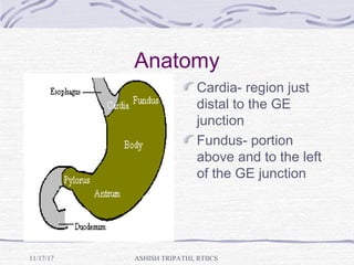
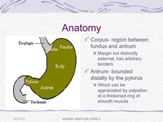
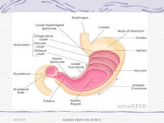
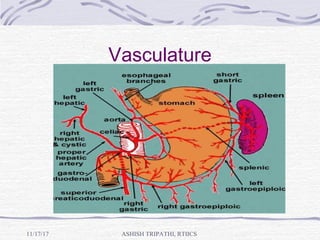
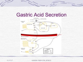


The document summarizes gastric anatomy and physiology. It describes the four regions of the stomach - cardia, fundus, corpus, and antrum. It discusses the stomach's position, vasculature, nerve supply, and microscopic anatomy including the three types of gastric glands. The physiology section covers the roles of gastrin and somatostatin in regulating acid secretion. It also describes the cephalic, gastric, and intestinal phases of acid secretion stimulated by histamine, gastrin, and acetylcholine. Gastric motility involves prolonged contractions in the proximal stomach and pacesetter potentials driving contractions in the distal stomach.


![Stomach by kp [autosaved]](https://cdn.slidesharecdn.com/ss_thumbnails/stomachbykpautosaved-140422141057-phpapp02-thumbnail.jpg?width=640&height=640&fit=bounds)


