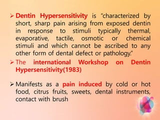
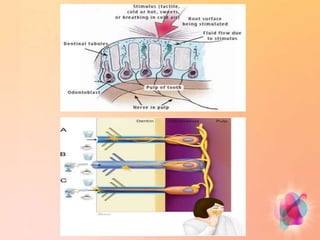
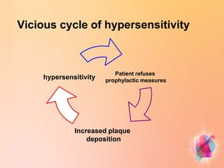
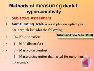
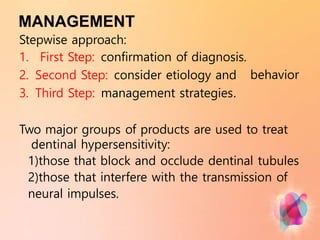
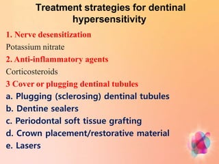
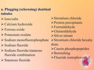
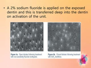
HYPERSENSITIVITY OF TEETH &
ITS MANAGEMENT
DR ASHWINI M PATIL
Reader
Navodaya dental college
Raichur
 Dentin Hypersensitivity is “characterized by
short, sharp pain arising from exposed dentin
in response to stimuli typically thermal,
evaporative, tactile, osmotic or chemical
stimuli and which cannot be ascribed to any
other form of dental defect or pathology.”
 The international Workshop on Dentin
Hypersensitivity(1983)
 Manifests as a pain induced by cold or hot
food, citrus fruits, sweets, dental instruments,
contact with brush
PREVALANCE
 14.3% of all dental patients.
 Periodontal patients - upto to 72-98%
 Adults in age group of 20-50 yrs ( Peaks in
30- 40 years)
 More common in cervical area of facial surface
of permanent teeth ( Canines and Premolars
commonly involved )
ETIOLOGY
 Scaling and root planing in periodontal
therapy
 Dietary factors: fruit, juice, yoghurt & wines.
 Acid in dental plaque, gastric reflux
 Agents in toothpaste like abrasive and
surfactant
 Psychological disorder (bruxism, abnormal
clenching habits)
Pathological conditions that cause dentinal
hypersensitivity
 Cracked teeth / chipped tooth
 Leaky restoration margin
 Gingival recession and Periodontal disease
 Deep Dentinal Caries
 Root caries
 Trauma from occlusion
 pulpitis
Mechanism of pain transmission – theories of
dentin hypersensitivity
 Direct neural stimulation theory
 Fluid/hydrodynamic theory (most accepted)
 Transduction theory
Direct innervation theory
 According to this theory , ‘ nerve fibers
present within the dentinal tubules initiates
impulses when they are injured and this
causes dentinal hypersensitivity’ .
Transduction theory
 This theory suggests that the odontoblasts or
their processes are damaged when external
stimuli are applied to exposed dentin. As a
result of this they conduct impulses to the
nerves in the predentin and underlying pulp
from where they proceed to the CNS.
Hydrodynamic Theory
 Proposed by Brannstrom M
 Most accepted theory
 Dentin has over 30,000 dentinal tubules /mm2
 These are filled with dentinal fluid which is the
intracellular fluid of the pulpal connective
tissue .
 Whenever exposed dentin is stimulated by
tactile , chemical, thermal or osmotic stimuli
there is rapid movement of the dentinal fluid
either towards the pulp or outward.
This can cause:
 Direct stimulation of the low threshold A-delta
nerve fibers in the pulp.
 Indirect stimulation of A- delta nerve fibers in
the pulp by displacing the odontoblastic cell
bodies.
 Such rapid displacement of the dentinal fluid
in thousands of dentinal tubules at the same
time produces a cumulative effect and this
causes hypersensitivity .
Vicious cycle of hypersensitivity
hypersensitivity
Patient refuses
prophylactic measures
Increased plaque
deposition
Symptoms
 Initial : sharp pain of rapid onset and
disappears once the stimulus is removed
 Severe: long standing –shorter or longer
periods of lingering, dull aching pain may be
provoked
 Even a minimal contact with the toothbrush
may elicit intense pain
Diagnosis
 History of nature, intensity, duration and
frequency of pain
 History- periodontal treatment, dietary habits.
 History of pain during brushing, h/o of trauma,
diurnal variation of pain.
 physical findings of tooth/teeth are essential to
rule out caries, cracked tooth, pulpitis, non
vital tooth
 Rule out any other cause of sharp pain.
Clinical examination
Patient often directs the operator toward
the hypersensitive area may be located by
gentle exploration with probe or cold air
(due to root surface exposure)
Tender on percussion
Clinical findings like attrition, fractured
cusp
Gingival recession/loss of attachment
Thermal test
Electrical test
Radiography
Methods of measuring dental
hypersensitivity
• Subjective Assessment
1. Verbal rating scale is a simple descriptive pain
scale which includes the following:
• 0 – No discomfort
• 1 – Mild discomfort
• 2 – Marked discomfort
• 3 – Marked discomfort that lasted for more than
10 seconds
Gillam and new Man (1993)
Objective Assessment
Mechanical / Tactile stimuli
1. Explorer probe
2. Mechanical pressure stimulation
3. Yeaple Probe
MANAGEMENT
behavior
Stepwise approach:
1. First Step: confirmation of diagnosis.
2. Second Step: consider etiology and
3. Third Step: management strategies.
Two major groups of products are used to treat
dentinal hypersensitivity:
1)those that block and occlude dentinal tubules
2)those that interfere with the transmission of
neural impulses.
Treatment strategies for dentinal
hypersensitivity
1. Nerve desensitization
Potassium nitrate
2. Anti-inflammatory agents
Corticosteroids
3 Cover or plugging dentinal tubules
a. Plugging (sclerosing) dentinal tubules
b. Dentine sealers
c. Periodontal soft tissue grafting
d. Crown placement/restorative material
e. Lasers
a. Plugging (sclerosing) dentinal
tubules
 Ions/salts
 Calcium hydroxide
 Ferrous oxide
 Potassium oxalate
 Sodium monofluorophosphate
 Sodium fluoride
 Sodium fluoride/stannous
fluoride combination
 Stannous fluoride
Strontium chloride
Protein precipitants
Formaldehyde
Glutaraldehyde
Silver nitrate
Strontium chloride hexahy
drate
Casein phosphopeptides
Burnishing
Fluoride iontophoresis
b. Dentine sealers
Glass ionomer cements
Composites
Resins
Varnishes
Sealants
Methyl methacrylate
 Dietary advice: citrus fruits, apple or any other
food or drink that acidic in nature should be
avoided .
 Plaque control
 Severe symptoms: use those agents which
block the tubular opening.
 In very severe case, remedy is achieved by
pulpectomy or root canal filling.
Information to patient
• Possibility of root hypersensitivity before
treatment is undertaken
• How to cope with the problem:
1. Hypersensitivity is inevitable if calculus and
plaque buried in the root is to be removed.
2. Disappears slowly over a few weeks.
3. Plaque control is important for its reduction.
4. Desensitizing agent should be used
continuously for at least 2 weeks.
Desensitizing agent
• Can be applied :
1.By the patient at home
2.By the dentist or hygienist in the dental office.
Clinical evaluation of different agent is difficult
because
 Measuring and comparing pain between different
persons is difficult.
 Hypersensitivity disappears by itself after a time and,
 Desensitizing agents usually take a few weeks to act.
Agent used at home
 Desensitizing toothpastes/dentifrices:
 The following dentifrices have been approved
by the American Dental Association for
desensitizing purposes:
 I) Sensodyne II)Thermodent
 which contain strontium chloride.
 Denquel, and Promise, which contain potassium
nitrate.
 Protect which contains sodium citrate
ACTIONS
 Potassium nitrate 5%-Blocks sensory nerve
activity at pulpal end of tubules by altering the
excitability of nerves
 Strontium chloride 0.4%-combines with
phosphate in dentinal fluid, strontium phosphate
crystals then binds to tubular matrix thus leading
gradual reduction of tubular radius and finally
leads to closure.
 sodium citrate : Act by precipitation of crystalline
salt on dentinal surface → block the dentinal
tubule
Agents used at dental office
1)Topically applied desensitizing agents:-
Fluoride( sod.fluoride, stannous fluoride) :-
possibly by precipitation of insoluble calcium
fluoride within the tubules. Which blocks fluid
movement within the dentin
Potassium nitrate:- potassium ions do reduce nerve
excitability
permeability and
Oxalate:- Oxalate products
occlude
reduce
tubules
dentin
more
consistently
Currently , potassium and ferric oxalate solutions
are the preferred agents. They form insoluble
calcium oxalate crystals that occlude the
dentinal tubules and prevent fluid movement.
ferric oxalate under the name Sensodyne
Sealant.
Calcium phosphates:- Calcium phosphates
occlude dentinal tubules in vitro and decrease
in vitro dentin permeability by 85%
2)Placement of restorations:
Glass ionomer or a composite resin restoration may
be placed to replace the lost tooth structure and
seal the exposed dentin .
3) Ionto-phoresis
• This procedure uses electricity to enhance
diffusion of ions into the tissues.
• Dental iontophoresis is used most often in
conjunction with fluoride pastes or solutions
(2% sodium fluoride)
• A 2% sodium fluoride is applied on the exposed
dentin and this is transferred deep into the dentin
on activation of the unit.
improve
treatment
4)Lasers
• Recently , attempts have been made to
the success and longevity of these
using lasers.
• Low- level laser “melting” of the dentin
surface appears to seal dentinal tubules without
damage to the pulp.
• Finally , in a combined treatment modality, the
Nd:YAG laser has been used to congeal fluoride
varnish on root surfaces.
 This in vitro study demonstrated
that the laser treated fluoride
varnish resisted removal by
electric tooth-brushing,
 with 90% of tubules remaining
blocked while in the controls
(no laser treatment) the fluoride
varnish was almost completely
brushed away.
 Further research is still on
progression.
Suggestions for patients:
 Avoid using large amounts of dentifrice or
reapplying it during brushing.
 Avoid medium- or hard-bristle toothbrushes.
 Avoid brushing teeth immediately after
ingesting acidic foods.
 Avoid over brushing with excessive pressure
or for an extended period of time.
 Avoid excessive flossing or improper use of
other interproximal cleaning devices.
 Avoid “picking” or scratching at the gumline or
using toothpicks inappropriately.
Suggestions for professionals:
 Avoid over instrumenting the root surfaces during
scaling and root planing, particularly in the cervical
area of the tooth.
 Avoid over polishing exposed dentin during stain
removal.
 Avoid violating the biologic width when placing
crown margins causing subsequent recession.
 Avoid burning the gingival tissues during in-office
tooth whitening or bleaching procedures.

DENTINAL HYPERSENSITIVITY..pptx

  • 1.
    HYPERSENSITIVITY OF TEETH& ITS MANAGEMENT DR ASHWINI M PATIL Reader Navodaya dental college Raichur
  • 2.
     Dentin Hypersensitivityis “characterized by short, sharp pain arising from exposed dentin in response to stimuli typically thermal, evaporative, tactile, osmotic or chemical stimuli and which cannot be ascribed to any other form of dental defect or pathology.”  The international Workshop on Dentin Hypersensitivity(1983)  Manifests as a pain induced by cold or hot food, citrus fruits, sweets, dental instruments, contact with brush
  • 3.
    PREVALANCE  14.3% ofall dental patients.  Periodontal patients - upto to 72-98%  Adults in age group of 20-50 yrs ( Peaks in 30- 40 years)  More common in cervical area of facial surface of permanent teeth ( Canines and Premolars commonly involved )
  • 4.
    ETIOLOGY  Scaling androot planing in periodontal therapy  Dietary factors: fruit, juice, yoghurt & wines.  Acid in dental plaque, gastric reflux  Agents in toothpaste like abrasive and surfactant  Psychological disorder (bruxism, abnormal clenching habits)
  • 5.
    Pathological conditions thatcause dentinal hypersensitivity  Cracked teeth / chipped tooth  Leaky restoration margin  Gingival recession and Periodontal disease  Deep Dentinal Caries  Root caries  Trauma from occlusion  pulpitis
  • 6.
    Mechanism of paintransmission – theories of dentin hypersensitivity  Direct neural stimulation theory  Fluid/hydrodynamic theory (most accepted)  Transduction theory
  • 7.
    Direct innervation theory According to this theory , ‘ nerve fibers present within the dentinal tubules initiates impulses when they are injured and this causes dentinal hypersensitivity’ .
  • 8.
    Transduction theory  Thistheory suggests that the odontoblasts or their processes are damaged when external stimuli are applied to exposed dentin. As a result of this they conduct impulses to the nerves in the predentin and underlying pulp from where they proceed to the CNS.
  • 9.
    Hydrodynamic Theory  Proposedby Brannstrom M  Most accepted theory  Dentin has over 30,000 dentinal tubules /mm2  These are filled with dentinal fluid which is the intracellular fluid of the pulpal connective tissue .  Whenever exposed dentin is stimulated by tactile , chemical, thermal or osmotic stimuli there is rapid movement of the dentinal fluid either towards the pulp or outward.
  • 10.
    This can cause: Direct stimulation of the low threshold A-delta nerve fibers in the pulp.  Indirect stimulation of A- delta nerve fibers in the pulp by displacing the odontoblastic cell bodies.  Such rapid displacement of the dentinal fluid in thousands of dentinal tubules at the same time produces a cumulative effect and this causes hypersensitivity .
  • 12.
    Vicious cycle ofhypersensitivity hypersensitivity Patient refuses prophylactic measures Increased plaque deposition
  • 13.
    Symptoms  Initial :sharp pain of rapid onset and disappears once the stimulus is removed  Severe: long standing –shorter or longer periods of lingering, dull aching pain may be provoked  Even a minimal contact with the toothbrush may elicit intense pain
  • 14.
    Diagnosis  History ofnature, intensity, duration and frequency of pain  History- periodontal treatment, dietary habits.  History of pain during brushing, h/o of trauma, diurnal variation of pain.  physical findings of tooth/teeth are essential to rule out caries, cracked tooth, pulpitis, non vital tooth  Rule out any other cause of sharp pain.
  • 15.
    Clinical examination Patient oftendirects the operator toward the hypersensitive area may be located by gentle exploration with probe or cold air (due to root surface exposure) Tender on percussion Clinical findings like attrition, fractured cusp Gingival recession/loss of attachment Thermal test Electrical test Radiography
  • 16.
    Methods of measuringdental hypersensitivity • Subjective Assessment 1. Verbal rating scale is a simple descriptive pain scale which includes the following: • 0 – No discomfort • 1 – Mild discomfort • 2 – Marked discomfort • 3 – Marked discomfort that lasted for more than 10 seconds Gillam and new Man (1993)
  • 17.
    Objective Assessment Mechanical /Tactile stimuli 1. Explorer probe 2. Mechanical pressure stimulation 3. Yeaple Probe
  • 18.
    MANAGEMENT behavior Stepwise approach: 1. FirstStep: confirmation of diagnosis. 2. Second Step: consider etiology and 3. Third Step: management strategies. Two major groups of products are used to treat dentinal hypersensitivity: 1)those that block and occlude dentinal tubules 2)those that interfere with the transmission of neural impulses.
  • 19.
    Treatment strategies fordentinal hypersensitivity 1. Nerve desensitization Potassium nitrate 2. Anti-inflammatory agents Corticosteroids 3 Cover or plugging dentinal tubules a. Plugging (sclerosing) dentinal tubules b. Dentine sealers c. Periodontal soft tissue grafting d. Crown placement/restorative material e. Lasers
  • 20.
    a. Plugging (sclerosing)dentinal tubules  Ions/salts  Calcium hydroxide  Ferrous oxide  Potassium oxalate  Sodium monofluorophosphate  Sodium fluoride  Sodium fluoride/stannous fluoride combination  Stannous fluoride Strontium chloride Protein precipitants Formaldehyde Glutaraldehyde Silver nitrate Strontium chloride hexahy drate Casein phosphopeptides Burnishing Fluoride iontophoresis
  • 21.
    b. Dentine sealers Glassionomer cements Composites Resins Varnishes Sealants Methyl methacrylate
  • 22.
     Dietary advice:citrus fruits, apple or any other food or drink that acidic in nature should be avoided .  Plaque control  Severe symptoms: use those agents which block the tubular opening.  In very severe case, remedy is achieved by pulpectomy or root canal filling.
  • 23.
    Information to patient •Possibility of root hypersensitivity before treatment is undertaken • How to cope with the problem: 1. Hypersensitivity is inevitable if calculus and plaque buried in the root is to be removed. 2. Disappears slowly over a few weeks. 3. Plaque control is important for its reduction. 4. Desensitizing agent should be used continuously for at least 2 weeks.
  • 24.
    Desensitizing agent • Canbe applied : 1.By the patient at home 2.By the dentist or hygienist in the dental office. Clinical evaluation of different agent is difficult because  Measuring and comparing pain between different persons is difficult.  Hypersensitivity disappears by itself after a time and,  Desensitizing agents usually take a few weeks to act.
  • 25.
    Agent used athome  Desensitizing toothpastes/dentifrices:  The following dentifrices have been approved by the American Dental Association for desensitizing purposes:  I) Sensodyne II)Thermodent  which contain strontium chloride.  Denquel, and Promise, which contain potassium nitrate.  Protect which contains sodium citrate
  • 27.
    ACTIONS  Potassium nitrate5%-Blocks sensory nerve activity at pulpal end of tubules by altering the excitability of nerves  Strontium chloride 0.4%-combines with phosphate in dentinal fluid, strontium phosphate crystals then binds to tubular matrix thus leading gradual reduction of tubular radius and finally leads to closure.  sodium citrate : Act by precipitation of crystalline salt on dentinal surface → block the dentinal tubule
  • 28.
    Agents used atdental office 1)Topically applied desensitizing agents:- Fluoride( sod.fluoride, stannous fluoride) :- possibly by precipitation of insoluble calcium fluoride within the tubules. Which blocks fluid movement within the dentin Potassium nitrate:- potassium ions do reduce nerve excitability
  • 29.
    permeability and Oxalate:- Oxalateproducts occlude reduce tubules dentin more consistently Currently , potassium and ferric oxalate solutions are the preferred agents. They form insoluble calcium oxalate crystals that occlude the dentinal tubules and prevent fluid movement. ferric oxalate under the name Sensodyne Sealant. Calcium phosphates:- Calcium phosphates occlude dentinal tubules in vitro and decrease in vitro dentin permeability by 85%
  • 30.
    2)Placement of restorations: Glassionomer or a composite resin restoration may be placed to replace the lost tooth structure and seal the exposed dentin . 3) Ionto-phoresis • This procedure uses electricity to enhance diffusion of ions into the tissues. • Dental iontophoresis is used most often in conjunction with fluoride pastes or solutions (2% sodium fluoride)
  • 31.
    • A 2%sodium fluoride is applied on the exposed dentin and this is transferred deep into the dentin on activation of the unit.
  • 32.
    improve treatment 4)Lasers • Recently ,attempts have been made to the success and longevity of these using lasers. • Low- level laser “melting” of the dentin surface appears to seal dentinal tubules without damage to the pulp. • Finally , in a combined treatment modality, the Nd:YAG laser has been used to congeal fluoride varnish on root surfaces.
  • 33.
     This invitro study demonstrated that the laser treated fluoride varnish resisted removal by electric tooth-brushing,  with 90% of tubules remaining blocked while in the controls (no laser treatment) the fluoride varnish was almost completely brushed away.  Further research is still on progression.
  • 34.
    Suggestions for patients: Avoid using large amounts of dentifrice or reapplying it during brushing.  Avoid medium- or hard-bristle toothbrushes.  Avoid brushing teeth immediately after ingesting acidic foods.  Avoid over brushing with excessive pressure or for an extended period of time.  Avoid excessive flossing or improper use of other interproximal cleaning devices.  Avoid “picking” or scratching at the gumline or using toothpicks inappropriately.
  • 35.
    Suggestions for professionals: Avoid over instrumenting the root surfaces during scaling and root planing, particularly in the cervical area of the tooth.  Avoid over polishing exposed dentin during stain removal.  Avoid violating the biologic width when placing crown margins causing subsequent recession.  Avoid burning the gingival tissues during in-office tooth whitening or bleaching procedures.