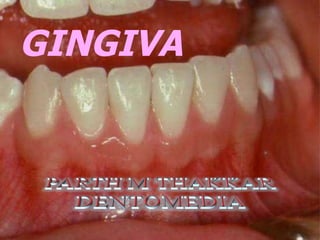
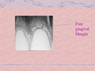
GINGIVA
Part of oral mucosa that covers the alveolar processes of the jaws and surrounds the necks of the teeth.
GINGIVA Marginal gingiva Attached Gingiva Gingival Sulcus Interdental Gingiva
 
 
Marginal Gingiva Terminal edge of the Gingiva surrounding the teeth in collar like  fashion.  1.5-2mm wide. Sluice Way.
Rounded free gingiva forming a sulcus between tooth and gingiva.  Gingival pocket seen while probing.
Gingival Sulcus V-Shaped shallow Crevice 1.8mm Presence of crevicular fluid
Interdental gingiva Occupy gingival embrasure Site of initial lesion on gingivitis Anterior region-Pyramidal form and flattened  over Molar region Scalloped Form Presence of Interdental COL
 
Attached Gingiva Functional Mucosa. Firm,resilient and tightly bound to alveolar Bone. Pale,pink and Stippled appearance Incisal Molars Maxilla 3.5-  4.5mm 1.9mm Mandible 3.3-3.9mm 1.8mm
Free gingival groove Muco-gingival junction Attached Gingiva
 
Free gingival Margin
Clinical Features Color :-Coral pink Size :-0-2mm for Marginal gingiva 0-9mm for attached gingiva Contour :-Scalloped Shape :-Goverend by contour of proximal tooth surface Consistency :- Firm & resilient Surface Texture :- Stippled(Absent over marginal gingiva)
Stippling Appearance
MICROSCOPIC FEATURES Gingival epithelium Oral   Epithelium Sulcular Epithelium Junctional epithelium
 
Oral Epithelium Keratinized,stratified,sqamous epithelium. Parakeratinized :-75% Orthokeratinized :-15% Non-Keratinized :-10%  Cells of epithelium
Keratinocytes (90%)   Melanocytes  Langerhans Cells  Merkel Cells  Cells Of Epithelium
Keratinized Epithelium
Layers of Oral Epithelium
Presence of KERATOHYALINE granules in S.Granulosum
Sulcular Epithelium Thin,nonkeratintzed,stratified Squamous Epithelium Extends From Coronal Limit of J.E to the crest of gingival margin
Junctional Epithelium Collar Like band of stratified,Non-keratinized Stratified Sqamous epithelium Extends from base of sulcus to cemento-enamel junctional-a distance of 2mm  Derived from reduced Enamel epithelium
 
Gingival Connective Tissue Lamina Propia Dense Collagen network Presence of glycoproteins  & proteoglycans Cells :-Fibroblast,cementocytes,macrophages ,mast cells,neutrophils,plasma cells & lymphocytes.
 
 
Blood Supply Supra-periosteal vessels Vessels from PDL Arterioles from Crest of interdental bone
 
 
Lymphatic Supply
Nerve Supply Mainly by Trigeminal Nerve And its branches
Gingival Fibers PRINICIPAL FIBERS SECONDARY GP.FIBERS Dentogingival Alveolar  Dentoperiosteal Transseptal Circular Periosteo – gingival Interpappilary Transgingival Intercircular Intergingival Semicircular
Nerve Supply Mainly by Trigeminal Nerve And its branches
Gingival  fibers

gingiva