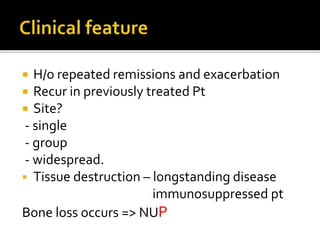
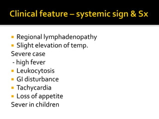
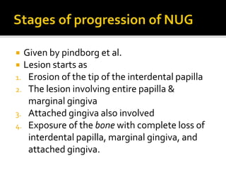
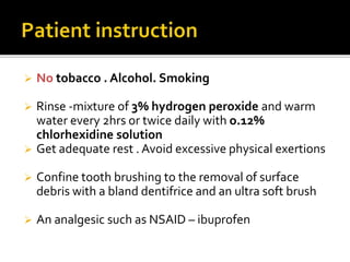
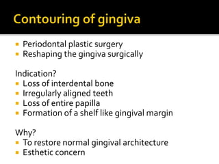
Keumyi Chin
 Introduction
 Clinical Feature
 Diagnosis
 Management
 Prognosis
 Referred books
 Definition
Microbial disease of the gingiva in the context
of an impaired host response.
Characterized by the death and sloughing of
gingival tissue
Presents with characteristics sign and
symptoms
 Severe necrosis of the free gingival margin,
the crest of the gingiva and interdental
papilla
VsA. B
 Also known as TRENCH MOUTH
because of its prevalence in the soldiers
working in trenches during WW1.
Vincent’s disease
Fusospirochetal gingivitis
 H/o repeated remissions and exacerbation
 Recur in previously treated Pt
 Site?
- single
- group
- widespread.
 Tissue destruction – longstanding disease
immunosuppressed pt
Bone loss occurs => NUP
 Punched out
 Crater like depressions at the crest of
interdental papillae, may extend up to
marginal gingiva
 Surface of gingival craters is covered by a
gray pseudo membrane + necrotic tissue
debris
 Age b/w 15 – 35yrs
 Pain, interdental ulceration, and gingival
bleeding are the diagnostic triad
 Interdental papillae - inflamed, edematous,
and hemorrhagic.
 Spontaneous gingival hemorrhage after
slight stimulation
 fetid odor and increased salivation
 Progressively destroy the gingiva and
periodontal tissue
 Constant radiating, gnawing pain
intensified by eating spicy or hot foods and
chewing
 Metallic taste to saliva
• Extremely sensitive to touch
• Excessive amount of pasty saliva
 Regional lymphadenopathy
 Slight elevation of temp.
Severe case
- high fever
 Leukocytosis
 GI disturbance
 Tachycardia
 Loss of appetite
Sever in children
 Given by pindborg et al.
 Lesion starts as
1. Erosion of the tip of the interdental papilla
2. The lesion involving entire papilla &
marginal gingiva
3. Attached gingiva also involved
4. Exposure of the bone with complete loss of
interdental papilla, marginal gingiva, and
attached gingiva.
 By Horning and Cohen
 1. necrosis of the tip of the interdental papilla
 2. necrosis of the entire papilla
 3. necrosis extending to gingival margin(NUP)
 4. necrosis extending to attached gingiva
 5. necrosis extending into buccal or labial
mucosa( necrotizing stomatitis)
 6. necrosis exposing alveolar bone
 7. necrosis perforating skin or cheek(noma)
 Based on clinical findings of gingival pain,
ulceration, and bleeding
 Microscopic examination of a bacterial smear or
biopsy specimen does not give specific picture.
 Histologic picture greatly resembles marginal
gingivitis, periodontal pockets, pericoronitis or
primary herpetic gingivostomatitis
 1. alleviation of acute inflammation by
reducing microbial load & removal of necrotic
tissue
 2. alleviation of genenralized sx – fever&
malaise
 3. correction of systemic conditions that
contributes to the initiation or progression of
the gingival change
 1) first visit
 Goal- reduce microbial load & remove
necrotic tissue
 Complete evaluation of the pt
 Treatment of acute lesion is primary goal.
 Topical anesthetic applied
 2-3min > gently swabbed. Remove pseudo
membrane and nonattached surface debris
 cleaning with warm water
 Ultrasonic scaling may be preferable, with
minimal pressure against the soft tissue
 Sub gingival scaling and curettage are C/I at
this time
 This may extend the infection to the deeper
tissues and cause bacteremia
 Amoxicillin 500mg O 6hr 10days
 Erythromycin 500mg 6hr
 Metronidazole 500mg twice daily 7days
 No tobacco . Alcohol. Smoking
 Rinse -mixture of 3% hydrogen peroxide and warm
water every 2hrs or twice daily with o.12%
chlorhexidine solution
 Get adequate rest . Avoid excessive physical exertions
 Confine tooth brushing to the removal of surface
debris with a bland dentifrice and an ultra soft brush
 An analgesic such as NSAID – ibuprofen
 2 days after the first visit
 Pt is evaluated for resolution of signs and Sx
 Lesion - erythematous without a superficial
pseudo membrane
 Shrinkage of the gingiva may expose
previously covered calculus, which is gently
removed.
 Instructions are given same as previously
 5 days after the second visit
- pt is evaluated for resolution of Sx, and a
comprehensive plan for the management of
the pt’s periodontal conditions is formulated
Hydrogen peroxide rinse – discontinued
Chlorhexidine mouthwash – continued 2
or3 wks
 Supportive therapy (e.g rest, appropriate
fluid intake, soft nutritious diet)
 Repeat scaling & root planning (if required)
 Reinstructed – plaque control measures
 Pt counseling – nutrition and smoking
cessation
 Appointments should be scheduled for t/t
1. Chronic gingivitis
2. Periodontal pockets
3. Pericoronal flap
4. Local irritants
Patient is reevaluated after 1 month.
1. Contouring of gingiva as adjunctive
procedure
2. nutritional supplement
 Periodontal plastic surgery
 Reshaping the gingiva surgically
Indication?
 Loss of interdental bone
 Irregularly aligned teeth
 Loss of entire papilla
 Formation of a shelf like gingival margin
Why?
 To restore normal gingival architecture
 Esthetic concern
 Carranza’s clinical periodontology vol. 1
( pg. 133 -138)
 Carranza’s clinical periodontology vol.2
( pg. 607- 610)
 Textbook of periodontology and oral
implantology (pg. 167-171)
ANUG

ANUG

  • 1.
  • 2.
     Introduction  ClinicalFeature  Diagnosis  Management  Prognosis  Referred books
  • 3.
     Definition Microbial diseaseof the gingiva in the context of an impaired host response. Characterized by the death and sloughing of gingival tissue Presents with characteristics sign and symptoms
  • 4.
     Severe necrosisof the free gingival margin, the crest of the gingiva and interdental papilla VsA. B
  • 5.
     Also knownas TRENCH MOUTH because of its prevalence in the soldiers working in trenches during WW1. Vincent’s disease Fusospirochetal gingivitis
  • 6.
     H/o repeatedremissions and exacerbation  Recur in previously treated Pt  Site? - single - group - widespread.  Tissue destruction – longstanding disease immunosuppressed pt Bone loss occurs => NUP
  • 7.
     Punched out Crater like depressions at the crest of interdental papillae, may extend up to marginal gingiva
  • 8.
     Surface ofgingival craters is covered by a gray pseudo membrane + necrotic tissue debris  Age b/w 15 – 35yrs  Pain, interdental ulceration, and gingival bleeding are the diagnostic triad  Interdental papillae - inflamed, edematous, and hemorrhagic.
  • 9.
     Spontaneous gingivalhemorrhage after slight stimulation  fetid odor and increased salivation  Progressively destroy the gingiva and periodontal tissue
  • 10.
     Constant radiating,gnawing pain intensified by eating spicy or hot foods and chewing  Metallic taste to saliva • Extremely sensitive to touch • Excessive amount of pasty saliva
  • 11.
     Regional lymphadenopathy Slight elevation of temp. Severe case - high fever  Leukocytosis  GI disturbance  Tachycardia  Loss of appetite Sever in children
  • 12.
     Given bypindborg et al.  Lesion starts as 1. Erosion of the tip of the interdental papilla 2. The lesion involving entire papilla & marginal gingiva 3. Attached gingiva also involved 4. Exposure of the bone with complete loss of interdental papilla, marginal gingiva, and attached gingiva.
  • 13.
     By Horningand Cohen  1. necrosis of the tip of the interdental papilla  2. necrosis of the entire papilla  3. necrosis extending to gingival margin(NUP)  4. necrosis extending to attached gingiva
  • 14.
     5. necrosisextending into buccal or labial mucosa( necrotizing stomatitis)  6. necrosis exposing alveolar bone  7. necrosis perforating skin or cheek(noma)
  • 15.
     Based onclinical findings of gingival pain, ulceration, and bleeding  Microscopic examination of a bacterial smear or biopsy specimen does not give specific picture.  Histologic picture greatly resembles marginal gingivitis, periodontal pockets, pericoronitis or primary herpetic gingivostomatitis
  • 16.
     1. alleviationof acute inflammation by reducing microbial load & removal of necrotic tissue  2. alleviation of genenralized sx – fever& malaise  3. correction of systemic conditions that contributes to the initiation or progression of the gingival change
  • 17.
     1) firstvisit  Goal- reduce microbial load & remove necrotic tissue  Complete evaluation of the pt  Treatment of acute lesion is primary goal.  Topical anesthetic applied  2-3min > gently swabbed. Remove pseudo membrane and nonattached surface debris  cleaning with warm water
  • 18.
     Ultrasonic scalingmay be preferable, with minimal pressure against the soft tissue  Sub gingival scaling and curettage are C/I at this time  This may extend the infection to the deeper tissues and cause bacteremia
  • 19.
     Amoxicillin 500mgO 6hr 10days  Erythromycin 500mg 6hr  Metronidazole 500mg twice daily 7days
  • 20.
     No tobacco. Alcohol. Smoking  Rinse -mixture of 3% hydrogen peroxide and warm water every 2hrs or twice daily with o.12% chlorhexidine solution  Get adequate rest . Avoid excessive physical exertions  Confine tooth brushing to the removal of surface debris with a bland dentifrice and an ultra soft brush  An analgesic such as NSAID – ibuprofen
  • 21.
     2 daysafter the first visit  Pt is evaluated for resolution of signs and Sx  Lesion - erythematous without a superficial pseudo membrane  Shrinkage of the gingiva may expose previously covered calculus, which is gently removed.  Instructions are given same as previously
  • 22.
     5 daysafter the second visit - pt is evaluated for resolution of Sx, and a comprehensive plan for the management of the pt’s periodontal conditions is formulated Hydrogen peroxide rinse – discontinued Chlorhexidine mouthwash – continued 2 or3 wks  Supportive therapy (e.g rest, appropriate fluid intake, soft nutritious diet)
  • 23.
     Repeat scaling& root planning (if required)  Reinstructed – plaque control measures  Pt counseling – nutrition and smoking cessation  Appointments should be scheduled for t/t 1. Chronic gingivitis 2. Periodontal pockets 3. Pericoronal flap 4. Local irritants Patient is reevaluated after 1 month.
  • 24.
    1. Contouring ofgingiva as adjunctive procedure 2. nutritional supplement
  • 25.
     Periodontal plasticsurgery  Reshaping the gingiva surgically Indication?  Loss of interdental bone  Irregularly aligned teeth  Loss of entire papilla  Formation of a shelf like gingival margin Why?  To restore normal gingival architecture  Esthetic concern
  • 26.
     Carranza’s clinicalperiodontology vol. 1 ( pg. 133 -138)  Carranza’s clinical periodontology vol.2 ( pg. 607- 610)  Textbook of periodontology and oral implantology (pg. 167-171)