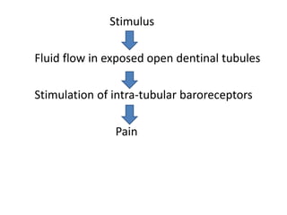
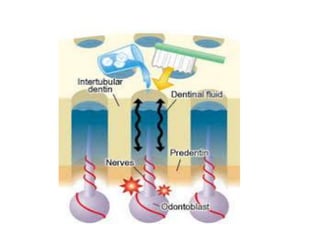
DENTIN HYPERSENSITIVITY
READER
DR PANKAJ PRIYADARSHI
DEPARTMENT OF CONSERVATIVE
DENTISTRY AND ENDODONTICS
Definition
According to Holland et al, dentin
hypersensitivity is characterized by short,
sharp pain arising from exposed dentin in
response to stimuli typically thermal,
evaporative, tactile, osmotic or chemical and
which cannot be ascribed to any other form of
dental defect or pathology.
Etiology
Dentin hypersensitivity is a multifactorial
condition and is not fully understood.
However, there is a definite change in the
structure of dentin in the affected area. These
areas contain a larger number of patent
dentinal tubules with a larger tubular
diameter than the normal unaffected areas.
The following three theories have been
proposed to explain this condition:
1. Direct Innervation Theory-
This theory postulates that direct mechanical
stimulation of exposed nerve endings at the
dentinoenamel junction (DEJ) is responsible
for dentinal hypersensitivity.
The major shortcoming of this theory is that
there is insufficient evidence to prove that
the outer dentin that is most prone to be
sensitive is well innervated.
2. Odontoblast Receptor Theory-
This theory proposes that the odontoblasts
themselves act as neural receptors and relay
the signal to the nerve terminal.
The major shortcoming of this theory is that
there is no evidence to demonstrate synapses
between odontoblasts and nerve terminals.
3. Hydrodynamic Theory-
This is the most accepted theory to explain
the mechanism behind dentin
hypersensitivity.
The hydrodynamic theory proposed by
Brannstorm suggests that dentin
hypersensitivity is due to the hydrodynamic
fluid movements occuring across exposed
dentin with open tubules which in turn
mechanically activates the nerves present at
the inner ends of the dentin tubules or in the
outer layers of the pulp.
The dentinal tubules, which are open an wide,
contain fluid. Various stimuli (i.e. thermal,
tactile, chemical or osmotic changes) displaces
the fluid in the dentinal tubules in either an
inwardly or outwardly direction. The
movement of this liquid stimulates the
odontoblastic processes, and the subsequent
mechanical disturbances stimulates
baroreceptors (a nerve receptor sensitive to
pressure) that leads to neural discharges
(depolarization). This neural pulpal activation
is perceived as pain by the patient.
Stimulus
Fluid flow in exposed open dentinal tubules
Stimulation of intra-tubular baroreceptors
Pain
Etiological and predisposing factors associated with
dentin hypersensitivity:
- Loss of enamel
- Gingival recession
- Attrition
- Erosion (intrinsic and extrinsic)
- Tooth malposition
- Denudation of cementum
- Thinning, fenestration, absent buccal alveolar bone
plate
- Abrasion
- Abfraction
- Periodontal disease and its treatment
- Periodontal surgery
- Patient habits
Clinical Features
1. Pain is the most common clinical feature
associated with dentin hypersensitivity.
2. The intensity of pain varies from mild
discomfort to severe sensitivity.
3. The pain is typically rapid in onset, sharp in
character and is of short duration.
4. External stimuli which can elicit the
expression of this condition include:
i. Thermal stimuli
a. Hot/cold food and beverages
b. Cold blast of air
ii. Osmotic stimuli
a. Sweet food
iii. Acidic stimuli
a. Citrus fruits
b. Acidic beverages
c. Medicines
iv. Mechanical stimuli
a. Toothbrush
b. Dental instruments
5. The most commonly involved teeth are
i. Buccal surfaces of premolars
ii. Facial surfaces of incisors
Management of
Dentin Hypersensitivity
Diagnosis:
1. A thorough patient history and clinical
examination
2. Diagnosis is confirmed by using a jet of air or
by using a diagnostic probe on dentin in a
mesiodistal direction in the area where the
patient complains of pain.
Clinical Management:
Removal of Etiological Factors-
The removal of the following etiological
factors can prevent dentin hypersensitivity
from recurring
1. Improper tooth brushing technique
2. Poor oral hygiene
3. Premature contacts
4. Gingival recession
5. Endogenous and Exogenous acids
Treatment Strategies-
The various clinical methods of management
of dentin hypersensitivity are:
1.Desensitizing the nerve
Daily use of 5% potassium nitrate containing
toothpaste for 4 weeks is an effective way to
desensitize dentin.
Potassium nitrate increases the extracellular
potassium ion concentration and thus
depolarizes the nerve. This prevents the
transmission of pain signals to the brain.
2. Occluding the dentinal tubules
Occluding the distal terminal ends of exposed
dentinal tubules is another method of
managing dentin hypersensitivity.
i. Salts or ions
a. 0.4% Stannous fluoride
b. 2% Sodium fluoride
c. Potassium oxalate
d. Strontium chloride
e. Calcium carbonate
ii. Protein precipitates
a. CPP-ACP
b. Silver nitrate
c. Glutaraldehyde
d. Zinc chloride
3. Dentin adhesives
i. Dentin bonding agents
ii. Composites
iii. Glass ionomers
4. Crown placement
5. Periodontal grafting
6. Lasers
Nd- YAG lasers have shown to effectively occlude
dentinal tubules.

DENTIN HYPERSENSITIVITY.pptx

  • 1.
    DENTIN HYPERSENSITIVITY READER DR PANKAJPRIYADARSHI DEPARTMENT OF CONSERVATIVE DENTISTRY AND ENDODONTICS
  • 2.
    Definition According to Hollandet al, dentin hypersensitivity is characterized by short, sharp pain arising from exposed dentin in response to stimuli typically thermal, evaporative, tactile, osmotic or chemical and which cannot be ascribed to any other form of dental defect or pathology.
  • 3.
    Etiology Dentin hypersensitivity isa multifactorial condition and is not fully understood. However, there is a definite change in the structure of dentin in the affected area. These areas contain a larger number of patent dentinal tubules with a larger tubular diameter than the normal unaffected areas.
  • 4.
    The following threetheories have been proposed to explain this condition: 1. Direct Innervation Theory- This theory postulates that direct mechanical stimulation of exposed nerve endings at the dentinoenamel junction (DEJ) is responsible for dentinal hypersensitivity. The major shortcoming of this theory is that there is insufficient evidence to prove that the outer dentin that is most prone to be sensitive is well innervated.
  • 5.
    2. Odontoblast ReceptorTheory- This theory proposes that the odontoblasts themselves act as neural receptors and relay the signal to the nerve terminal. The major shortcoming of this theory is that there is no evidence to demonstrate synapses between odontoblasts and nerve terminals.
  • 6.
    3. Hydrodynamic Theory- Thisis the most accepted theory to explain the mechanism behind dentin hypersensitivity. The hydrodynamic theory proposed by Brannstorm suggests that dentin hypersensitivity is due to the hydrodynamic fluid movements occuring across exposed dentin with open tubules which in turn mechanically activates the nerves present at the inner ends of the dentin tubules or in the outer layers of the pulp.
  • 8.
    The dentinal tubules,which are open an wide, contain fluid. Various stimuli (i.e. thermal, tactile, chemical or osmotic changes) displaces the fluid in the dentinal tubules in either an inwardly or outwardly direction. The movement of this liquid stimulates the odontoblastic processes, and the subsequent mechanical disturbances stimulates baroreceptors (a nerve receptor sensitive to pressure) that leads to neural discharges (depolarization). This neural pulpal activation is perceived as pain by the patient.
  • 9.
    Stimulus Fluid flow inexposed open dentinal tubules Stimulation of intra-tubular baroreceptors Pain
  • 11.
    Etiological and predisposingfactors associated with dentin hypersensitivity: - Loss of enamel - Gingival recession - Attrition - Erosion (intrinsic and extrinsic) - Tooth malposition - Denudation of cementum - Thinning, fenestration, absent buccal alveolar bone plate - Abrasion - Abfraction - Periodontal disease and its treatment - Periodontal surgery - Patient habits
  • 12.
    Clinical Features 1. Painis the most common clinical feature associated with dentin hypersensitivity. 2. The intensity of pain varies from mild discomfort to severe sensitivity. 3. The pain is typically rapid in onset, sharp in character and is of short duration. 4. External stimuli which can elicit the expression of this condition include:
  • 13.
    i. Thermal stimuli a.Hot/cold food and beverages b. Cold blast of air ii. Osmotic stimuli a. Sweet food iii. Acidic stimuli a. Citrus fruits b. Acidic beverages c. Medicines iv. Mechanical stimuli a. Toothbrush b. Dental instruments 5. The most commonly involved teeth are i. Buccal surfaces of premolars ii. Facial surfaces of incisors
  • 14.
  • 15.
    Diagnosis: 1. A thoroughpatient history and clinical examination 2. Diagnosis is confirmed by using a jet of air or by using a diagnostic probe on dentin in a mesiodistal direction in the area where the patient complains of pain.
  • 16.
    Clinical Management: Removal ofEtiological Factors- The removal of the following etiological factors can prevent dentin hypersensitivity from recurring 1. Improper tooth brushing technique 2. Poor oral hygiene 3. Premature contacts 4. Gingival recession 5. Endogenous and Exogenous acids
  • 17.
    Treatment Strategies- The variousclinical methods of management of dentin hypersensitivity are: 1.Desensitizing the nerve Daily use of 5% potassium nitrate containing toothpaste for 4 weeks is an effective way to desensitize dentin. Potassium nitrate increases the extracellular potassium ion concentration and thus depolarizes the nerve. This prevents the transmission of pain signals to the brain.
  • 18.
    2. Occluding thedentinal tubules Occluding the distal terminal ends of exposed dentinal tubules is another method of managing dentin hypersensitivity. i. Salts or ions a. 0.4% Stannous fluoride b. 2% Sodium fluoride c. Potassium oxalate d. Strontium chloride e. Calcium carbonate
  • 19.
    ii. Protein precipitates a.CPP-ACP b. Silver nitrate c. Glutaraldehyde d. Zinc chloride 3. Dentin adhesives i. Dentin bonding agents ii. Composites iii. Glass ionomers 4. Crown placement 5. Periodontal grafting 6. Lasers Nd- YAG lasers have shown to effectively occlude dentinal tubules.