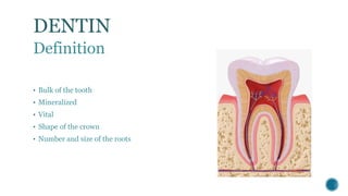
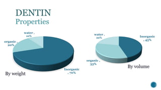
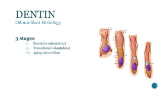
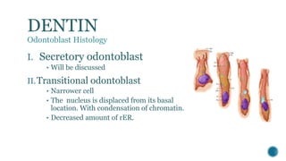
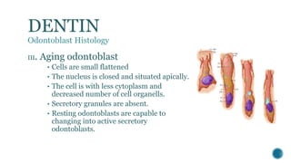
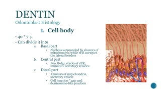
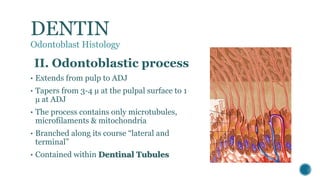
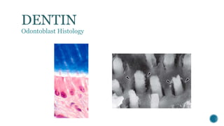
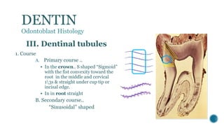
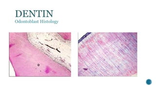
DENTIN
Menatalla M. Elhindawy
DENTIN
Definition
Properties
• Physical
• Chemical
Dentin formation
• Odontoblast
A. Differentiation
B. Histology
• Dentinogenesis
A. Matrix formation
B. Mineralization
DENTIN
Definition
• Bulk of the tooth
• Mineralized
• Vital
• Shape of the crown
• Number and size of the roots
 Color… yellowish “ light yellow in deciduous and pale yellow in permanent, gets
darker with age.
 Hardness.. Elastic, less than enamel and harder then cementum, harder in permanent
than deciduous.
 Thickness… 3-10mm.
 Radio-opacity.. Less radio-opaque than enamel.
 Permeability … permeable, more permeable at cervical region, permeability decreases
with age.
DENTIN
Properties
DENTIN
Properties
Inorganic
, 70%
organic ,
20%
water ,
10%
By weight
Inorganic
, 45%
organic ,
33%
water ,
22%
By volume
DENTIN
Properties
 In-organic component in the form of hydroxy-apatite crystals, smaller
in size than enamel crystals.
 Organic component in the form of collagen type I “ some of type III” &
ground substance “ muco-polysaccharides, glycose-aminoglycans &
proteoglycans”
DENTIN
Dentin formation
• Odontoblast
A. Differentiation
B. Histology
• Dentinogenesis
A. Matrix formation
B. Mineralization
 Highly differentiated
 Highly polarized
 Post-mitotic cells
 Originated from neural crest cells.
 Cell body & process
DENTIN
Odontoblast
DENTIN
Odontoblast Differentiation
 At early bell stage, the IEE changes from short to tall columnar cells leading to gradual
obliteration of the cell free zone.
 Cells of dental papilla become in contact with IEE separated only by basal lamina,
cells of dental papilla undergo the last mitotic division “asymmetrical one” producing
2 types of cells
A. Large cells in contact with dental lamina become pre-odontoblasts
B. Smaller cells with stem cell potentials grouped in “Hoehl cell layer
 IEE release some factors that help in the organization of odontoblast cytoskeleton
assembly which is important for relocation of organelles during transformation from
pre-odontoblast to odontoblast.
DENTIN
Odontoblast Differentiation
 Pre-odontoblasts differentiate into columnar odontoblast, cells become polarized and
capable to secret the first layer of dentin.
 As the first layer of dentine is formed ameloblasts are differentiated and the first
formed layer of enamel is formed and the ADJ is established .
 As dentin formation precedes, odontoblasts retreats toward the pulp and
odontoblastic process is formed.
DENTIN
Odontoblast Differentiation
DENTIN
Odontoblast Differentiation
3 stages
I. Secretory odontoblast
II. Transitional odontoblast
III. Aging odontoblast
DENTIN
Odontoblast Histology
I. Secretory odontoblast
• Will be discussed
II.Transitional odontoblast
• Narrower cell
• The nucleus is displaced from its basal
location. With condensation of chromatin.
• Decreased amount of rER.
DENTIN
Odontoblast Histology
III. Aging odontoblast
• Cells are small flattened
• The nucleus is closed and situated apically.
• The cell is with less cytoplasm and
decreased number of cell organells.
• Secretory granules are absent.
• Resting odontoblasts are capable to
changing into active secretory
odontoblasts.
DENTIN
Odontoblast Histology
DENTIN
Odontoblast Histology
I. Cell body
• 40 * 7 µ
• Can divide it into
a. Basal part
• Nucleus surrounded by clusters of
mitochondria while rER occupies
the lateral borders
b. Central part
• Few Golgi, stacks of rER,
immature secretory vesicles
c. Distal part
• Clusters of mitochondria,
secretory vesicle
• Cell junction “ gap and
desmosome-like junction
DENTIN
Odontoblast Histology
II. Odontoblastic process
• Extends from pulp to ADJ
• Tapers from 3-4 µ at the pulpal surface to 1
µ at ADJ
• The process contains only microtubules,
microfilaments & mitochondria
• Branched along its course “lateral and
terminal”
• Contained within Dentinal Tubules
DENTIN
Odontoblast Histology
DENTIN
Odontoblast Histology
Near pulp Mid- dentin ADJ
DENTIN
Odontoblast Histology
III. Dentinal tubules
1. Course
A. Primary course ..
 In the crown.. S shaped “Sigmoid”
with the fist convexity toward the
root in the middle and cervical
13s & straight under cup tip or
incisal edge.
 In in root straight
B. Secondary course..
“Sinusoidal” shaped
DENTIN
Odontoblast Histology
DENTIN
Odontoblast Histology
III. Dentinal tubules
2. Ratio  unite are “pulpADJ”
 5:1
 DT are closely packed near the pulp
and further apart near the ADJ
3. Diameter “pulpADJ”
 4:1
 At the pulpal side 3-4 µ to reach the
ADJ as 1µ
4. Number unite area
 crown>root
DENTIN
Odontoblast Histology
III. Dentinal tubules
5. Branches
 Terminal
 Lateral
Note … DTs may cross the ADJ
and become “Enamel spindle”
DENTIN
 Definition
 Properties
• Physical
• Chemical
Dentin formation
• Odontoblast
A. Differentiation
B. Histology
• Dentinogenesis
A. Matrix formation
B. Mineralization
DENTIN
Dentinogenesis
DENTIN
Dentinogenesis
MATRIX
FORMATION
Mantle Dentin
Circumpulpal
Dentin
Mineralization
I. Mantle Dentin MATRIX
formation
 5 - 20 µ in thickness.
 Collagen fibers are
• Von Kroff’s collagen type
III fibers
• Thick 0.1 -0.2 µ
• Directed perpendicular
to ADJ
 Ground substance
• Preexisting from the cell
fee zone.
DENTIN
Matrix Formation
Mantle D.
II. Circumpulpal Dentin MATRIX
formation
 It represents the entire thickness of
D.
 Collagen fibers are
• Type I fibers
• Thin 0.05 µ
• Directed parallel to ADJ &
perpendicular to DT
 Ground substance
• Newly formed by odontoblasts
DENTIN
Matrix Formation
Mantle D.
Ciucumpulpal
D.
Pre-dentin
Constant feature of dentin.
Un-mineralized zone between
odontoblasts & mineralized D
 2-6 µ in thickness.
Formation and mineralization
begins at the tip of cusps or
incisal ridge and proceed
cervically.
DENTIN
Mineralization
DENTIN
Mineralization
 Mineralization occurs by globular, or calcospheric calcification and it
involves the deposition of the crystals in certain areas.
 Initially mineralization starts by deposition of very fine plates of
hydroxyapatite on the surface of collagen fibrils and the ground
substance, later crystals are deposited within the fibrils themselves.
 Crystals are arranged parallel to the collagen fibers long axis.
Globular “calcospheric”
calcification
 Matrix vesicles
 First mineral crystals
 Continued crystal growth
 Globular masses which enlarge
to form single calcified mass
DENTIN
Mineralization
Less mineralized & fewer
defects
Globular “calcospheric” calcification
Or
Linear calcification
Mantle Dentin circumpulpal Dentin
DENTIN
Mantle Dentin Circumpulpal Dentin
Thickness 5- 20µ Bulk of the tooth
Diameter of
collagen fibers
Large (0.1-0.2 µ) Small (0.05µ)
Direction of
collagen fibers
Perpendicular to ADJ and parallel to the
dentinal tubules
Parallel to ADJ and perpendicular to the
dentinal tubules
Ground substance
Pre- existing from dental papilla cell free
zone and the odontoblasts.
Formed by the odontoblasts only
Mineralization Globular Globular or linear
Dentin
Dentin

Dentin

  • 2.
  • 4.
    DENTIN Definition Properties • Physical • Chemical Dentinformation • Odontoblast A. Differentiation B. Histology • Dentinogenesis A. Matrix formation B. Mineralization
  • 5.
    DENTIN Definition • Bulk ofthe tooth • Mineralized • Vital • Shape of the crown • Number and size of the roots
  • 6.
     Color… yellowish“ light yellow in deciduous and pale yellow in permanent, gets darker with age.  Hardness.. Elastic, less than enamel and harder then cementum, harder in permanent than deciduous.  Thickness… 3-10mm.  Radio-opacity.. Less radio-opaque than enamel.  Permeability … permeable, more permeable at cervical region, permeability decreases with age. DENTIN Properties
  • 7.
    DENTIN Properties Inorganic , 70% organic , 20% water, 10% By weight Inorganic , 45% organic , 33% water , 22% By volume
  • 8.
    DENTIN Properties  In-organic componentin the form of hydroxy-apatite crystals, smaller in size than enamel crystals.  Organic component in the form of collagen type I “ some of type III” & ground substance “ muco-polysaccharides, glycose-aminoglycans & proteoglycans”
  • 9.
    DENTIN Dentin formation • Odontoblast A.Differentiation B. Histology • Dentinogenesis A. Matrix formation B. Mineralization
  • 10.
     Highly differentiated Highly polarized  Post-mitotic cells  Originated from neural crest cells.  Cell body & process DENTIN Odontoblast
  • 11.
    DENTIN Odontoblast Differentiation  Atearly bell stage, the IEE changes from short to tall columnar cells leading to gradual obliteration of the cell free zone.  Cells of dental papilla become in contact with IEE separated only by basal lamina, cells of dental papilla undergo the last mitotic division “asymmetrical one” producing 2 types of cells A. Large cells in contact with dental lamina become pre-odontoblasts B. Smaller cells with stem cell potentials grouped in “Hoehl cell layer  IEE release some factors that help in the organization of odontoblast cytoskeleton assembly which is important for relocation of organelles during transformation from pre-odontoblast to odontoblast.
  • 12.
    DENTIN Odontoblast Differentiation  Pre-odontoblastsdifferentiate into columnar odontoblast, cells become polarized and capable to secret the first layer of dentin.  As the first layer of dentine is formed ameloblasts are differentiated and the first formed layer of enamel is formed and the ADJ is established .  As dentin formation precedes, odontoblasts retreats toward the pulp and odontoblastic process is formed.
  • 13.
  • 14.
  • 15.
    3 stages I. Secretoryodontoblast II. Transitional odontoblast III. Aging odontoblast DENTIN Odontoblast Histology
  • 16.
    I. Secretory odontoblast •Will be discussed II.Transitional odontoblast • Narrower cell • The nucleus is displaced from its basal location. With condensation of chromatin. • Decreased amount of rER. DENTIN Odontoblast Histology
  • 17.
    III. Aging odontoblast •Cells are small flattened • The nucleus is closed and situated apically. • The cell is with less cytoplasm and decreased number of cell organells. • Secretory granules are absent. • Resting odontoblasts are capable to changing into active secretory odontoblasts. DENTIN Odontoblast Histology
  • 18.
    DENTIN Odontoblast Histology I. Cellbody • 40 * 7 µ • Can divide it into a. Basal part • Nucleus surrounded by clusters of mitochondria while rER occupies the lateral borders b. Central part • Few Golgi, stacks of rER, immature secretory vesicles c. Distal part • Clusters of mitochondria, secretory vesicle • Cell junction “ gap and desmosome-like junction
  • 19.
    DENTIN Odontoblast Histology II. Odontoblasticprocess • Extends from pulp to ADJ • Tapers from 3-4 µ at the pulpal surface to 1 µ at ADJ • The process contains only microtubules, microfilaments & mitochondria • Branched along its course “lateral and terminal” • Contained within Dentinal Tubules
  • 20.
  • 21.
  • 22.
    DENTIN Odontoblast Histology III. Dentinaltubules 1. Course A. Primary course ..  In the crown.. S shaped “Sigmoid” with the fist convexity toward the root in the middle and cervical 13s & straight under cup tip or incisal edge.  In in root straight B. Secondary course.. “Sinusoidal” shaped
  • 23.
  • 24.
    DENTIN Odontoblast Histology III. Dentinaltubules 2. Ratio unite are “pulpADJ”  5:1  DT are closely packed near the pulp and further apart near the ADJ 3. Diameter “pulpADJ”  4:1  At the pulpal side 3-4 µ to reach the ADJ as 1µ 4. Number unite area  crown>root
  • 25.
    DENTIN Odontoblast Histology III. Dentinaltubules 5. Branches  Terminal  Lateral Note … DTs may cross the ADJ and become “Enamel spindle”
  • 26.
    DENTIN  Definition  Properties •Physical • Chemical Dentin formation • Odontoblast A. Differentiation B. Histology • Dentinogenesis A. Matrix formation B. Mineralization
  • 27.
  • 28.
  • 29.
    I. Mantle DentinMATRIX formation  5 - 20 µ in thickness.  Collagen fibers are • Von Kroff’s collagen type III fibers • Thick 0.1 -0.2 µ • Directed perpendicular to ADJ  Ground substance • Preexisting from the cell fee zone. DENTIN Matrix Formation Mantle D.
  • 30.
    II. Circumpulpal DentinMATRIX formation  It represents the entire thickness of D.  Collagen fibers are • Type I fibers • Thin 0.05 µ • Directed parallel to ADJ & perpendicular to DT  Ground substance • Newly formed by odontoblasts DENTIN Matrix Formation Mantle D. Ciucumpulpal D.
  • 31.
    Pre-dentin Constant feature ofdentin. Un-mineralized zone between odontoblasts & mineralized D  2-6 µ in thickness. Formation and mineralization begins at the tip of cusps or incisal ridge and proceed cervically. DENTIN Mineralization
  • 32.
    DENTIN Mineralization  Mineralization occursby globular, or calcospheric calcification and it involves the deposition of the crystals in certain areas.  Initially mineralization starts by deposition of very fine plates of hydroxyapatite on the surface of collagen fibrils and the ground substance, later crystals are deposited within the fibrils themselves.  Crystals are arranged parallel to the collagen fibers long axis.
  • 33.
    Globular “calcospheric” calcification  Matrixvesicles  First mineral crystals  Continued crystal growth  Globular masses which enlarge to form single calcified mass DENTIN Mineralization Less mineralized & fewer defects Globular “calcospheric” calcification Or Linear calcification Mantle Dentin circumpulpal Dentin
  • 34.
    DENTIN Mantle Dentin CircumpulpalDentin Thickness 5- 20µ Bulk of the tooth Diameter of collagen fibers Large (0.1-0.2 µ) Small (0.05µ) Direction of collagen fibers Perpendicular to ADJ and parallel to the dentinal tubules Parallel to ADJ and perpendicular to the dentinal tubules Ground substance Pre- existing from dental papilla cell free zone and the odontoblasts. Formed by the odontoblasts only Mineralization Globular Globular or linear