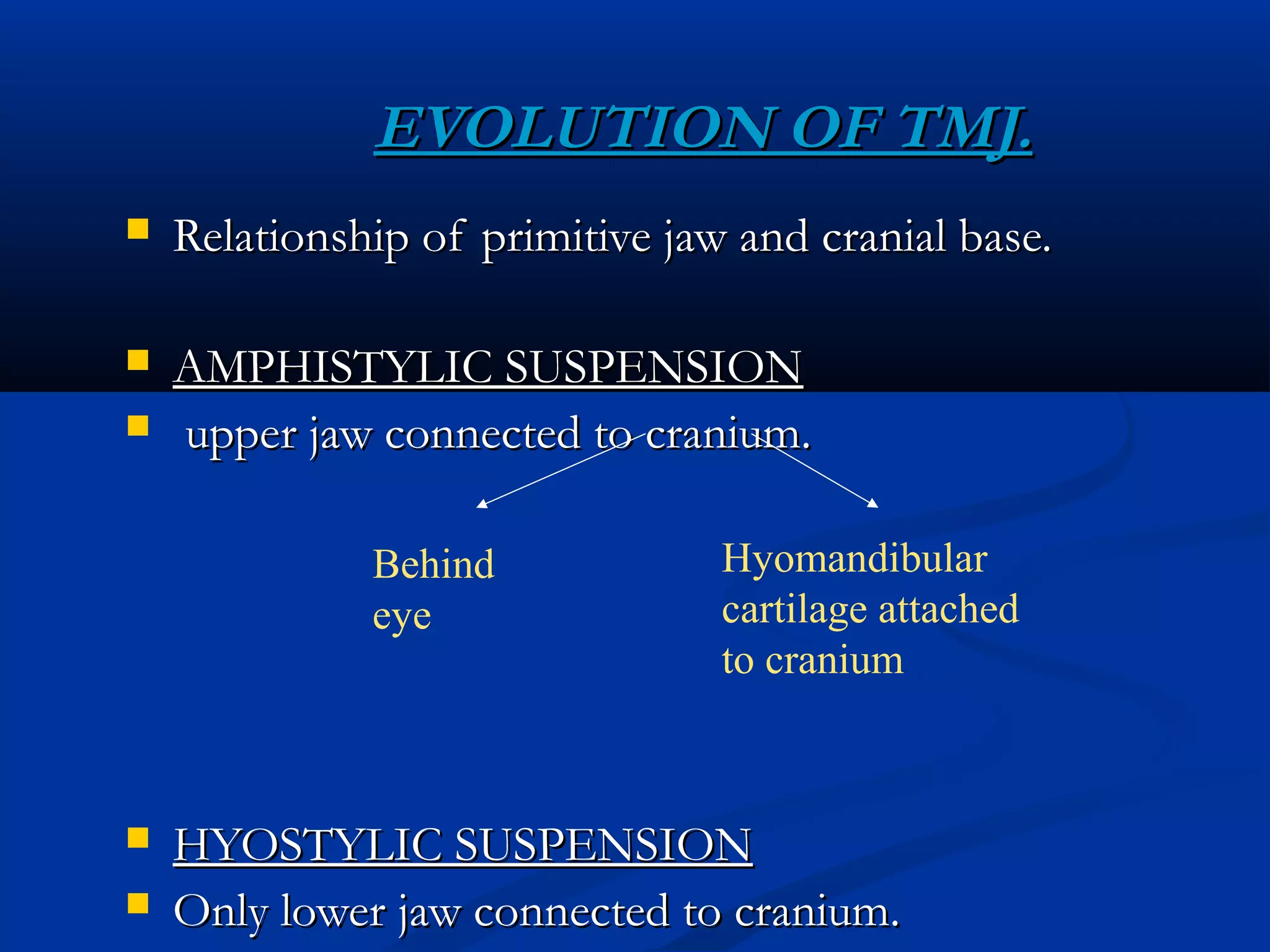
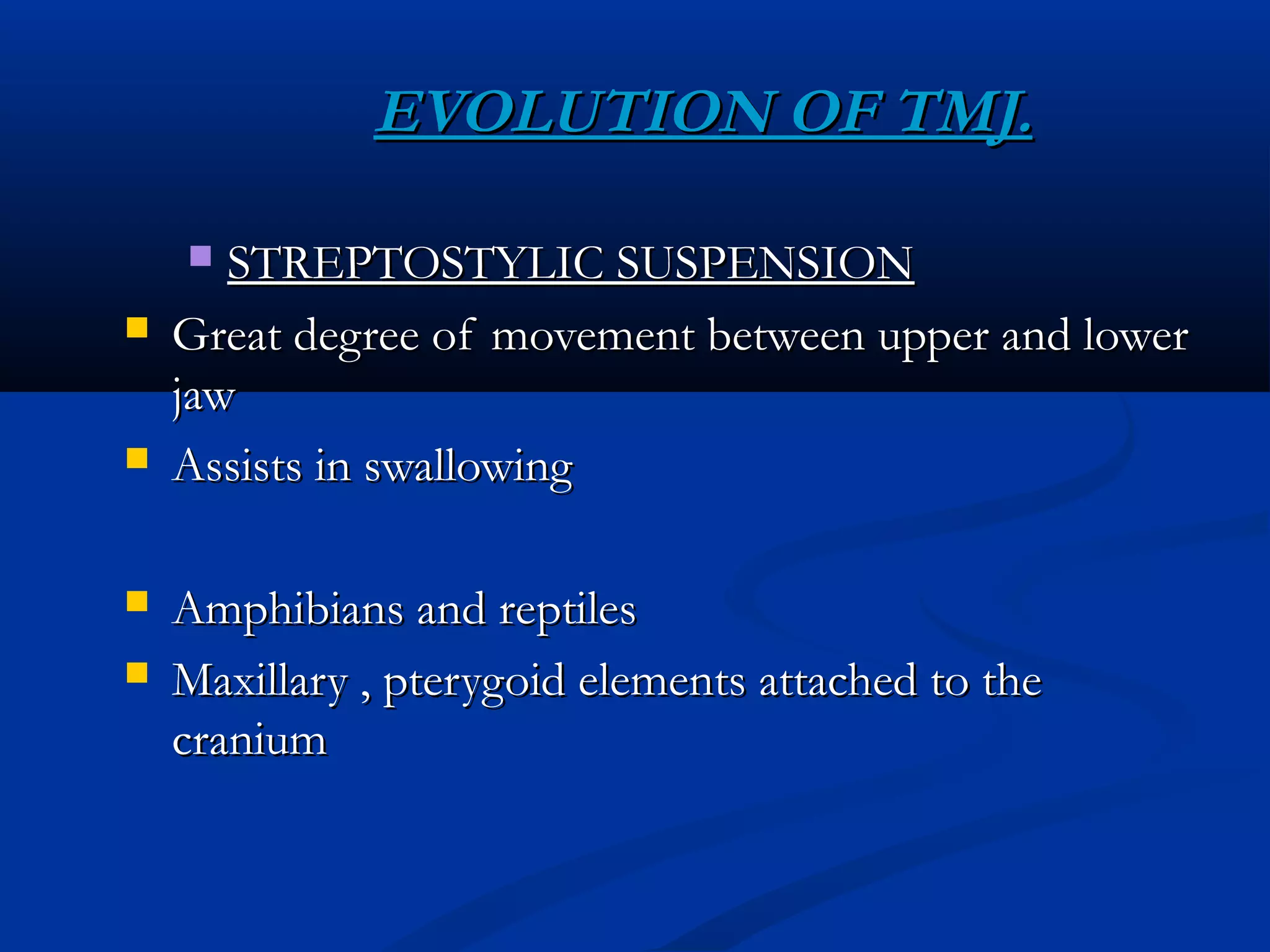
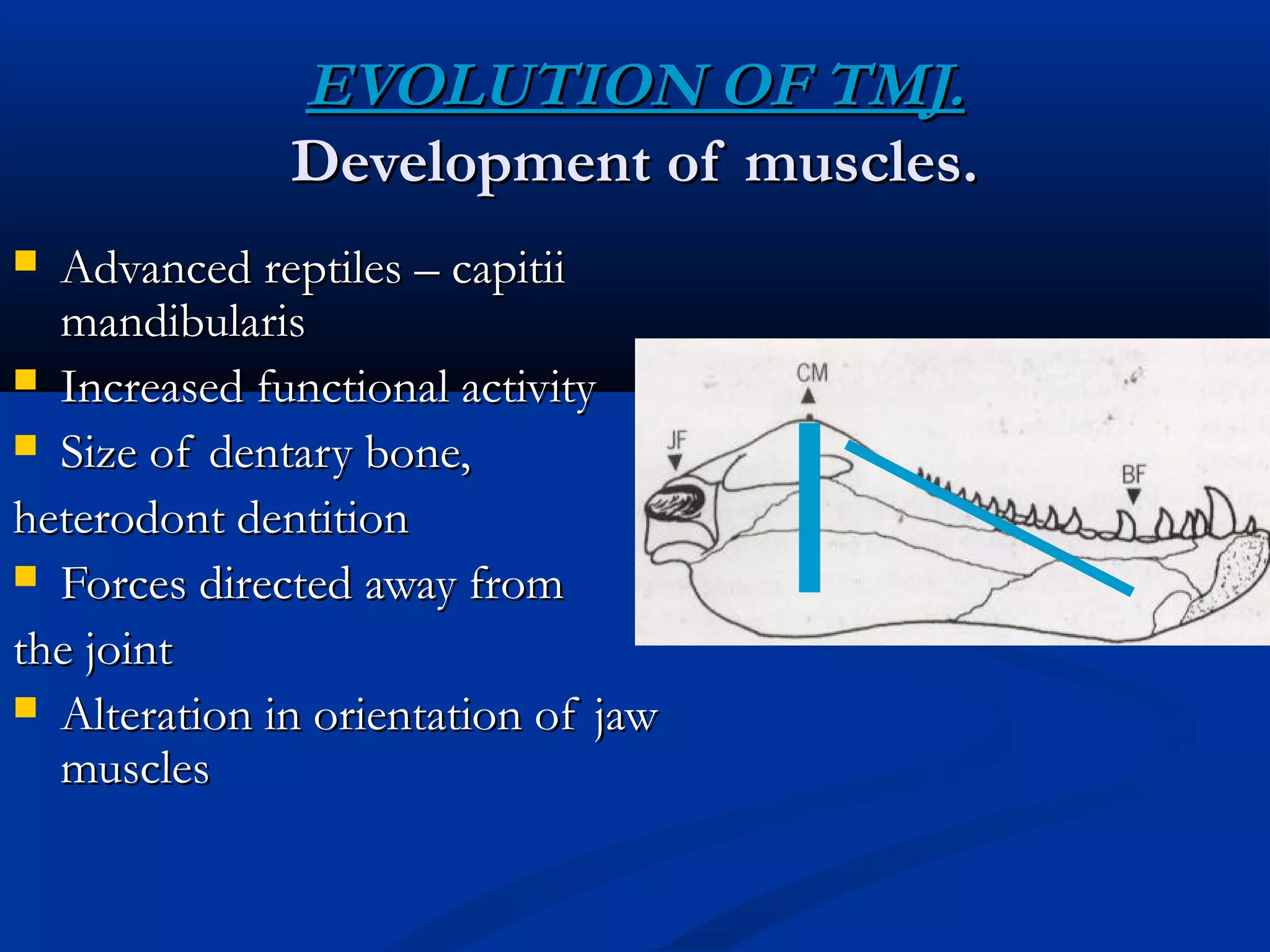
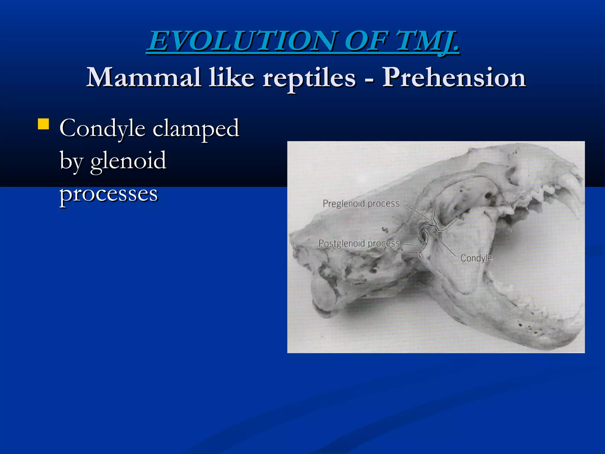
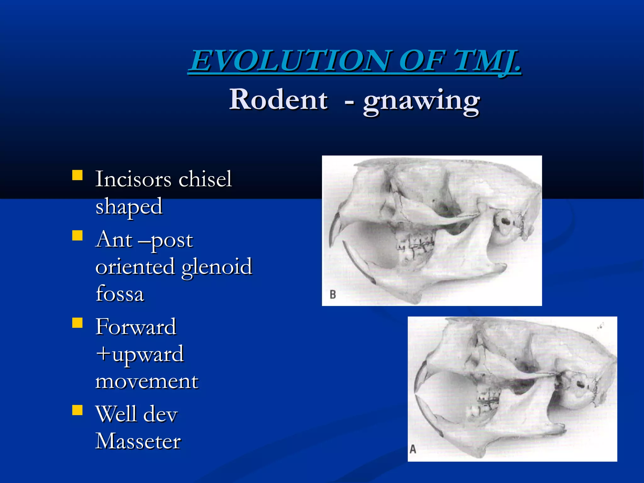
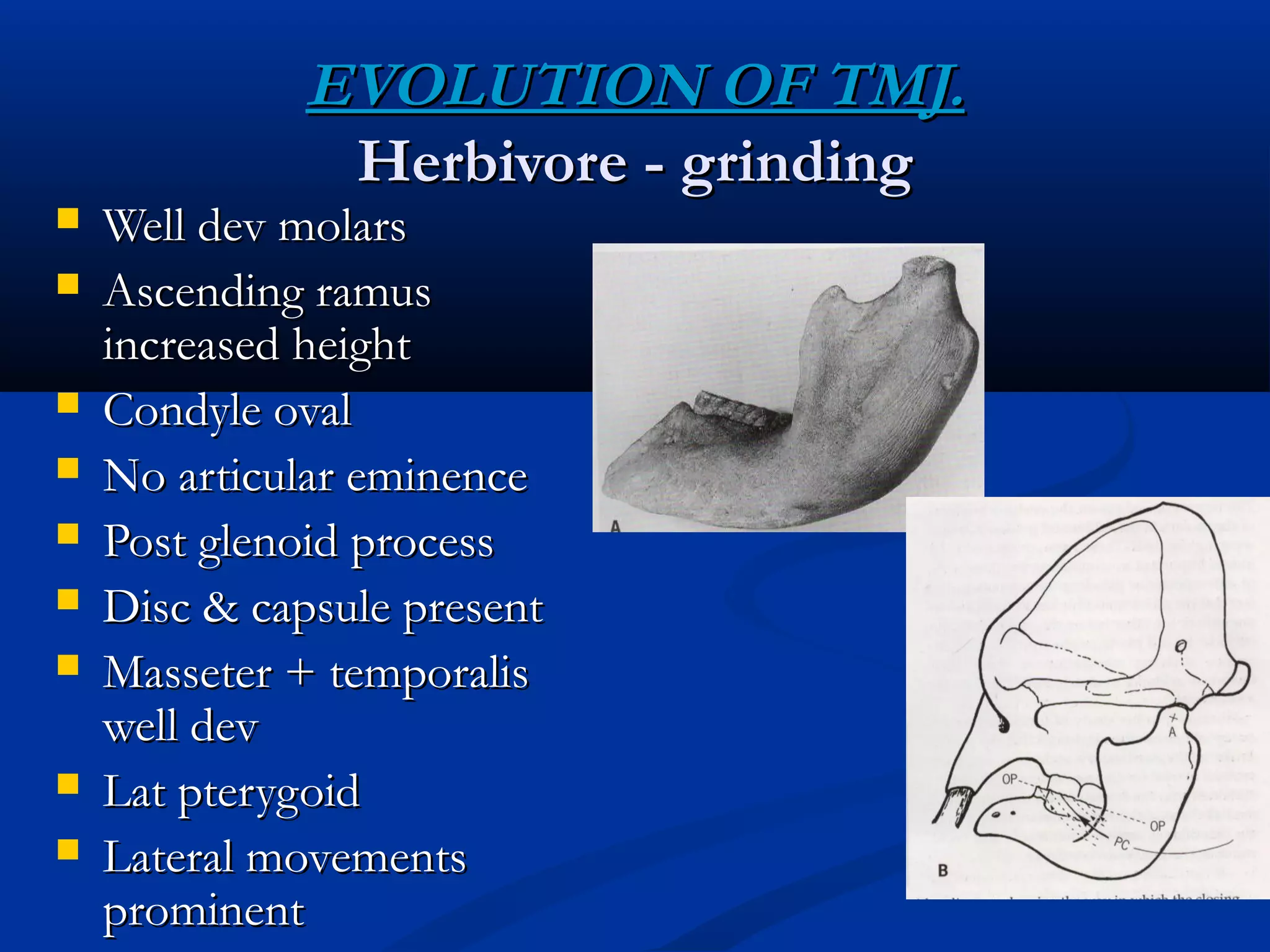
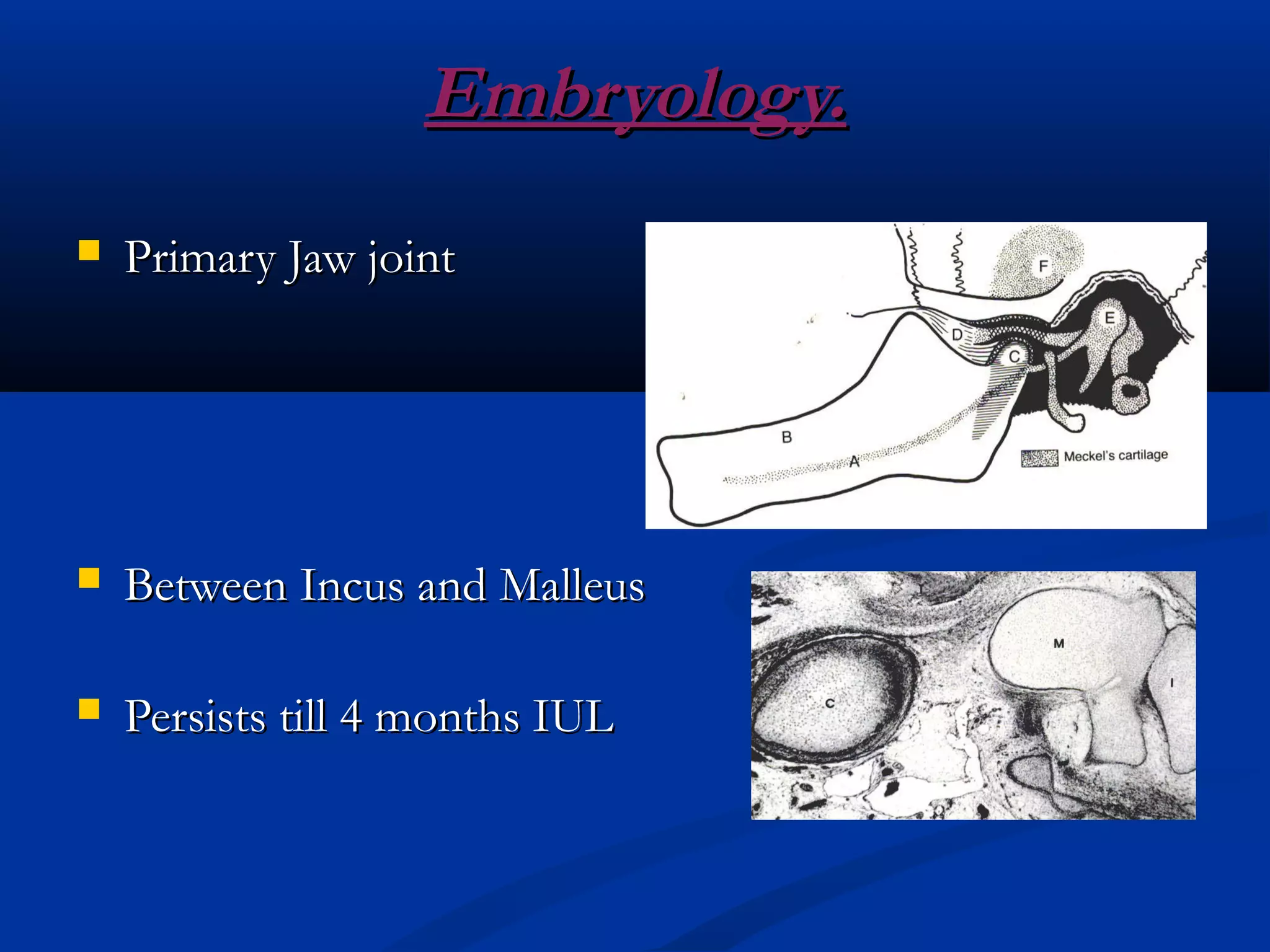
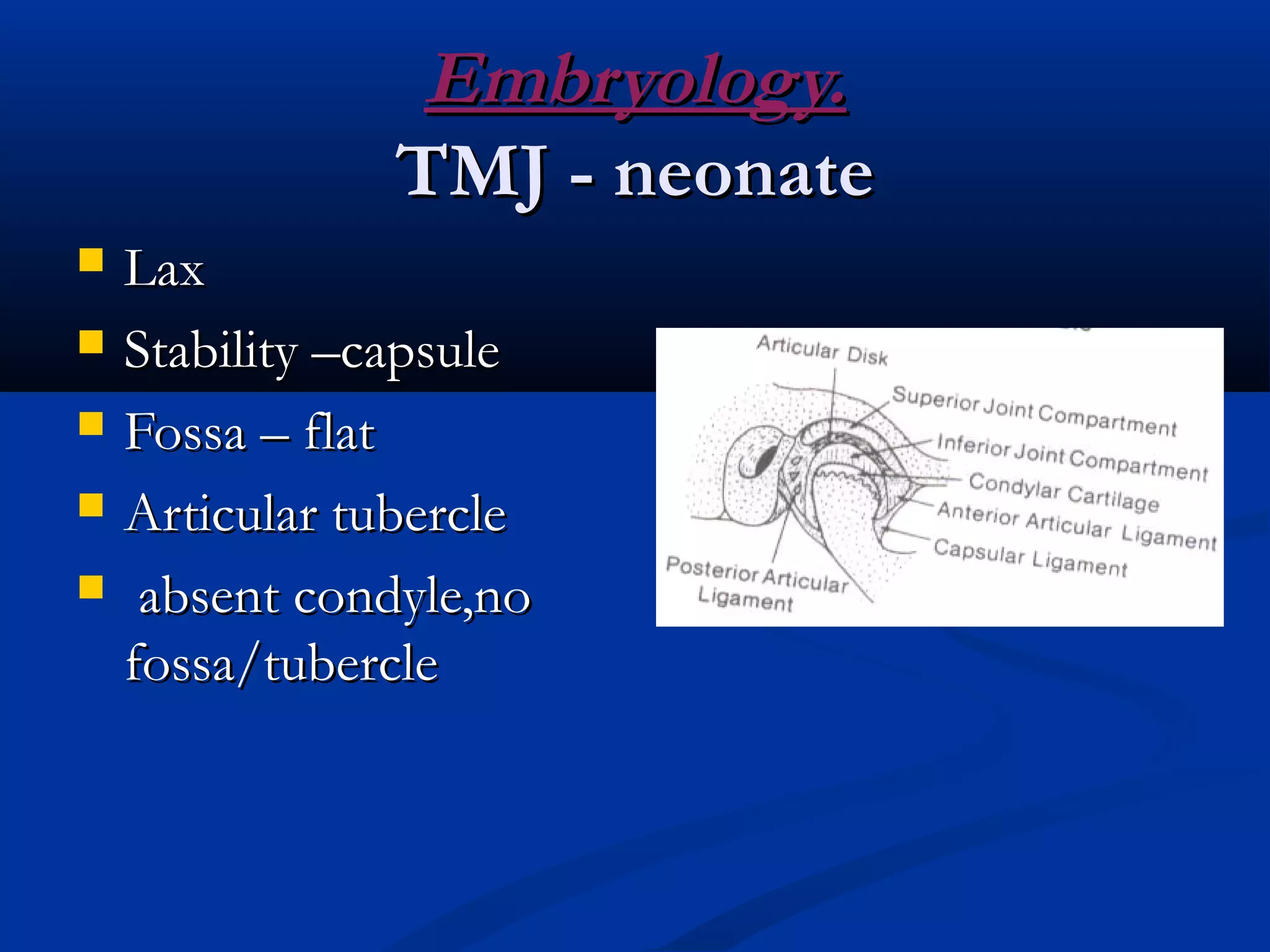
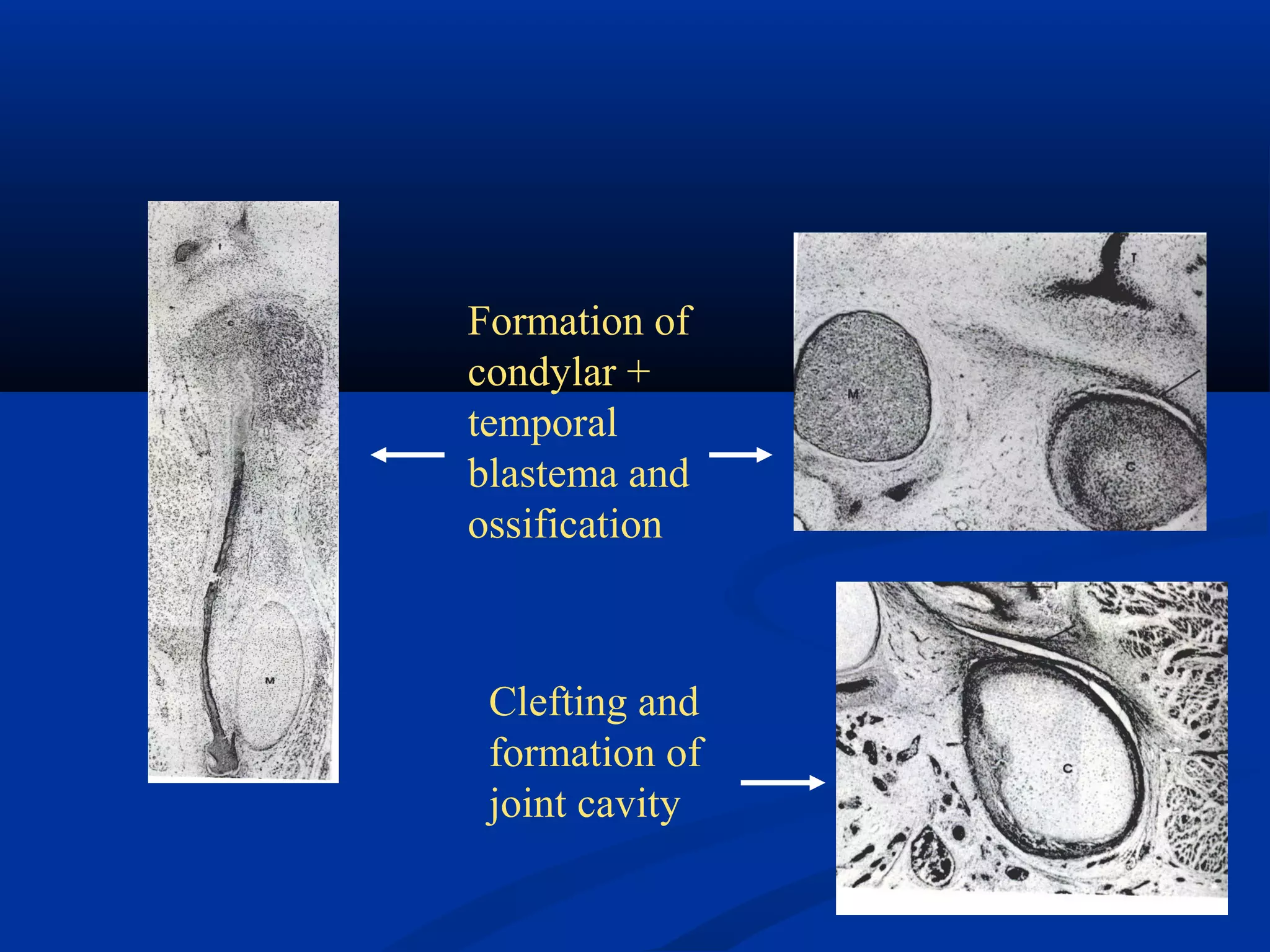
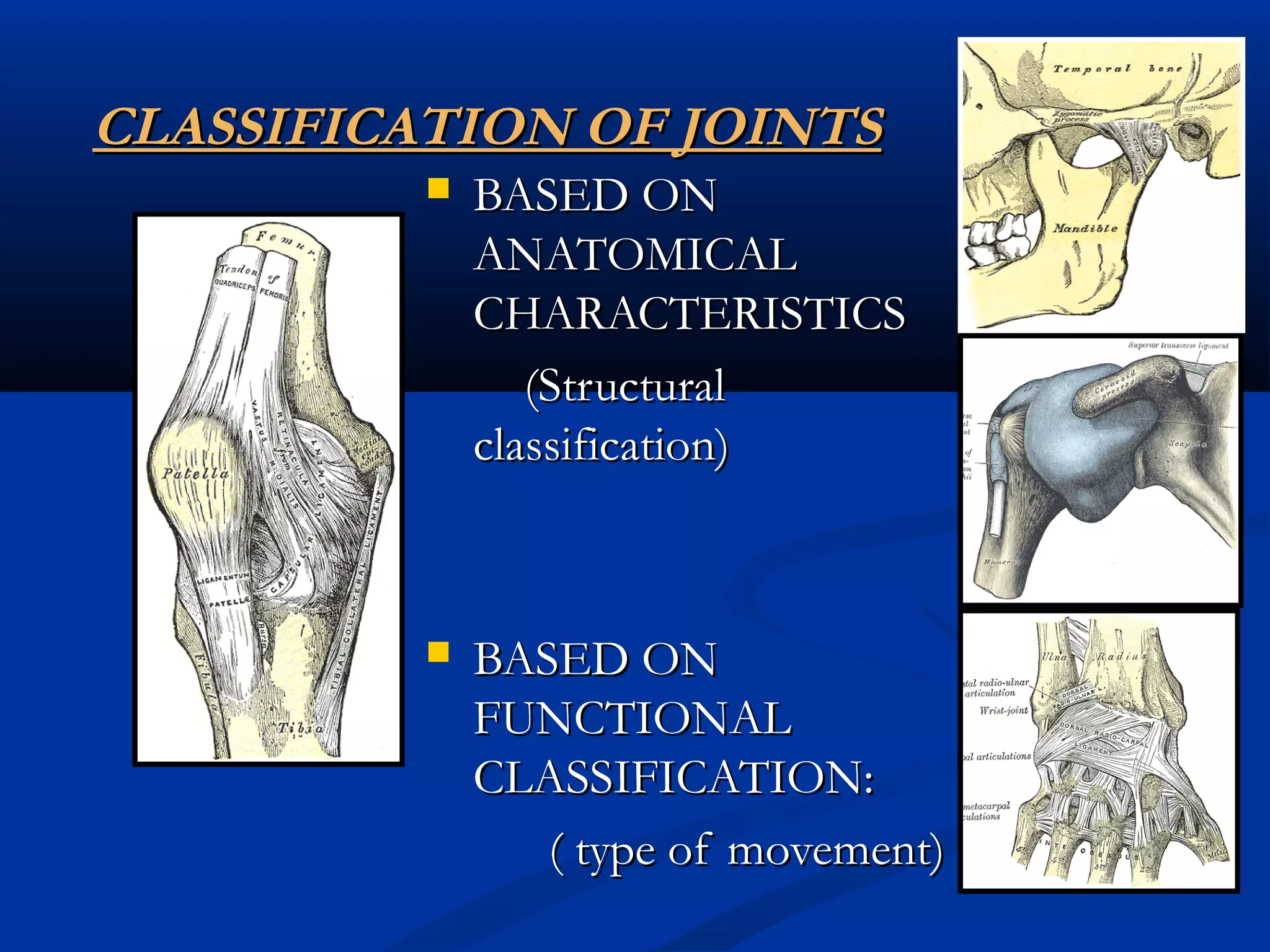
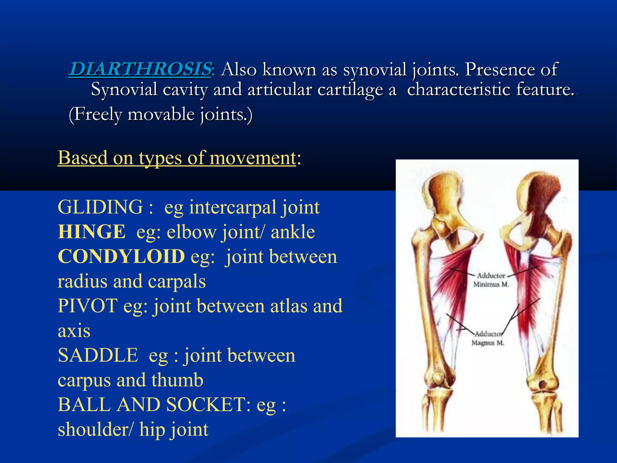
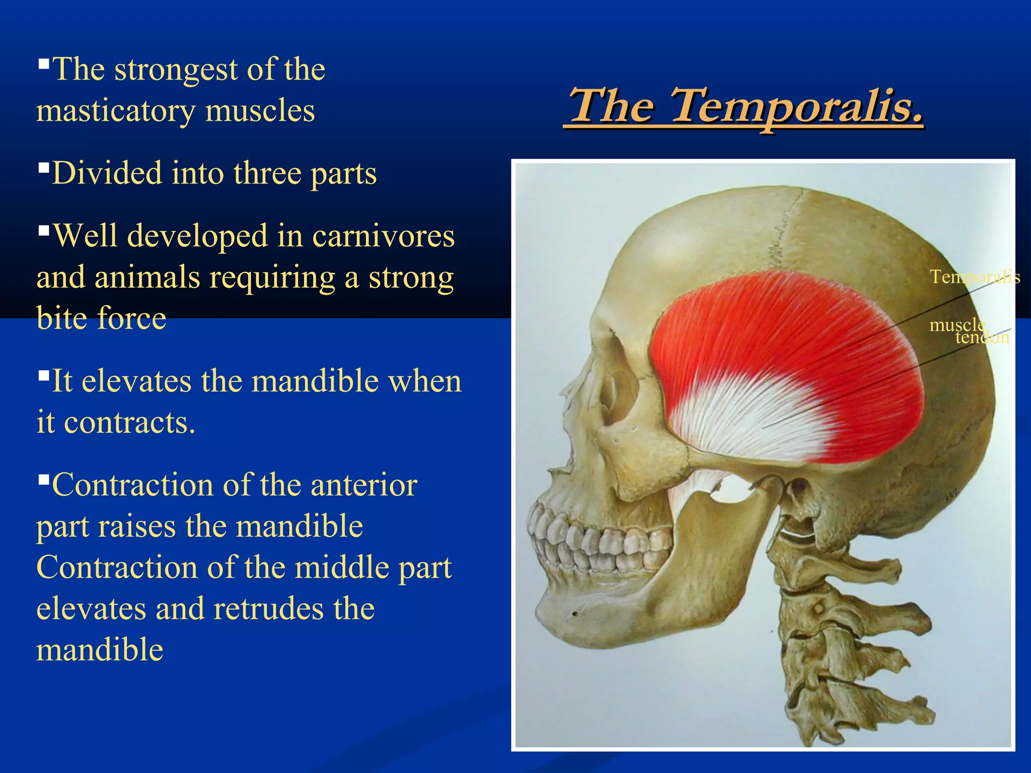
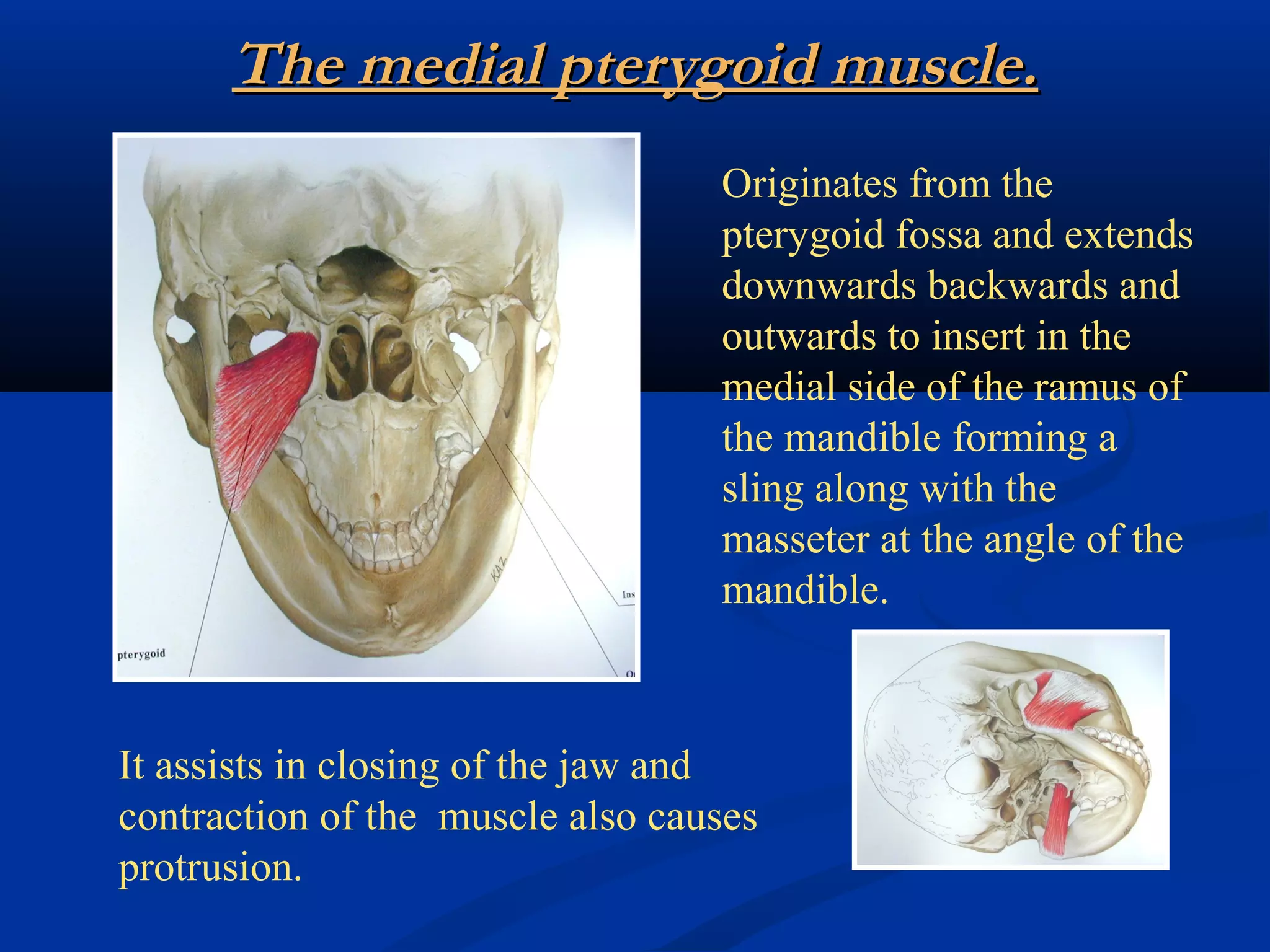
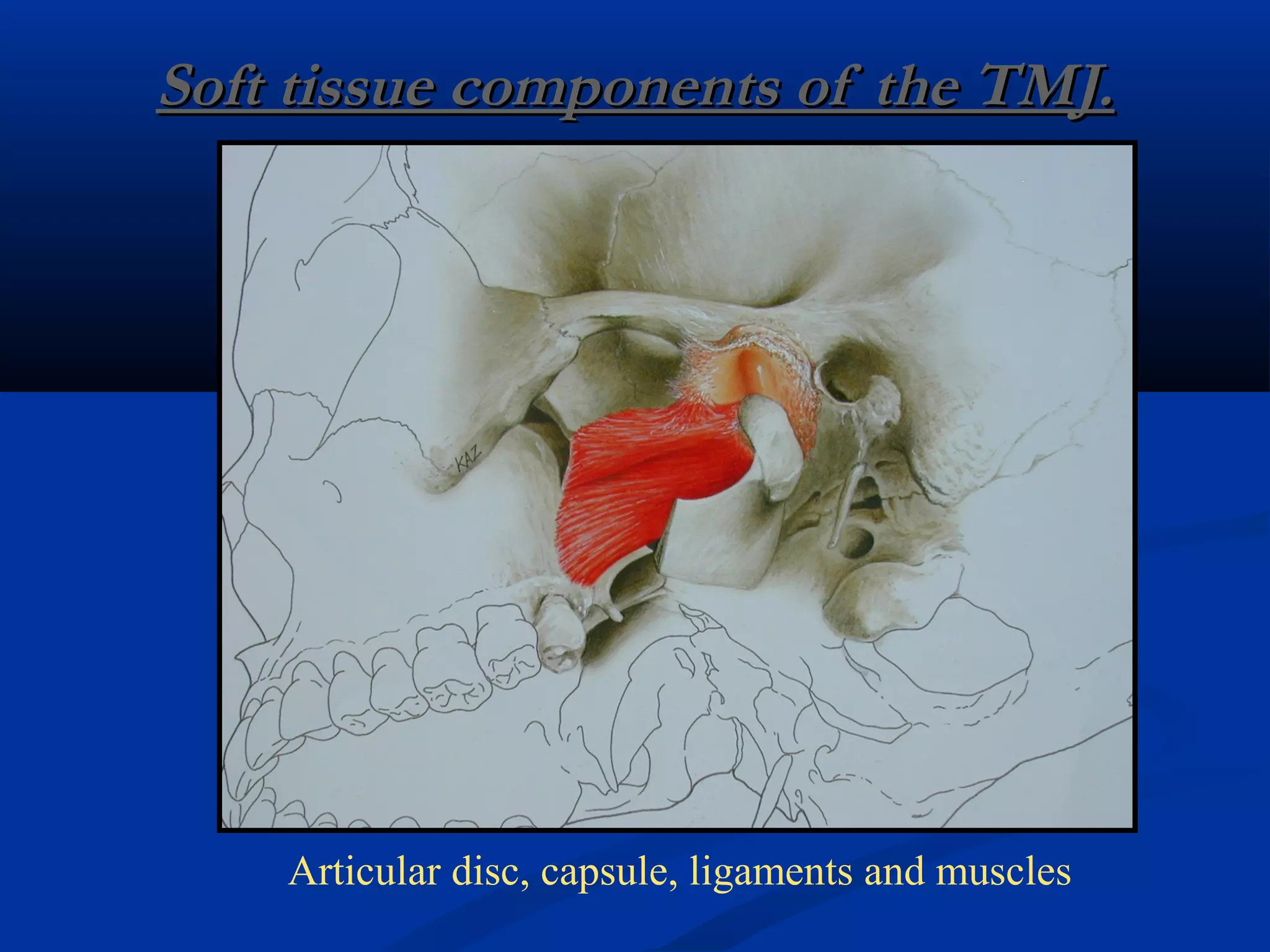
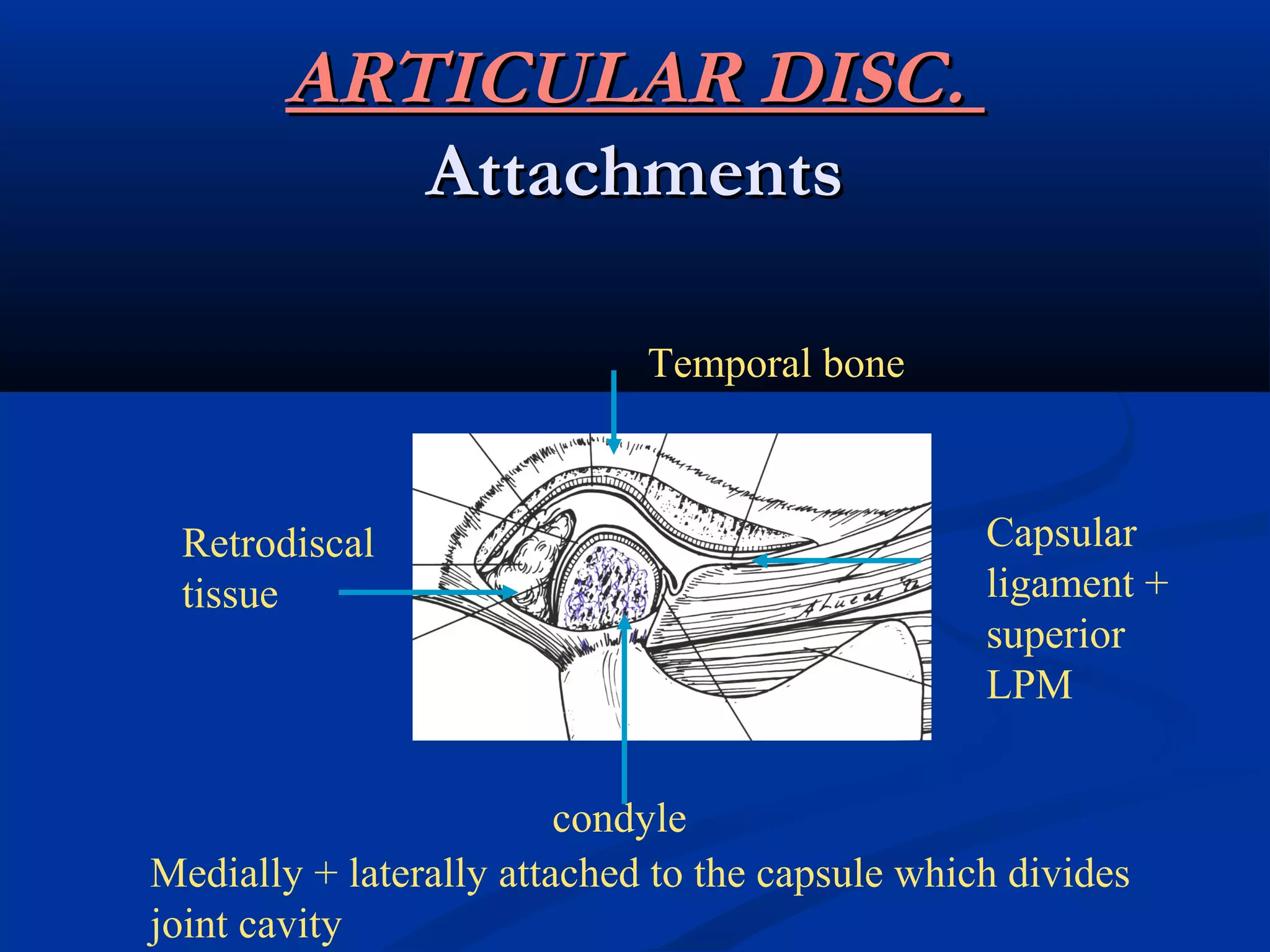
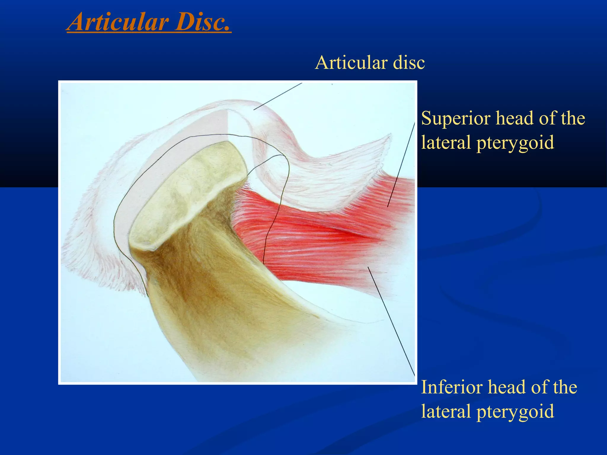
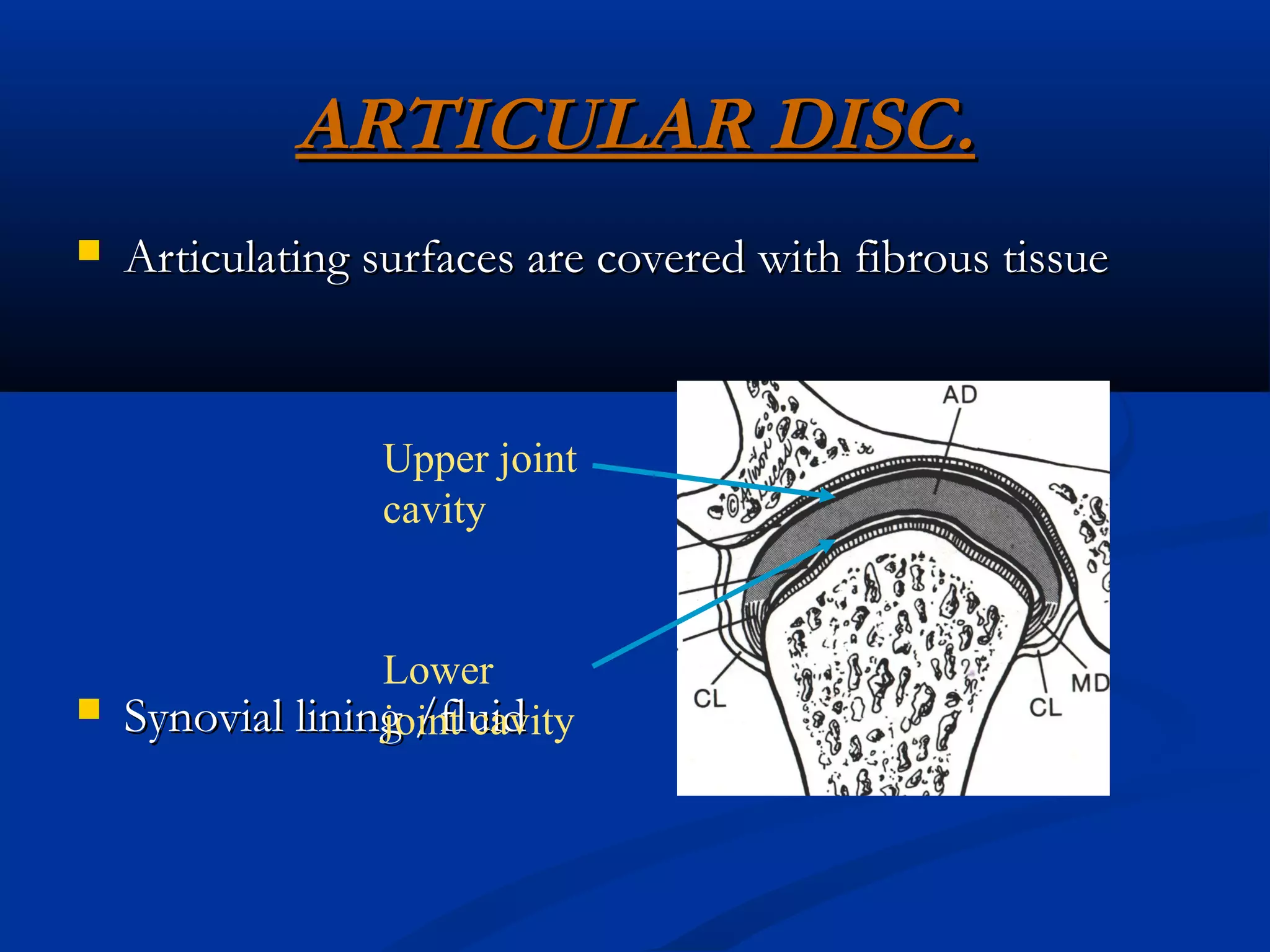
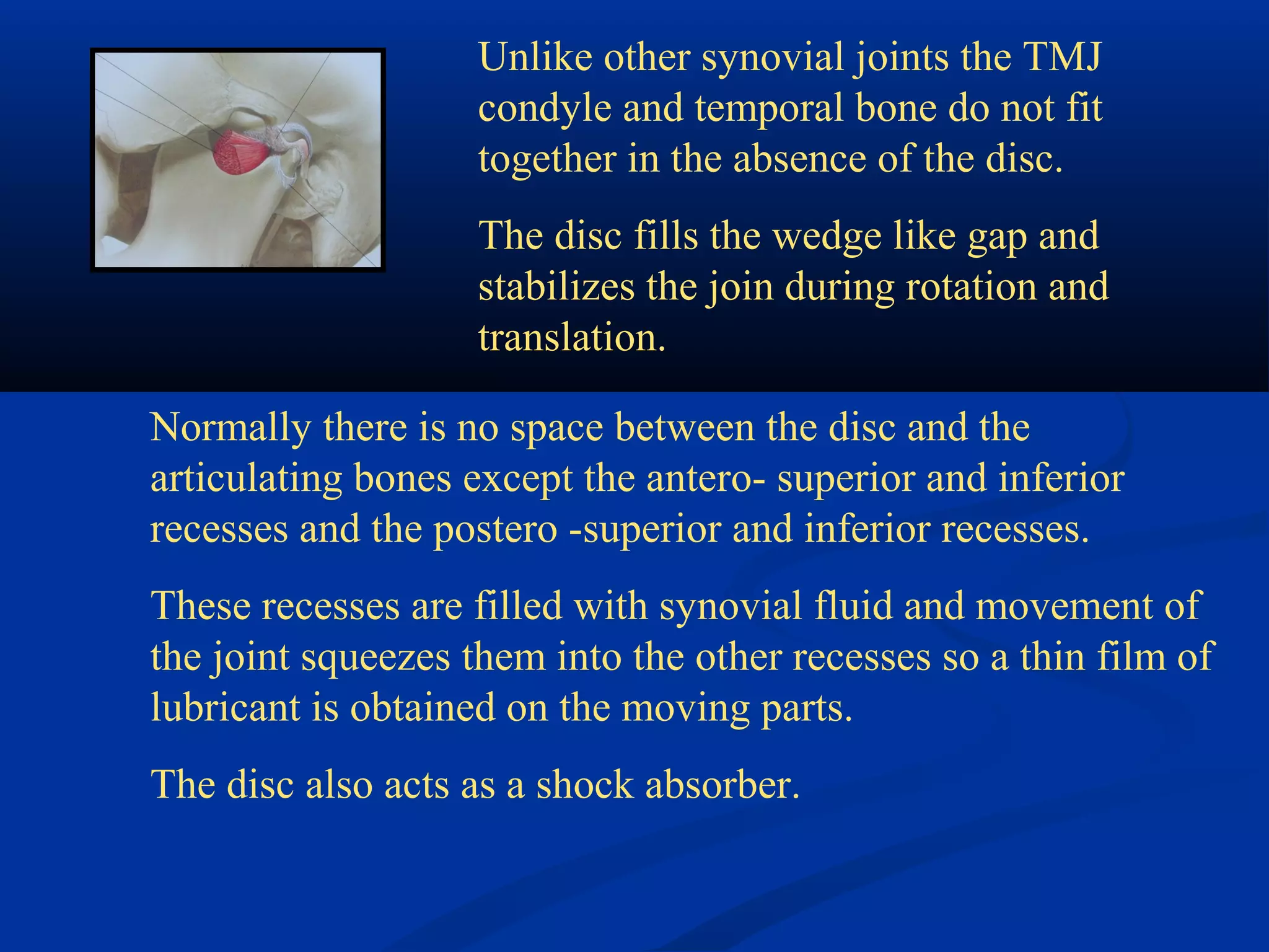
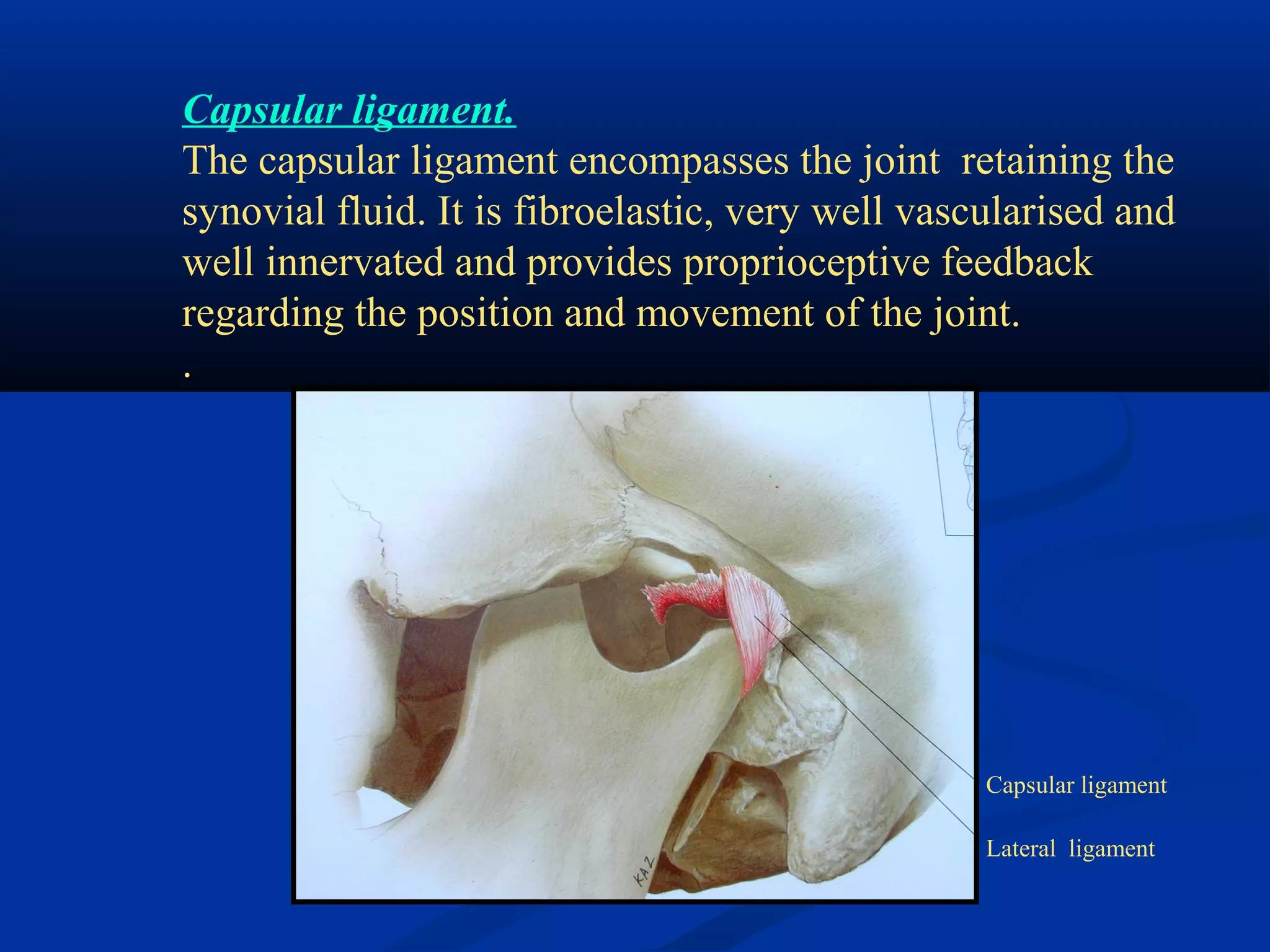
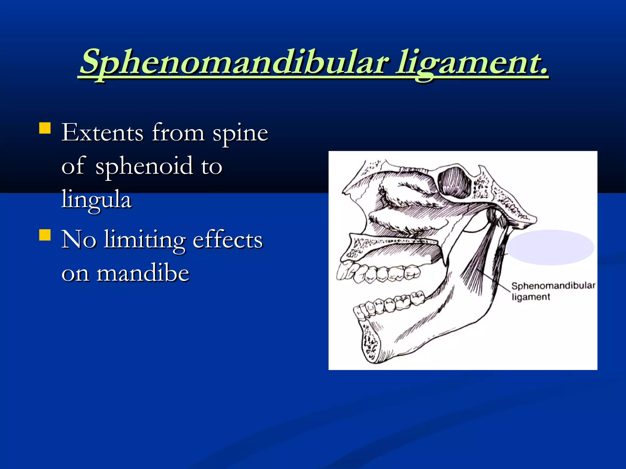
SEMINAR
ON
DEVELOPMENT & ANATOMY OF T.M.J
by
Dr. Sangeeta Poriya
MDS 1ST
YEAR
Prosthodontics
Temporomandibular joint.
 IntroductionIntroduction
 DefinationDefination
 EvolutionEvolution
 EmbryologyEmbryology
 Classification of jointsClassification of joints
 AnatomyAnatomy
 Muscles of masticationMuscles of mastication
 References.References.
INTRODUCTIONINTRODUCTION
 ““FUNCTION MUST BE UNDERSTOODFUNCTION MUST BE UNDERSTOOD
BEFORE DYSFUNCTION CAN HAVEBEFORE DYSFUNCTION CAN HAVE
MEANING”MEANING”
 Masticatory system a functional unit of bodyMasticatory system a functional unit of body
includes TMJ .includes TMJ .
 Is responsible for chewing, speaking, swallowingIs responsible for chewing, speaking, swallowing
etcetc
 It is most complex joint of body.It is most complex joint of body.
 Hence its functions should be understoodHence its functions should be understood
before resulting in its dysfunction.
TMJ DefinationTMJ Defination
 The articulation between the temporal bone andThe articulation between the temporal bone and
the mandible. It is diarthoidal,bilateral ginglymusthe mandible. It is diarthoidal,bilateral ginglymus
joint (GPT).joint (GPT).
 The area where craniomandibular articulationThe area where craniomandibular articulation
occurs is called the temporomandibular joint.occurs is called the temporomandibular joint.
 The TMJ provides hinging movement in oneThe TMJ provides hinging movement in one
plane and is considered as a ginglymoid joint.plane and is considered as a ginglymoid joint.
 It also provides gliding movements, whichIt also provides gliding movements, which
classifies it as an arthrodial jointclassifies it as an arthrodial joint
EVOLUTION OF TMJ.EVOLUTION OF TMJ.
 Primitive vertebratesPrimitive vertebrates
Collection of food led to dev of jaws along withCollection of food led to dev of jaws along with
fins.fins.
 Amphibians and reptilesAmphibians and reptiles
Greater demand on jaw mechanics to adjust toGreater demand on jaw mechanics to adjust to
new habitat.new habitat.
EVOLUTION OF TMJ.EVOLUTION OF TMJ.
 Increasing efficiency of the feeding mechanism-Increasing efficiency of the feeding mechanism-
critical factor in vertebrate evolution.critical factor in vertebrate evolution.
 Earliest functional activity of TMJ.Earliest functional activity of TMJ.
prehension Control size of mass
of food entering
alimentary tract
EVOLUTION OF TMJ.EVOLUTION OF TMJ.
 MammalsMammals
 Greater benefit from food sourceGreater benefit from food source
 Modification in jaws, joint, dentitionModification in jaws, joint, dentition
 HumansHumans
 Upright posture / bipedal locomotionUpright posture / bipedal locomotion
 Food collection shifted to jaws.Food collection shifted to jaws.
EVOLUTION OF TMJ.EVOLUTION OF TMJ.
 Primitive jaw joint –Primitive jaw joint –
reptilian jointreptilian joint
1.1. DentaryDentary
2.2. Quardate(incus)Quardate(incus)
3.3. Articulare(malleus)Articulare(malleus)
 Dentary –Dentary –
squamosalsquamosal
joint/mammalianjoint/mammalian
jaw jointjaw joint
EVOLUTION OF TMJ.EVOLUTION OF TMJ.
 Relationship of primitive jaw and cranial base.Relationship of primitive jaw and cranial base.
 AMPHISTYLIC SUSPENSIONAMPHISTYLIC SUSPENSION
 upper jaw connected to cranium.upper jaw connected to cranium.
 HYOSTYLIC SUSPENSIONHYOSTYLIC SUSPENSION
 Only lower jaw connected to cranium.Only lower jaw connected to cranium.
Behind
eye
Hyomandibular
cartilage attached
to cranium
EVOLUTION OF TMJ.EVOLUTION OF TMJ.
 STREPTOSTYLIC SUSPENSIONSTREPTOSTYLIC SUSPENSION
 Great degree of movement between upper and lowerGreat degree of movement between upper and lower
jawjaw
 Assists in swallowingAssists in swallowing
 Amphibians and reptilesAmphibians and reptiles
 Maxillary , pterygoid elements attached to theMaxillary , pterygoid elements attached to the
craniumcranium
EVOLUTION OF TMJ.EVOLUTION OF TMJ.
Development of muscles.Development of muscles.
 Advanced reptiles – capitiiAdvanced reptiles – capitii
mandibularismandibularis
 Increased functional activityIncreased functional activity
 Size of dentary bone,Size of dentary bone,
heterodont dentitionheterodont dentition
 Forces directed away fromForces directed away from
the jointthe joint
 Alteration in orientation of jawAlteration in orientation of jaw
musclesmuscles
EVOLUTION OF TMJ.EVOLUTION OF TMJ.
Mammal like reptiles - PrehensionMammal like reptiles - Prehension
 Condyle clampedCondyle clamped
by glenoidby glenoid
processesprocesses
EVOLUTION OF TMJ.EVOLUTION OF TMJ.
Carnivore - cuttingCarnivore - cutting
 Well dev caninesWell dev canines
 Coronoid – largeCoronoid – large
 Condyle encircled in fossaCondyle encircled in fossa
 Disc presentDisc present
 Masseter +temporalis –Masseter +temporalis –
well devwell dev
 No forwardNo forward
 Minimum lateralMinimum lateral
EVOLUTION OF TMJ.EVOLUTION OF TMJ.
Rodent - gnawingRodent - gnawing
 Incisors chiselIncisors chisel
shapedshaped
 Ant –postAnt –post
oriented glenoidoriented glenoid
fossafossa
 ForwardForward
+upward+upward
movementmovement
 Well devWell dev
MasseterMasseter
EVOLUTION OF TMJ.EVOLUTION OF TMJ.
Herbivore - grindingHerbivore - grinding
 Well dev molarsWell dev molars
 Ascending ramusAscending ramus
increased heightincreased height
 Condyle ovalCondyle oval
 No articular eminenceNo articular eminence
 Post glenoid processPost glenoid process
 Disc & capsule presentDisc & capsule present
 Masseter + temporalisMasseter + temporalis
well devwell dev
 Lat pterygoidLat pterygoid
 Lateral movementsLateral movements
prominentprominent
Embryology.Embryology.
 Primary Jaw jointPrimary Jaw joint
 Between Incus and MalleusBetween Incus and Malleus
 Persists till 4 months IULPersists till 4 months IUL
Embryology.Embryology.
 Secondary jaw jointSecondary jaw joint
 Between the condyle and temporal boneBetween the condyle and temporal bone
 Differs from other synovial jointsDiffers from other synovial joints
Embryology.Embryology.
TMJ - neonateTMJ - neonate
 LaxLax
 Stability –capsuleStability –capsule
 Fossa – flatFossa – flat
 Articular tubercleArticular tubercle
 absent condyle,noabsent condyle,no
fossa/tuberclefossa/tubercle
DEVELOPMENTDEVELOPMENT
• Meckel’s cartilage provide skeletal support forMeckel’s cartilage provide skeletal support for
development of lower jaw.development of lower jaw.
• The primary jaw jointThe primary jaw joint
 - Articulation of Meckel’s cartilage with- Articulation of Meckel’s cartilage with
 Incal cartilageIncal cartilage
 - exists for 4 months till cartilages ossify- exists for 4 months till cartilages ossify
 & are incorporated in middle ear.& are incorporated in middle ear.
Secondary jaw joint-TMJ begins to form at 3Secondary jaw joint-TMJ begins to form at 3
 months of gestationmonths of gestation
• Most synovial joints have appeared by 7Most synovial joints have appeared by 7thth
wk inwk in
utero as a cavitation.utero as a cavitation.
• TMJ arises from 2 widely separated centers thatTMJ arises from 2 widely separated centers that
grow towards each other.grow towards each other.
• Regions of mesenchymal condensation –Regions of mesenchymal condensation –
temporal & condylar blastemas.temporal & condylar blastemas.
• Meniscus is formed- pair of clefts develop inMeniscus is formed- pair of clefts develop in
interposed condensation of mesenchyma whichinterposed condensation of mesenchyma which
form upper and lower joint cavities.form upper and lower joint cavities.
• 12 wks in utero- condylar blastema differentiates12 wks in utero- condylar blastema differentiates
into a condylar cartilage.into a condylar cartilage.
• The cartilage is replaced by membraneous bone &The cartilage is replaced by membraneous bone &
it’s posterior part persists in the condyle – site ofit’s posterior part persists in the condyle – site of
active growth.active growth.
• Further adaptation of condylar morphology-Further adaptation of condylar morphology-
functional & environmental changes.functional & environmental changes.
• Capsule develops by condensation ofCapsule develops by condensation of
surrounding mesenchyma- isolates the joint.surrounding mesenchyma- isolates the joint.
Clefting and
formation of
joint cavity
Formation of
condylar +
temporal
blastema and
ossification
CLASSIFICATION OF JOINTSCLASSIFICATION OF JOINTS
 BASED ONBASED ON
ANATOMICALANATOMICAL
CHARACTERISTICSCHARACTERISTICS
(Structural(Structural
classification)classification)
 BASED ONBASED ON
FUNCTIONALFUNCTIONAL
CLASSIFICATION:CLASSIFICATION:
( type of movement)( type of movement)
STRUCTURAL CLASSIFICATION:
Based on presence or absence of joint cavity:
FIBROUS JOINT:
CARTILAGENOUS JOINT:
SYNOVIAL JOINT
FUNCTIONAL CLASSIFICATION:
SYNARTHROSIS: Immovable joints
AMPHIARTHROSES: Slightly movable joints
DIARTHROSIS: Freely movable joints:
Types of joints.Types of joints.
SUTURE: sutura = seam
Fibrous joint composed of thin layer of dense fibrous
connective tissue that unites bones of the skull
GOMPHOSIS: to bolt together
Cone shaped peg fits into a socket eg tooth into alveolar
bone through periodontal ligament
SYNCHRONDROSIS: syn= together ,
chondros=cartilage
Cartilaginous joint in which connective material is hyaline
cartilage eg epiphyseal plate
SYNARTHROSIS: Immovable joints
AMPHIARTHROSES: (Slightly movable joints)
SYNDESMOSIS:band /ligament
fibrous joint in which there is considerably more fibrous
connective tissue than in a suture. The fit of the bones is
not so tight. Some amount of flexible movement. EG
distal articulation between fibula and tibia
SYMPHYSIS:growing together
Cartilaginous joint in which connecting material is broad
flat disc of fibro cartilage EG intervertebral discs.pubic
symphysis
DIARTHROSISDIARTHROSIS:: Also known as synovial joints. Presence ofAlso known as synovial joints. Presence of
Synovial cavity and articular cartilage a characteristic feature.Synovial cavity and articular cartilage a characteristic feature...
(Freely movable joints.)(Freely movable joints.)
Based on types of movement:
GLIDING : eg intercarpal joint
HINGE eg: elbow joint/ ankle
CONDYLOID eg: joint between
radius and carpals
PIVOT eg: joint between atlas and
axis
SADDLE eg : joint between
carpus and thumb
BALL AND SOCKET: eg :
shoulder/ hip joint
BILATERAL SYNOVIAL
GINGLYMOID (DIARTHRODIAL )
COMPOUND JOINT
Synovial joints.Synovial joints.
 Permits significant movementPermits significant movement
 Synovial cavitySynovial cavity
 Synovial membraneSynovial membrane
 Synovial fluidSynovial fluid
 hyaline cartilagehyaline cartilage
 capsulecapsule
Synovial joints.Synovial joints.
 Uniaxial/biaxial /multiaxialUniaxial/biaxial /multiaxial
 Planar /ginglymoid /pivot/condyloid/saddle/ball andPlanar /ginglymoid /pivot/condyloid/saddle/ball and
socketsocket
 Hiltons lawHiltons law
Muscles acting upon a joint have same nerve supply asMuscles acting upon a joint have same nerve supply as
jointjoint
SYNOVIAL JOINT:
Specialized endothelial cells form a synovial lining and forms the
synovial fluid which fills both joint cavities and performs two functions:
 Medium for metabolic exchange: as the articular surfaces are
avascular
Lubricant during function:
Two mechanisms by which lubrication occurs:
BOUNDARY LUBRICATION:
primary mechanism;synovial fluid forced from one region to
another by movement of the joint itself.
WEEPING LUBRICATION:
The articular surfaces itself absorb some amount of synovial fluid
which due to the pressure during function is forced in and out of the
articular tissues and provided the medium for metabolic exchange. This
Functions of synovial fluid.Functions of synovial fluid.
 NutritionNutrition
 LubricationLubrication
1.1. Boundary lubricationBoundary lubrication
2.2. Weeping lubricationWeeping lubrication
• Minimizes frictionMinimizes friction
COMPONENTS OF THE TMJ:
CONDYLAR HEAD
GLENOID FOSSA
ARTICULAR EMINENCE
MUSCLES OF THE TMJ:
MUSCLES OF MASTICATION
SOFT TISSUE COMPONENTS:
ARTICULAR DISC
JOINT CAPSULE
LIGAMENTS
ARTERIAL AND NERVE SUPPLY TO THE JOINT
CONDYLAR HEADCONDYLAR HEAD
 The oval condylar head is shaped like a rugby ballThe oval condylar head is shaped like a rugby ball
 The lateral pole is slightly at a a lower level to the medial poleThe lateral pole is slightly at a a lower level to the medial pole
 The long axis makes a line of 140 degrees with the line joining theThe long axis makes a line of 140 degrees with the line joining the
external acoustic meatusexternal acoustic meatus
The cartilage layer is thicker
laterally and posteriorly
suggesting the growth
direction is more active in
these areas.
Anteromedially the cartilage
becomes thin early and bone
forms in this region of
attachment of the lateral
pterygoid
The glenoid fossa:
Shallow oval depression in the infratemporal area
Bone of the deepest part is quite thin and shows that this part
of the joint is not designed to play an active functional role in
the joint.
The articular
eminence:
The two slopes of the
articular eminence are
considered to be a
functional part of the
joint.
The posterior slope
resorbs with
edentulism
Muscles of mastication:
mastication is a a harmonious and skillful activity which requires the
presence and co ordination of not only the muscles of mastication but
also the supra infrahyoid muscles, and the facial muscles
Muscles of mastication.Muscles of mastication.
 Masseter.Masseter.
 Temporalis.Temporalis.
 Medial pterygoid.Medial pterygoid.
 Lateral pterygoid.Lateral pterygoid.
 Digastric.Digastric.
The Temporalis.The Temporalis.
The strongest of the
masticatory muscles
Divided into three parts
Well developed in carnivores
and animals requiring a strong
bite force
It elevates the mandible when
it contracts.
Contraction of the anterior
part raises the mandible
Contraction of the middle part
elevates and retrudes the
mandible
Temporalis
muscle
tendon
The masseter.The masseter.
Powerful elevator muscle
Superficial muscle helps in
protruding the mandible
Deep portion helps in
stabilizing the condyle against
the articular eminence when
biting in a protruded position.
Unilateral movement helps in
lateral movement of the
mandible
Well developed in ruminants
Deep portion
Superficial portion
The medial pterygoid muscle.The medial pterygoid muscle.
Originates from the
pterygoid fossa and extends
downwards backwards and
outwards to insert in the
medial side of the ramus of
the mandible forming a
sling along with the
masseter at the angle of the
mandible.
It assists in closing of the jaw and
contraction of the muscle also causes
protrusion.
The lateral pterygoid muscle.The lateral pterygoid muscle.
Superior lateral pterygoid muscle
Originates form the infratemporal
surface of the greater wing of the
sphenoid, extends almost
horizontally backward and outward
to insert on the articular capsule the
disc and the neck of the condyle.
60 to 70% of the fibres attach to the
condyle and the rest to the disc.
Plays an active role not during
opening but during closing (power
stroke)
Originates at the outer surface of
the lateral pterygoid plate and
extends backward upward and
outward to insert into the neck of
the condyle.
When there is bilateral contraction
the condyles are pulled down the
articular eminences and the
mandible is protruded.
Unilateral contraction causes a
mediotrusive movement of that
condyle and lateral movement of
the mandible to the other side
. The inferior lateral pterygoid is
active during opening in contrast
to the superior part.
Other muscles coordinatingOther muscles coordinating
Mandibular movements.Mandibular movements.
Summary ofSummary of
mandibularmandibular
movements &movements &
muscles involvedmuscles involved
Soft tissue components of the TMJ.Soft tissue components of the TMJ.
Articular disc, capsule, ligaments and muscles
ARTICULAR DISC.ARTICULAR DISC.
AttachmentsAttachments
Retrodiscal
tissue
Temporal bone
condyle
Capsular
ligament +
superior
LPM
Medially + laterally attached to the capsule which divides
joint cavity
Articular disc
Superior head of the
lateral pterygoid
Inferior head of the
lateral pterygoid
Articular Disc.
ARTICULAR DISC.ARTICULAR DISC.
ANTERIOR
BORDER
INTERMEDIATE
ZONE
POSTERIOR
BORDER
SAGITTAL
VIEW
ANTERIOR
VIEW
ARTICULAR DISC.ARTICULAR DISC.
 Articulating surfaces are covered with fibrous tissueArticulating surfaces are covered with fibrous tissue
 Synovial lining /fluidSynovial lining /fluid
Upper joint
cavity
Lower
joint cavity
ARTICULAR DISC.ARTICULAR DISC.
 Devoid of blood vessels and nerveDevoid of blood vessels and nerve
 Morphology maintainedMorphology maintained
 Flexible and adaptable to functional demandsFlexible and adaptable to functional demands
ARTICULAR DISC.
Divides the joint into two compartments
According to Rees divided into 4 parts:
Anterior band –thickened
Intermediate band- narrow and thin
Posterior band – again thick
Bilaminar zone-
upper part having ELASTIC fibres and
attaching to the posterior margin of the glenoid
fossa and the tympano squamous fissure,
forms the posterior border of the upper
compartment.
lower part: mainly collagen fibres and attached
to neck of condyle.
Posterior border of the lower compartment.
Articular disc
Joint capsule
Lateral ligament of the
TMJ
Articular disc has the shape of a laterally wide ovoid.
In frontal section the disc is wedge shaped thicker medially
and thinner laterally
Articular disc
Joint capsule
The intermediate band: It is the functional zone of the disc
Blood vessels are rarely found here in the intermediate part of
the disc
Unlike other synovial joints the TMJ
condyle and temporal bone do not fit
together in the absence of the disc.
The disc fills the wedge like gap and
stabilizes the join during rotation and
translation.
Normally there is no space between the disc and the
articulating bones except the antero- superior and inferior
recesses and the postero -superior and inferior recesses.
These recesses are filled with synovial fluid and movement of
the joint squeezes them into the other recesses so a thin film of
lubricant is obtained on the moving parts.
The disc also acts as a shock absorber.
They are made up of collagenous connective tissue which do
not stretch and do not actively participate in the normal function
They act as guide wires restricting certain movements while
permitting certain others.
They restrict movement mechanically as well as through neuro
muscular reflex activity.
Ligaments do not stretch. They can be elongated by traction
forces but once they have been elongated joint activity is usually
compromised.
LIGAMENTS:LIGAMENTS:
Ligaments of TMJ.Ligaments of TMJ.
 Collateral ligaments(Discal).Collateral ligaments(Discal).
 Capsular ligamentsCapsular ligaments
 Temporomandibular ligamentsTemporomandibular ligaments
 Sphenomandibular ligamentsSphenomandibular ligaments
 stylomandibular ligamentsstylomandibular ligaments
Functional
ligaments
Accessory
ligaments
The collateral ligaments (discal).
Attach the medial and lateral poles of the articular disc to the
poles of the condyle. They are two in number:- medial and
lateral .
They function in allowing the disc to move passively with the
condyle as it glides anteriorly and posteriorly.
They also allow the disc to be rotated anteriorly and
posteriorly on the articular surface of the condyle..
Thus these ligaments are responsible for the hinging
movements of the condyle which occurs in the lower
compartment.
Capsular ligament.
The capsular ligament encompasses the joint retaining the
synovial fluid. It is fibroelastic, very well vascularised and
well innervated and provides proprioceptive feedback
regarding the position and movement of the joint.
.
Capsular ligament
Lateral ligament
Temporomandibular ligament.Temporomandibular ligament.
 Reinforces the lateralReinforces the lateral
aspect of capsularaspect of capsular
ligamentligament
 Outer oblique portionOuter oblique portion
 Inner horizontalInner horizontal
portionportion
 OOP- PreventsOOP- Prevents
excessive dropping ofexcessive dropping of
condyle / limits extentcondyle / limits extent
of mouth openingof mouth opening
IHP
TM ligament.
It has two parts outer oblique and an inner horizontal.
The outer part extends from the articular tubercle to the neck of the
condyle
The outer oblique part has the following functions:
 It restricts excessive dropping of the condyle and therefore
limits the normal opening of the mandible.
Secondly during opening of the mouth, the condyle rotates
till this ligament becomes tight as its point of insertion is
rotated posteriorly. When it becomes taut the neck cannot
rotate any further.
This unique feature is found only in humans preventing excessive
rotation for the mandible from impinging on the vital mandibular
and retromandibular structures behind the jaw.
the inner horizontal part extends backwards from the
articular tubercle to insert into to the lateral pole of the
condyle and posterior part of the articular disc.
Function:
The inner horizontal portion protects the posterior
retrodiscal tissues from trauma and also prevents the lateral
pterygoid muscle from over lengthening.
Accessory ligaments:
Sphenomandibular ligament
Stylomandibular ligament.
It becomes taut
when the mandible is protruded
and thus limits the excessive
protrusive movements of the
mandible.
Sphenomandibular ligament.Sphenomandibular ligament.
 Extents from spineExtents from spine
of sphenoid toof sphenoid to
lingulalingula
 No limiting effectsNo limiting effects
on mandibeon mandibe
Stylomandibular ligament.Stylomandibular ligament.
 Extends from theExtends from the
styloid process tostyloid process to
the angle + postthe angle + post
border of ramusborder of ramus
 Limits excessiveLimits excessive
protrusiveprotrusive
movementmovement
Vascular supply:
Superficial temporal artery
Middle meningeal artery
Maxillary artery
Innervation:
Auriculotemporal nerve
Deep temporal and
masseteric nerve
Others : deep auricular ,
anterior tympanic,
ascending pharyngeal
VASCULAR SUPPLY
AND INNERVATION
OF THE JOINT
 Blood SupplyBlood Supply
 Branches from superficial temporal andBranches from superficial temporal and
maxillary arterymaxillary artery
 Nerve supplyNerve supply
 a. Auriculotemporal nervea. Auriculotemporal nerve
 b. Masseteric nerveb. Masseteric nerve
RefrencesRefrences
 Jeffrey P .Okeson, Management ofJeffrey P .Okeson, Management of
temporomandibular disorder and occlusion,temporomandibular disorder and occlusion,
55thth
edition;8-26; 258-74.edition;8-26; 258-74.
 Gray’s Anatomy 34Gray’s Anatomy 34thth
edition;459-61.edition;459-61.
 White & Pharoah; Oral radiology principles andWhite & Pharoah; Oral radiology principles and
interpretation,5interpretation,5thth
edition;538-49.edition;538-49.
 UdoStratmann, et al Journal of ProsthodonticUdoStratmann, et al Journal of Prosthodontic
Dentistry 2000 (3):548-54;Dentistry 2000 (3):548-54; CLINICALCLINICAL
ANATOMY AND PALPABILITY OF THEANATOMY AND PALPABILITY OF THE
INFERIOR LATERAL PTERYGOID MUSCLEINFERIOR LATERAL PTERYGOID MUSCLE
temporomandibular joint-development and anatomy

temporomandibular joint-development and anatomy

  • 1.
    SEMINAR ON DEVELOPMENT & ANATOMYOF T.M.J by Dr. Sangeeta Poriya MDS 1ST YEAR Prosthodontics
  • 2.
    Temporomandibular joint.  IntroductionIntroduction DefinationDefination  EvolutionEvolution  EmbryologyEmbryology  Classification of jointsClassification of joints  AnatomyAnatomy  Muscles of masticationMuscles of mastication  References.References.
  • 3.
    INTRODUCTIONINTRODUCTION  ““FUNCTION MUSTBE UNDERSTOODFUNCTION MUST BE UNDERSTOOD BEFORE DYSFUNCTION CAN HAVEBEFORE DYSFUNCTION CAN HAVE MEANING”MEANING”  Masticatory system a functional unit of bodyMasticatory system a functional unit of body includes TMJ .includes TMJ .  Is responsible for chewing, speaking, swallowingIs responsible for chewing, speaking, swallowing etcetc  It is most complex joint of body.It is most complex joint of body.  Hence its functions should be understoodHence its functions should be understood before resulting in its dysfunction.
  • 4.
    TMJ DefinationTMJ Defination The articulation between the temporal bone andThe articulation between the temporal bone and the mandible. It is diarthoidal,bilateral ginglymusthe mandible. It is diarthoidal,bilateral ginglymus joint (GPT).joint (GPT).  The area where craniomandibular articulationThe area where craniomandibular articulation occurs is called the temporomandibular joint.occurs is called the temporomandibular joint.  The TMJ provides hinging movement in oneThe TMJ provides hinging movement in one plane and is considered as a ginglymoid joint.plane and is considered as a ginglymoid joint.  It also provides gliding movements, whichIt also provides gliding movements, which classifies it as an arthrodial jointclassifies it as an arthrodial joint
  • 5.
    EVOLUTION OF TMJ.EVOLUTIONOF TMJ.  Primitive vertebratesPrimitive vertebrates Collection of food led to dev of jaws along withCollection of food led to dev of jaws along with fins.fins.  Amphibians and reptilesAmphibians and reptiles Greater demand on jaw mechanics to adjust toGreater demand on jaw mechanics to adjust to new habitat.new habitat.
  • 6.
    EVOLUTION OF TMJ.EVOLUTIONOF TMJ.  Increasing efficiency of the feeding mechanism-Increasing efficiency of the feeding mechanism- critical factor in vertebrate evolution.critical factor in vertebrate evolution.  Earliest functional activity of TMJ.Earliest functional activity of TMJ. prehension Control size of mass of food entering alimentary tract
  • 7.
    EVOLUTION OF TMJ.EVOLUTIONOF TMJ.  MammalsMammals  Greater benefit from food sourceGreater benefit from food source  Modification in jaws, joint, dentitionModification in jaws, joint, dentition  HumansHumans  Upright posture / bipedal locomotionUpright posture / bipedal locomotion  Food collection shifted to jaws.Food collection shifted to jaws.
  • 8.
    EVOLUTION OF TMJ.EVOLUTIONOF TMJ.  Primitive jaw joint –Primitive jaw joint – reptilian jointreptilian joint 1.1. DentaryDentary 2.2. Quardate(incus)Quardate(incus) 3.3. Articulare(malleus)Articulare(malleus)  Dentary –Dentary – squamosalsquamosal joint/mammalianjoint/mammalian jaw jointjaw joint
  • 9.
    EVOLUTION OF TMJ.EVOLUTIONOF TMJ.  Relationship of primitive jaw and cranial base.Relationship of primitive jaw and cranial base.  AMPHISTYLIC SUSPENSIONAMPHISTYLIC SUSPENSION  upper jaw connected to cranium.upper jaw connected to cranium.  HYOSTYLIC SUSPENSIONHYOSTYLIC SUSPENSION  Only lower jaw connected to cranium.Only lower jaw connected to cranium. Behind eye Hyomandibular cartilage attached to cranium
  • 10.
    EVOLUTION OF TMJ.EVOLUTIONOF TMJ.  STREPTOSTYLIC SUSPENSIONSTREPTOSTYLIC SUSPENSION  Great degree of movement between upper and lowerGreat degree of movement between upper and lower jawjaw  Assists in swallowingAssists in swallowing  Amphibians and reptilesAmphibians and reptiles  Maxillary , pterygoid elements attached to theMaxillary , pterygoid elements attached to the craniumcranium
  • 11.
    EVOLUTION OF TMJ.EVOLUTIONOF TMJ. Development of muscles.Development of muscles.  Advanced reptiles – capitiiAdvanced reptiles – capitii mandibularismandibularis  Increased functional activityIncreased functional activity  Size of dentary bone,Size of dentary bone, heterodont dentitionheterodont dentition  Forces directed away fromForces directed away from the jointthe joint  Alteration in orientation of jawAlteration in orientation of jaw musclesmuscles
  • 12.
    EVOLUTION OF TMJ.EVOLUTIONOF TMJ. Mammal like reptiles - PrehensionMammal like reptiles - Prehension  Condyle clampedCondyle clamped by glenoidby glenoid processesprocesses
  • 13.
    EVOLUTION OF TMJ.EVOLUTIONOF TMJ. Carnivore - cuttingCarnivore - cutting  Well dev caninesWell dev canines  Coronoid – largeCoronoid – large  Condyle encircled in fossaCondyle encircled in fossa  Disc presentDisc present  Masseter +temporalis –Masseter +temporalis – well devwell dev  No forwardNo forward  Minimum lateralMinimum lateral
  • 14.
    EVOLUTION OF TMJ.EVOLUTIONOF TMJ. Rodent - gnawingRodent - gnawing  Incisors chiselIncisors chisel shapedshaped  Ant –postAnt –post oriented glenoidoriented glenoid fossafossa  ForwardForward +upward+upward movementmovement  Well devWell dev MasseterMasseter
  • 15.
    EVOLUTION OF TMJ.EVOLUTIONOF TMJ. Herbivore - grindingHerbivore - grinding  Well dev molarsWell dev molars  Ascending ramusAscending ramus increased heightincreased height  Condyle ovalCondyle oval  No articular eminenceNo articular eminence  Post glenoid processPost glenoid process  Disc & capsule presentDisc & capsule present  Masseter + temporalisMasseter + temporalis well devwell dev  Lat pterygoidLat pterygoid  Lateral movementsLateral movements prominentprominent
  • 16.
    Embryology.Embryology.  Primary JawjointPrimary Jaw joint  Between Incus and MalleusBetween Incus and Malleus  Persists till 4 months IULPersists till 4 months IUL
  • 17.
    Embryology.Embryology.  Secondary jawjointSecondary jaw joint  Between the condyle and temporal boneBetween the condyle and temporal bone  Differs from other synovial jointsDiffers from other synovial joints
  • 18.
    Embryology.Embryology. TMJ - neonateTMJ- neonate  LaxLax  Stability –capsuleStability –capsule  Fossa – flatFossa – flat  Articular tubercleArticular tubercle  absent condyle,noabsent condyle,no fossa/tuberclefossa/tubercle
  • 19.
    DEVELOPMENTDEVELOPMENT • Meckel’s cartilageprovide skeletal support forMeckel’s cartilage provide skeletal support for development of lower jaw.development of lower jaw. • The primary jaw jointThe primary jaw joint  - Articulation of Meckel’s cartilage with- Articulation of Meckel’s cartilage with  Incal cartilageIncal cartilage  - exists for 4 months till cartilages ossify- exists for 4 months till cartilages ossify  & are incorporated in middle ear.& are incorporated in middle ear. Secondary jaw joint-TMJ begins to form at 3Secondary jaw joint-TMJ begins to form at 3  months of gestationmonths of gestation
  • 20.
    • Most synovialjoints have appeared by 7Most synovial joints have appeared by 7thth wk inwk in utero as a cavitation.utero as a cavitation. • TMJ arises from 2 widely separated centers thatTMJ arises from 2 widely separated centers that grow towards each other.grow towards each other. • Regions of mesenchymal condensation –Regions of mesenchymal condensation – temporal & condylar blastemas.temporal & condylar blastemas. • Meniscus is formed- pair of clefts develop inMeniscus is formed- pair of clefts develop in interposed condensation of mesenchyma whichinterposed condensation of mesenchyma which form upper and lower joint cavities.form upper and lower joint cavities.
  • 21.
    • 12 wksin utero- condylar blastema differentiates12 wks in utero- condylar blastema differentiates into a condylar cartilage.into a condylar cartilage. • The cartilage is replaced by membraneous bone &The cartilage is replaced by membraneous bone & it’s posterior part persists in the condyle – site ofit’s posterior part persists in the condyle – site of active growth.active growth. • Further adaptation of condylar morphology-Further adaptation of condylar morphology- functional & environmental changes.functional & environmental changes. • Capsule develops by condensation ofCapsule develops by condensation of surrounding mesenchyma- isolates the joint.surrounding mesenchyma- isolates the joint.
  • 23.
    Clefting and formation of jointcavity Formation of condylar + temporal blastema and ossification
  • 24.
    CLASSIFICATION OF JOINTSCLASSIFICATIONOF JOINTS  BASED ONBASED ON ANATOMICALANATOMICAL CHARACTERISTICSCHARACTERISTICS (Structural(Structural classification)classification)  BASED ONBASED ON FUNCTIONALFUNCTIONAL CLASSIFICATION:CLASSIFICATION: ( type of movement)( type of movement)
  • 25.
    STRUCTURAL CLASSIFICATION: Based onpresence or absence of joint cavity: FIBROUS JOINT: CARTILAGENOUS JOINT: SYNOVIAL JOINT FUNCTIONAL CLASSIFICATION: SYNARTHROSIS: Immovable joints AMPHIARTHROSES: Slightly movable joints DIARTHROSIS: Freely movable joints: Types of joints.Types of joints.
  • 26.
    SUTURE: sutura =seam Fibrous joint composed of thin layer of dense fibrous connective tissue that unites bones of the skull GOMPHOSIS: to bolt together Cone shaped peg fits into a socket eg tooth into alveolar bone through periodontal ligament SYNCHRONDROSIS: syn= together , chondros=cartilage Cartilaginous joint in which connective material is hyaline cartilage eg epiphyseal plate SYNARTHROSIS: Immovable joints
  • 27.
    AMPHIARTHROSES: (Slightly movablejoints) SYNDESMOSIS:band /ligament fibrous joint in which there is considerably more fibrous connective tissue than in a suture. The fit of the bones is not so tight. Some amount of flexible movement. EG distal articulation between fibula and tibia SYMPHYSIS:growing together Cartilaginous joint in which connecting material is broad flat disc of fibro cartilage EG intervertebral discs.pubic symphysis
  • 28.
    DIARTHROSISDIARTHROSIS:: Also knownas synovial joints. Presence ofAlso known as synovial joints. Presence of Synovial cavity and articular cartilage a characteristic feature.Synovial cavity and articular cartilage a characteristic feature... (Freely movable joints.)(Freely movable joints.) Based on types of movement: GLIDING : eg intercarpal joint HINGE eg: elbow joint/ ankle CONDYLOID eg: joint between radius and carpals PIVOT eg: joint between atlas and axis SADDLE eg : joint between carpus and thumb BALL AND SOCKET: eg : shoulder/ hip joint
  • 29.
  • 30.
    Synovial joints.Synovial joints. Permits significant movementPermits significant movement  Synovial cavitySynovial cavity  Synovial membraneSynovial membrane  Synovial fluidSynovial fluid  hyaline cartilagehyaline cartilage  capsulecapsule
  • 31.
    Synovial joints.Synovial joints. Uniaxial/biaxial /multiaxialUniaxial/biaxial /multiaxial  Planar /ginglymoid /pivot/condyloid/saddle/ball andPlanar /ginglymoid /pivot/condyloid/saddle/ball and socketsocket  Hiltons lawHiltons law Muscles acting upon a joint have same nerve supply asMuscles acting upon a joint have same nerve supply as jointjoint
  • 32.
    SYNOVIAL JOINT: Specialized endothelialcells form a synovial lining and forms the synovial fluid which fills both joint cavities and performs two functions:  Medium for metabolic exchange: as the articular surfaces are avascular Lubricant during function: Two mechanisms by which lubrication occurs: BOUNDARY LUBRICATION: primary mechanism;synovial fluid forced from one region to another by movement of the joint itself. WEEPING LUBRICATION: The articular surfaces itself absorb some amount of synovial fluid which due to the pressure during function is forced in and out of the articular tissues and provided the medium for metabolic exchange. This
  • 33.
    Functions of synovialfluid.Functions of synovial fluid.  NutritionNutrition  LubricationLubrication 1.1. Boundary lubricationBoundary lubrication 2.2. Weeping lubricationWeeping lubrication • Minimizes frictionMinimizes friction
  • 34.
    COMPONENTS OF THETMJ: CONDYLAR HEAD GLENOID FOSSA ARTICULAR EMINENCE MUSCLES OF THE TMJ: MUSCLES OF MASTICATION SOFT TISSUE COMPONENTS: ARTICULAR DISC JOINT CAPSULE LIGAMENTS ARTERIAL AND NERVE SUPPLY TO THE JOINT
  • 35.
    CONDYLAR HEADCONDYLAR HEAD The oval condylar head is shaped like a rugby ballThe oval condylar head is shaped like a rugby ball  The lateral pole is slightly at a a lower level to the medial poleThe lateral pole is slightly at a a lower level to the medial pole  The long axis makes a line of 140 degrees with the line joining theThe long axis makes a line of 140 degrees with the line joining the external acoustic meatusexternal acoustic meatus The cartilage layer is thicker laterally and posteriorly suggesting the growth direction is more active in these areas. Anteromedially the cartilage becomes thin early and bone forms in this region of attachment of the lateral pterygoid
  • 36.
    The glenoid fossa: Shallowoval depression in the infratemporal area Bone of the deepest part is quite thin and shows that this part of the joint is not designed to play an active functional role in the joint. The articular eminence: The two slopes of the articular eminence are considered to be a functional part of the joint. The posterior slope resorbs with edentulism
  • 37.
    Muscles of mastication: masticationis a a harmonious and skillful activity which requires the presence and co ordination of not only the muscles of mastication but also the supra infrahyoid muscles, and the facial muscles
  • 38.
    Muscles of mastication.Musclesof mastication.  Masseter.Masseter.  Temporalis.Temporalis.  Medial pterygoid.Medial pterygoid.  Lateral pterygoid.Lateral pterygoid.  Digastric.Digastric.
  • 39.
    The Temporalis.The Temporalis. Thestrongest of the masticatory muscles Divided into three parts Well developed in carnivores and animals requiring a strong bite force It elevates the mandible when it contracts. Contraction of the anterior part raises the mandible Contraction of the middle part elevates and retrudes the mandible Temporalis muscle tendon
  • 40.
    The masseter.The masseter. Powerfulelevator muscle Superficial muscle helps in protruding the mandible Deep portion helps in stabilizing the condyle against the articular eminence when biting in a protruded position. Unilateral movement helps in lateral movement of the mandible Well developed in ruminants Deep portion Superficial portion
  • 41.
    The medial pterygoidmuscle.The medial pterygoid muscle. Originates from the pterygoid fossa and extends downwards backwards and outwards to insert in the medial side of the ramus of the mandible forming a sling along with the masseter at the angle of the mandible. It assists in closing of the jaw and contraction of the muscle also causes protrusion.
  • 42.
    The lateral pterygoidmuscle.The lateral pterygoid muscle. Superior lateral pterygoid muscle Originates form the infratemporal surface of the greater wing of the sphenoid, extends almost horizontally backward and outward to insert on the articular capsule the disc and the neck of the condyle. 60 to 70% of the fibres attach to the condyle and the rest to the disc. Plays an active role not during opening but during closing (power stroke)
  • 43.
    Originates at theouter surface of the lateral pterygoid plate and extends backward upward and outward to insert into the neck of the condyle. When there is bilateral contraction the condyles are pulled down the articular eminences and the mandible is protruded. Unilateral contraction causes a mediotrusive movement of that condyle and lateral movement of the mandible to the other side . The inferior lateral pterygoid is active during opening in contrast to the superior part.
  • 44.
    Other muscles coordinatingOthermuscles coordinating Mandibular movements.Mandibular movements.
  • 45.
    Summary ofSummary of mandibularmandibular movements&movements & muscles involvedmuscles involved
  • 46.
    Soft tissue componentsof the TMJ.Soft tissue components of the TMJ. Articular disc, capsule, ligaments and muscles
  • 47.
    ARTICULAR DISC.ARTICULAR DISC. AttachmentsAttachments Retrodiscal tissue Temporalbone condyle Capsular ligament + superior LPM Medially + laterally attached to the capsule which divides joint cavity
  • 48.
    Articular disc Superior headof the lateral pterygoid Inferior head of the lateral pterygoid Articular Disc.
  • 49.
  • 50.
    ARTICULAR DISC.ARTICULAR DISC. Articulating surfaces are covered with fibrous tissueArticulating surfaces are covered with fibrous tissue  Synovial lining /fluidSynovial lining /fluid Upper joint cavity Lower joint cavity
  • 51.
    ARTICULAR DISC.ARTICULAR DISC. Devoid of blood vessels and nerveDevoid of blood vessels and nerve  Morphology maintainedMorphology maintained  Flexible and adaptable to functional demandsFlexible and adaptable to functional demands
  • 52.
    ARTICULAR DISC. Divides thejoint into two compartments According to Rees divided into 4 parts: Anterior band –thickened Intermediate band- narrow and thin Posterior band – again thick Bilaminar zone- upper part having ELASTIC fibres and attaching to the posterior margin of the glenoid fossa and the tympano squamous fissure, forms the posterior border of the upper compartment. lower part: mainly collagen fibres and attached to neck of condyle. Posterior border of the lower compartment.
  • 53.
    Articular disc Joint capsule Lateralligament of the TMJ Articular disc has the shape of a laterally wide ovoid. In frontal section the disc is wedge shaped thicker medially and thinner laterally
  • 54.
    Articular disc Joint capsule Theintermediate band: It is the functional zone of the disc Blood vessels are rarely found here in the intermediate part of the disc
  • 55.
    Unlike other synovialjoints the TMJ condyle and temporal bone do not fit together in the absence of the disc. The disc fills the wedge like gap and stabilizes the join during rotation and translation. Normally there is no space between the disc and the articulating bones except the antero- superior and inferior recesses and the postero -superior and inferior recesses. These recesses are filled with synovial fluid and movement of the joint squeezes them into the other recesses so a thin film of lubricant is obtained on the moving parts. The disc also acts as a shock absorber.
  • 56.
    They are madeup of collagenous connective tissue which do not stretch and do not actively participate in the normal function They act as guide wires restricting certain movements while permitting certain others. They restrict movement mechanically as well as through neuro muscular reflex activity. Ligaments do not stretch. They can be elongated by traction forces but once they have been elongated joint activity is usually compromised. LIGAMENTS:LIGAMENTS:
  • 57.
    Ligaments of TMJ.Ligamentsof TMJ.  Collateral ligaments(Discal).Collateral ligaments(Discal).  Capsular ligamentsCapsular ligaments  Temporomandibular ligamentsTemporomandibular ligaments  Sphenomandibular ligamentsSphenomandibular ligaments  stylomandibular ligamentsstylomandibular ligaments Functional ligaments Accessory ligaments
  • 58.
    The collateral ligaments(discal). Attach the medial and lateral poles of the articular disc to the poles of the condyle. They are two in number:- medial and lateral . They function in allowing the disc to move passively with the condyle as it glides anteriorly and posteriorly. They also allow the disc to be rotated anteriorly and posteriorly on the articular surface of the condyle.. Thus these ligaments are responsible for the hinging movements of the condyle which occurs in the lower compartment.
  • 59.
    Capsular ligament. The capsularligament encompasses the joint retaining the synovial fluid. It is fibroelastic, very well vascularised and well innervated and provides proprioceptive feedback regarding the position and movement of the joint. . Capsular ligament Lateral ligament
  • 60.
    Temporomandibular ligament.Temporomandibular ligament. Reinforces the lateralReinforces the lateral aspect of capsularaspect of capsular ligamentligament  Outer oblique portionOuter oblique portion  Inner horizontalInner horizontal portionportion  OOP- PreventsOOP- Prevents excessive dropping ofexcessive dropping of condyle / limits extentcondyle / limits extent of mouth openingof mouth opening IHP
  • 61.
    TM ligament. It hastwo parts outer oblique and an inner horizontal. The outer part extends from the articular tubercle to the neck of the condyle The outer oblique part has the following functions:  It restricts excessive dropping of the condyle and therefore limits the normal opening of the mandible. Secondly during opening of the mouth, the condyle rotates till this ligament becomes tight as its point of insertion is rotated posteriorly. When it becomes taut the neck cannot rotate any further. This unique feature is found only in humans preventing excessive rotation for the mandible from impinging on the vital mandibular and retromandibular structures behind the jaw.
  • 62.
    the inner horizontalpart extends backwards from the articular tubercle to insert into to the lateral pole of the condyle and posterior part of the articular disc. Function: The inner horizontal portion protects the posterior retrodiscal tissues from trauma and also prevents the lateral pterygoid muscle from over lengthening.
  • 63.
    Accessory ligaments: Sphenomandibular ligament Stylomandibularligament. It becomes taut when the mandible is protruded and thus limits the excessive protrusive movements of the mandible.
  • 64.
    Sphenomandibular ligament.Sphenomandibular ligament. Extents from spineExtents from spine of sphenoid toof sphenoid to lingulalingula  No limiting effectsNo limiting effects on mandibeon mandibe
  • 65.
    Stylomandibular ligament.Stylomandibular ligament. Extends from theExtends from the styloid process tostyloid process to the angle + postthe angle + post border of ramusborder of ramus  Limits excessiveLimits excessive protrusiveprotrusive movementmovement
  • 66.
    Vascular supply: Superficial temporalartery Middle meningeal artery Maxillary artery Innervation: Auriculotemporal nerve Deep temporal and masseteric nerve Others : deep auricular , anterior tympanic, ascending pharyngeal VASCULAR SUPPLY AND INNERVATION OF THE JOINT
  • 67.
     Blood SupplyBloodSupply  Branches from superficial temporal andBranches from superficial temporal and maxillary arterymaxillary artery  Nerve supplyNerve supply  a. Auriculotemporal nervea. Auriculotemporal nerve  b. Masseteric nerveb. Masseteric nerve
  • 68.
    RefrencesRefrences  Jeffrey P.Okeson, Management ofJeffrey P .Okeson, Management of temporomandibular disorder and occlusion,temporomandibular disorder and occlusion, 55thth edition;8-26; 258-74.edition;8-26; 258-74.  Gray’s Anatomy 34Gray’s Anatomy 34thth edition;459-61.edition;459-61.  White & Pharoah; Oral radiology principles andWhite & Pharoah; Oral radiology principles and interpretation,5interpretation,5thth edition;538-49.edition;538-49.  UdoStratmann, et al Journal of ProsthodonticUdoStratmann, et al Journal of Prosthodontic Dentistry 2000 (3):548-54;Dentistry 2000 (3):548-54; CLINICALCLINICAL ANATOMY AND PALPABILITY OF THEANATOMY AND PALPABILITY OF THE INFERIOR LATERAL PTERYGOID MUSCLEINFERIOR LATERAL PTERYGOID MUSCLE

Editor's Notes

  • #2 SEMINAR ON ANATOMY