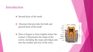
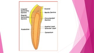
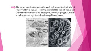
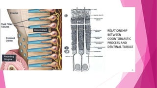
2. contents
introduction
physical properties
composition
dentinogenesis
types of dentin
formation of pulp
vasculature and lymphatic supply of pulp
innervation of dentin-pulp complex
3. clinical considerations of dentin
developmental anaomolies of dentin
clinical considerations of pulp
age changes in dentin and pulp
4. Introduction
Second layer of the tooth.
Structure that provides the bulk and
general form of the tooth
Since it begins to form slightly before the
enamel, it determines the shape of the
crown, including the cusps and ridges and
also the number and size of the roots.
5. Physically and chemically, it closely resembles bone
Said to be a living tissue since the tubules present in it contains
processes of specialized cells, the odontoblasts.
Main morphologic difference between bone and dentin is that
some of the osteoblasts that form bone marrow are enclosed
within its matrix substance as osteocytes, whereas the dentin
contains only the processes of the cells that form it.
6. Physical Properties
Color
Light yellowish in color becomes darker with age
Consistency
Elastic and resilient
Harder than bone but softer than enamel
Tensile strength :
40mpa
Compressive strength :
266mpa
8. The organic matrix of dentin is collagenous
The principle inorganic component of dentin is hydroxyapatite
crystals .The high mineral content of dentin makes it harder
than bone and cementum but softer than enamel
Lower content of mineral salts in dentin renders it more
radiolucent than enamel.
Provides resilience to the crown which is necessary to
withstand the forces of mastication.
9. Organic substances:
Type I collagenous fibrils
Type V collagenous fibrils (minor)
Non collagenous proteins:
•Dentin phosphoprotien (DPP)
•Dentin matrix protein 1 (DMP1)
•Dentin sialoprotein (DSP)
•Bone sialoprotein (BSP)
•Osteopontin, Osteocalcin
Proteoglycans
Phospholipids
Growth factors:
•Bone morphogenetic proteins (BMP)
•Insulin like growth factors (IGFs)
•Transforming growth factors β (TGF- β)
10. Inorganic substances:
Calcium hydroxy appatite crystals.
Type I collagen is the principal type of collagen found in dentine
Inorganic crystals are plate shaped and much smaller than hydroxyl
apatite crystals in enamel
Dentin also contains small amount of sulphates, phosphates and
carbonates.
11. BUD STAGE (Initiation) CAP STAGE (PROLIFERATION) BELL STAGE (HISTODIFFERENTITION)
ADVANCED BELL STAGE
(MORPHODIFFERENTIATION) ROOT FORMATION
12. DENTINOGENESIS
The process of formation of dentin is called dentinogenesis.
- The cells that form dentin – ODONTOBLASTS, derived from dental papilla
{ectomesenchymal componenet of tooth germ.}
and is directed outside inwards
- PRIMARY TEETH : Occurs around 14th week of fetal development.
- PERMANENT TEETH : 3 months old.
- AFTER THAT : continues at a very slow pace throughout the life.
14. These cells develop rich cytoplasmic organelles for protein synthesis and the nucleus shift
from the center to the basal region.
These cells form odontoblast layer that deposit dentin later.
The differentiation of odontoblasts occurs under the organizing influence of IEE
In early bell stage ,the cells of dental papilla adjacent to inner enamel epithelium align to
form a distinct layer .
Initially , these cells become cuboidal and later turn to columnar cells utilizing the
acellular space between IEE and dental papilla.
15. Hertwig’s Epithelial Root Sheath
• The development of Hertwig’s root sheath begins
with the formation of a bilayered extension of the
inner and outer dental epithelium from the cervical
loop of the enamel organ.
• Hertwig epithelial root sheath initiates the
formation of dentin in the root of a tooth by
causing the differentiation of odontoblasts from the
dental papilla.
• The root sheath eventually disintegrates with the
periodontal ligament, but residual pieces that do
not completely disappear are seen as epithelial cell
rests of Malassez (ERM).
16. Formation of dentin involves two steps :
1. Deposition of matrix which includes collagen and ground substance .
2. Mineralization .
17. Matrix deposition
Mantle dentin :
During the initial stage of dentin deposition , odontoblasts
are not grown to its full size and have space in between,
containing ground substance of dental papilla.
To this pre existing ground substance of dental papilla,
odontoblasts deposit collagen which together form the
organic matrix of the first formed dentin, referred as
“MANTLE DENTIN.”
18. COLLAGEN FIBERS – deposited : Large (0.1 -0.2 micrometer), discrete,
and arranged perpendicular to the basement membrane.
AFTER the deposition of collagen odontoblasts leave out many matrix
vesicles which help in initiation of mineralization.
As the deposition of dentin matrix proceeds the odontoblasts move
inwards pulpally, leaving behind its cytoplasmic process, referred to as
“TOMES’ PROCESS”. These cytoplasmic extensions can be seen in
mineralized dentin as odontoblast processes in dentinal tubules.
19. Circumpulpal dentin
After deposition of mantle dentin the odontoblast enlarge and get fully
differentiated, obliterating the space between them.
This makes it essential for the odontoblasts to secrete both collagen and ground
substance to form organic matrix.
These collagen fibers deposited are small diameter(50-200 nm), arranged in closely
packed bundles which are parallel to basement membrane.
Odontoblasts do not release matrix vesicles rather secrete further components such
as lipids, phosphoproteins, etc. to the matrix which may have role in
mineralization.
20. The dentin formation proceeds in the similar manner throughout the life of
the tooth.
The rate of dentin formation is around 4 microns/day till the crown
completion which slows down to 1 microns/day.
Afterwards it becomes a slow process which continues throughout a life.
21. Mineralization
For proper mineralization of dentin, three components are necessary ,
namely
COLLAGEN – forms a scaffold
NON COLLAGENOUS proteins- bind to the collagen template and
function as a mineral nucleator.
CRYSTALLINE CALCIUM PHOSPHATE deposited in an ordered
manner.
Non collagenous dentin matrix proteins 1 and 2 and dentin sialoprotein
are important ,and are capable of inducing the formation of
hydroxyapatite and inhibit mineral growth and regulate crystal size.
22. 3 types of patterns seen –
LINEAR
GLOBULAR (calcospheric calcification) – Mantle dentin and circumpulpal dentin found
just below mantle dentin. { failure of which leads to formation of interglobular dentin }
A COMBINATION OF TWO – rest of the circumpulpal dentin.
23. Types of Dentin
Dentin
Primary physiologic
dentin
Secondary physiologic
dentin
Tertiary dentin or
reparative dentin or
reactionary dentin or
irregular secondary dentin
Mantle
dentin
Circumpulpal
dentin
Peritubular
dentin
Intertubular
dentin
24. Types of Dentin
Primary dentin: is the dentin formed in a tooth before the
completion of the apical foramen of the root. Primary dentin is
noted for its regular pattern of tubules
Mantle dentin: the first predentin that forms and matures
within the tooth. It is the outermost layer of the dentin.
Circumpulpal dentin: the layer of dentin , which outlines the
pulp chamber.
Peritubular (intratubular) dentin: dentin that creates the wall
of the dentinal tubule.
Intertubular dentin: dentin found between the tubules.
25. Secondary dentin:
• It is the dentin that is formed after the completion of the
apical foramen and continues to form throughout the life
of the tooth .
•The tubules of the secondary dentin sclerose more readily
than that of primary dentin . Contains fewer dentin
compared to primary dentin
Tertiary dentin : Also referred to as reactive or reparative
dentin.
•It is produced in reaction to various stimuli , such as attrition ,
caries or a restorative dental procedure.
•The cells forming the tertiary dentin line its surface or become
included in the dentin , later it is referred as Osteodentin.
26.
27. PULP
Nutrition: blood supply for pulp and dentin.
Sensory: changes in temp., vibration and chemical that affect the
dentin and pulp.
Formative: the pulp involve in the support, maintenance and
continued formation of dentin.
Defensive: triggering of inflammatory and immune response.
Protective: Development and formation of secondary and tertiary
dentin which increase the coverage of the pulp.
The dental pulp is a soft connective tissue that supports the dentin
Functions of the Dental Pulp
28. The pulp organ is extensively vascular with vessels arising from
the external carotids to the superior or inferior alveolar arteries. It
drain by the same vein.
Blood flow is more rapid in the pulp than in most area of the
body, and the blood pressure is quite high.
The walls of the pulpal vessels become very thin as their enter
the pulp.
VASCULATURE AND LYMPHATIC SUPPLY OF PULP
29. Lymphatic vessels ; they arise as small, blind,thin-walled
vessels in the coronal region of the pulp and pass apically
through the middle and radicular regions of the pulp to exit
via one or two larger vessels through the apical foramen.
30. INNERVATION OF THE DENTIN -PULP COMPLEX
The dental pulp is innervated richly. Nerves enter the pulp
through the apical foramen, along with afferent blood vessels,
and together form the neurovascular bundle
31. The branches contribute to an extensive plexus of nerves in the
cell-free zone of Weil just below the cell bodies of the
odontoblasts in the crown portion of the tooth. This plexus of
nerves, which is called the subodontoblastic plexus of Raschkow.
.
In the root, no corresponding plexus exists.
32. The nerve bundles that enter the tooth pulp consist principally of
sensory afferent nerves of the trigeminal (fifth cranial) nerve and
sympathetic branches from the superior cervical ganglion. Each
bundle contains myelinated and unmyelinated axons
TEM SEM
33. As the nerve bundles ascend coronally, the myelinated axons
gradually lose their myelin coating so that a proportional increase
in the number of unmyelinated axons occurs in the more coronal
aspect of the tooth.
Although most of the nerve bundles terminate in the
subodontoblastic plexus as free, unmyelinated nerve endings, a
small number of axons pass between the odontoblasts and
sometimes extend into dentinal tubules .
No organized junction or synaptic relationship has been noted
between axons and the odontoblast process.
34. INNERVATION OF DENTIN
Nerve fibres were shown to accompany 30-70% of the
odontoblastic process and these are referred to as intratubular
nerves
These nerves and their terminals are found in close association
with the odontoblasts process within the tubules.
35. HISTOLOGY OF DENTIN
Microscopically, several structural features can be
identified in dentin :
o Dentinal tubules
o Peritubular dentin
o Sclerotic dentin
o Intertubular dentin
o Interglobular dentin
o Incremental growth lines
o Tomes granular layer
36. •Dentinal tubules extend through
the entire thickness of the dentin
from the DEJ.
•The tubules follow an S-shaped
path from the outer surface of the
dentin to the perimeter of the pulp
in coronal dentin.
•S- Shape curvature is least
pronounced beneath the incisal
edges and cusps.
•Dentinal tubules are larger near
the pulp and thinnest at the DEJ
37. DEAD TRACT:
Tubules in carious lesions may fill with bacteria and appear
darkly stained in histologic sections.
The processes in these tubules may disintegrate or retract ,
leaving behind an empty tubule , referred to as a dead tract.
Appears black in transmitted light and white in reflected
light.
39. PERITUBULAR DENTIN :
•The dentin that immediately surrounds the dentinal tubules
is termed as peritubular dentin
•Highly mineralised than intertubular dentin
•Twice as thick in outer dentin ( approx. 75 microns) than
inner dentin (approx 0.4 microns )
40. SCLEROTIC DENTIN :
•The dentinal tubules that have
become occluded with calcified
material.
•When this occurs in several
tubules in the same area, the
dentin assumes a glassy
appearance and becomes
translucent.
•The amount of sclerotic dentin
increases with age and is most
common in the apical third of the
root and in the crown midway
between the DEJ and the surface
of the pulp.
41. INTERTUBULAR DENTIN :
•It is considered as the main body of the dentin.Dentin located
between the dentinal tubules is called intertubular dentin
•Intertubular dentin represents the primary formative product of
the odontoblasts and consists of a tightly interwoven network of
type I collagen fibrils
•The fibrils are arranged randomly in a plane at roughly right
angles to the dentinal tubules.
•The ground substance consists of noncollagenous matrix
proteins and some plasma proteins.
42. Peritubular dentin seen in ground section by (A) light
microscopy and (B) scanning electron microscopy.The dark
central spots are empty dentinal tubules surrounded by a well-
defined collar of peritubular dentin.
43. INTERGLOBULAR DENTIN:
•Areas of unmineralized or hypomineralized dentin where
globular zones of mineralization (calcospherites) have failed to
fuse into a homogeneous mass within mature dentin .
•These areas are especially prevalent in human teeth in which the
person has had a deficiency in vitamin D or exposure to high
levels of fluoride at the time of dentin formation.
•Interglobular dentin is most common in the circumpulpal dentin
just below the mantle dentin
44. A, Ground section.
B, Demineralized section stained with hematoxylin-eosin.
C, Demineralized section stained with silver nitrate
45. INCREMENTAL LINES OF
VON EBNER:
•They appear as small lines or
striations in dentin.Run at right
angles to the dentin tubules
•These lines reflect the daily
rythmic, recurrent deposition of
dentinal matrix
•The organic matrix of primary
dentin is deposited incrementally at
a daily rate of approximately 4 µm.
46. NEONATAL LINE :
•The prenatal and postnatal dentin is separated by accentuated
contour line , termed as neonatal line.
•Seen in the decidious teeth and first permanent molars
•This line reflects the abrupt change in the environment that
occurs at birth
47. GRANULAR LAYER OF
TOMES :
•It is a zone of granules seen at the
peripheral layer of the radicular
dentin adjacent to the cementum
•It is seem to be caused by
coalescing or looping of the
terminal portion of dentinal tubules.
•Slightly increases in amount from
CEJ to the root apex
•Hypomineralised area.
48.
49. HISTOLOGY OF THE PULP
When pulp is examined histologically , it can be distinguished
into four different zones from the periphery to the centre of
the pulp :
50. Zones-from outer to inner zone Description
Odontoblastic layer Lines the outer pulpal wall and
consists of the cell bodies of
odontoblast. Secondary dentin may
form in this area from the apposition
of odontoblast.
Cell-free zone Fewer cells than odontoblastic layer.
Nerve and capillary plexus located
here
Cell-rich zone Increased density of cells as compared
to cell-free zone and also a more
extensive vascular system
Pulpal-core Located in the center of the pulp
chamber, which has many cells and an
extensice vascular supply, similar to
cell-rich zone
Microscopic Zones in Pulp
51. PRINICIPLE CELLS OF THE PULP
1) Odontoblasts
2) Fibroblasts
3) Undiffrentiated Mesenchymal Cells
4) Dental Pulp Stem Cells
5) Inflammatory Cells
6) Matrix and Ground Substance
52. 1)ODONTOBLASTS :
•These most distinctive cells of the dental pulp and the most easily
recognized.
•Odontoblasts form a layer lining the periphery of the pulp and
have a process extending into the dentin.
• The odontoblasts in the crown are larger than odontoblasts
in the root.
• In the crown of the fully developed tooth, the cell bodies of
odontoblasts are columnar and measure approximately 50 µm in
height, whereas in the midportion of the pulp, they are more
cuboid and in the apical part more flattened.
53. 2) FIBROBLAST
•The cells occurring in greatest numbers in the pulp are
fibroblasts. Fibroblasts are particularly numerous in the coronal
portion of the pulp, where they form the cell-rich zone.
•The function of fibroblasts is to form and maintain the pulp
matrix, which consists of collagen and ground substance.
•In young pulps the fibroblasts are actively synthesizing matrix
and have a plump cytoplasm and extensive amounts of all the
usual organelles associated with synthesis and secretion.
•With age the need for synthesis diminishes, and the fibroblasts
appear as flattened spindle-shaped cells with dense nuclei.
54. 3) Undiffrentiated Mesenchymal Cells
•These cells represent the pool from which connective tissue
cells of the pulp are derived.
• Depending on the stimulus, these cells may give rise to
odontoblasts and fibroblasts.
•The cells appear as large polyhedral cells possessing a large,
lightly stained, centrally placed nucleus.
• In older pulps the number of undifferentiated mesenchymal
cells diminishes, along with the number of other cells in the
pulp core, hence reduces the regenerative potential of the pulp.
55. 4) Dental Pulp Stem Cells
•These cells have a self-renewal capability and, under
appropriate environmental conditions,can differentiate into
odontoblasts,chondrocytes,adipocytes,and neurons.
•It has also been shown that these cells have the capacity to
give rise to osteoblasts and may therefore be a promising tool
for bone regeneration.
56. 5) Inflammatory Cells
•Macrophages tend to be located throughout the pulp center.
They appear as large oval or sometimes elongated cells that
under the light microscope exhibit a dark-stained nucleus.
• Pulp macrophages, are involved in the elimination of dead
cells, the presence of which further indicates that turnover of
dental pulp fibroblasts occurs.
•In normal pulps, T lymphocytes are found, but B
lymphocytes are scarce. There are also some leukocytes
(neutrophils and eosinophils), which increase substantially
during infection.
57. 6)Matrix and Ground Substance
•The extracellular compartment of the pulp, or matrix, consists
of collagen fibers and ground substance.
•The fibers are principally type I and type III collagen.
• In young pulps, single fibrils of collagen are found scattered
between the pulp cells. Whereas the overall collagen content of
the pulp increases with age, the ratio between types I and III
remains stable .
•The greatest concentration of collagen generally occurs in the
most apical portion of the pulp.
58. CLINICAL CONSIDERATIONS OF DENTIN :
• Exposure of dentin
When1 mm of dentin is exposed, about 30,000 living cells are
damaged.
59.
60. Dentinal hypersensitivity
Dentin hypersensitivity has been defined as the pain
arising from exposed dentin, typically in response to
chemical, thermal, tactile or osmotic stimuli that
cannot be explained as arising from any other form of
dental defect or pathology
66. Infected dentin Affected dentin
Outer carious dentin
Soft
Dark brown
Large no. of microbes
Collagen is irreversibly
denatured
Cannot be remineralized
Should be removed
Inner carious dentin
Hard
Light brown
Small no. of microbes
Collagen is reversibly
denatured
Can be remineralized
Can be retained
67. Types of Dentin
Dentin
Primary physiologic
dentin
Secondary physiologic
dentin
Tertiary dentin or
reparative dentin or
reactionary dentin or
irregular secondary dentin
Mantle
dentin
Circumpulpal
dentin
Peritubular
dentin
Intertubular
dentin
68. Secondary dentin
Formed after root completion
Appears in greater amount on the roof and floor of the coronal
pulp chamber where it protects the pulp from exposure in older
teeth
Not formed in response to any external stimuli
70. Reactionary dentin
Odontoblasts become hyperplastic
and deposit excess tissue in
response to an external stimuli as
protective mechanism
Has tubular structure that is
continuous with primary and
secondary dentin
Reparative dentin
Formed in cases where external
trauma leads to death of
odontoblasts
Undifferianted mesenchymal cells
from the pulp then change into
new odontoblasts and secrete
reparative tissue called reparative
tertiary dentin
72. Clinical considerations of pulp
Reversible pulpitis
Momentary pain –short
duration
Sharp pain usually caused by
cold
Stimuli like cold food
Regresses on removal of
stimulas
Can progress into irreversible
pulpitis
Restoration
Irreversible pulpitis
Long duration pain-
prolonged pain
Dull, long lasting caused by
cold and hot food
Lingers for a while
Noctural pain
Root canal treatment
73.
74. PULP STONES
• Discrete calcified structures found in dental pulp.
• They have calcium and phosphorous similar to that of dentin
and can be seen in healthy , diseased or even unerupted tooth.
• They are found more at the orifice of the pulp chamber or
within the root canal.
75. AGE CHANGES IN DENTIN-PULP
COMPLEX
In Pulp :
• The most conspicuous
change is the decreasing
volume of the pulp chamber
and root canal brought about
by continued dentin
deposition.
76. •Another age change is the occurrence of irregular areas of
dystrophic calcification, especially in the central pulp
77. In Dentin :
•Within dentin the deposition of intratubular dentin continues,
resulting in a gradual reduction of the tubule diameter. This
continued deposition often leads to complete closure of the
tubule