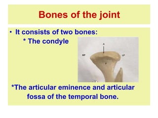
Professor of Oral Biology
Faculty of Dentistry
Mansoura University
TEMPORO-MANDIBULAR JOINT
Definition of a joint.
A joint is the location at which
two or more bones make
contact. They are constructed
to allow movement and provide
mechanical support.
Types of joints
• 1- Fibrous joints
• 2-Cartilaginous joints
• 3- Synovial joints
Fibrous joints (permit no or little movement)
Two bones connected with fibrous tissue .
Examples:
a) Suture (little or no movement).
b) Gomphosis (periodontal ligament).
c) Syndesmosis (fibula & tibia, radius & ulna and interosseous ligament between
them).
Cartilaginous joints(permit no or little movement)
Two subtypes:
a) Primary: (Bone-cartilage) e.g.: (costochondral joint).
b) Secondary: bone-cartilage-fibrous tissue-cartilage-bone.e.g.: (pubic symphysis).
Synovial joints (Permit significant movement)
Two bones covered by a hyaline cartilage
Surrounded by a capsule
Filled with synovial fluid formed by synovial membrane
Can be divided by articular disk
Ligaments are associated e.g.: (TMJ).
• IT is the area where the mandible
articulates with the cranium.
• IT IS DESCRIBED AS A COMPLEX,
MULTIAXIAL, SYNOVIAL, BICONDYLAR
AND GINGLIMOARTHROIDAL JOINT.
•The TMJ is a ginglymoarthrodial joint, a term
that is derived from ginglymus, meaning a hinge
joint, allowing motion only backward and forward
in one plane, and arthrodia, meaning a joint of
which permits a gliding motion of the surface
• TMJ IS ALSO KNOWN AS CRANIO
MANDIBULAR JOINT/ ARTICULATION
WHAT IS TEMPOROMANDIBULAR JOINT ?
Tempromandibular joint
• Bilateral synovial joint
• It is the articulation of the head of the mandible with the
articular fossa and articular eminence of the temporal
bone. The most important functions of the
temporomandibular joint (TMJ) are mastication and
speech.
TMJ is consisted of:
• 1-Bones of the joint
*Mandibular condyle
*Temporal bone……articular fossa & articular
eminence
• 2-Articular disk
• 3-Capsular ligament
• 4-Joint cavity ( synovial membrane)
Tempromandibular joint
Development of the joint
• At 12 w.i.u appearance of:
• Mesenchymal cells condensation(blastema)
followed by appearance of 2 clefts w become
upper and lower joint cavities
• Mesenchyme in between become joint disc,
surrounded by fibrous capsule.
Bones of the joint
• It consists of two bones:
* The condyle
*The articular eminence and articular
fossa of the temporal bone.
A- The condyle
1- Condylar head
• Composed of spongy bone
covered by thin layer of
compact bone.
• The trabeculae are grouped in such a
way that they radiate from the neck of
the condyle and reach cortex at right
anglegive max.strength to the
condyle.red marrow(myloid or
cellular)replaced by fatty by age.
• Marrow spaces decrease by age with
thickening of bone trabeculai.
Spongy
bone
Compact bone
Histological Structure of TMJ
Trabeculae radiate from the center of condyle and reach the surface at right angles
2-The fibrocartilage covering of the
condyle
• Its superficial layer consists of:
Network of strong collagenous f. & very occasionally
elastic f. & fibroblasts & cartilage cells
(chondrocytes) may be present ……increase in
number by age.
• The deeper layer consists of:
UMC as long as hyaline cartilage is present.
* By E/M……………….lamina splendens layer
(1-2 microns thick)
covering the fibrocartilage surface as
very smooth layer facing the joint cavity.
3-Cartilageonus Plate
During the period of growth
• Underneath the fibrous covering……… There is a
layer of hyaline cartilage which serves as an active
growth center till the age of 20 years
Fibrocartilage
Layer (fibrous
covering)
Articular surface
of mandibular condyle
(young)
Articular surface
of mandibular condyle
(adult)
B-temporal bone
• Articular eminence
• Articular (glenoid) fossa
• The articular eminence is composed of spongy bone.
• The eminence and fossa are covered by a thin layer of
compact bone.
• The fibrous layer is thin in the g.fossa and thickened
rapidly on the posterior slope of the articular eminence.
• In adult the deepest layer show thin zone of calcification.
The condyle
The disc
The articular
em.
• In this region (post. slope of
art.em.) the fibrous tissue is
arranged in 3 zones:
• inner layer - collagen fibers
perpendicular to the surface
• Intermediate transitional layer-
fibers run in complex fashion
• outer layer - fibers parallel to
the surface.
• Fibroblasts & chondrocytes
(single or gathered in groups)
form a type of
fibrocartilage..become thin
toward g.f……it disappears at
the tip of g.f……here a layer
of fibrous layer only is present
• Fibroblasts are flattened with long
processes,give appearance of endothelial
cs.
• Chondrocytes are present either isolated
or gathered in small groups,thus form
fibrocartilage in deepest layer.
Articular disk .The disc is divided the joint cavity
into upper larger compartment for gliding
movement and lower smaller compartment for
hinge movement.
The disc is oval in shape generally, saddle shape
from upper surface and concave shape from lower
surface.
The disc divided into three bands:
Anterior band – split into 2 lamellae
• -Upper lamella attached to articular eminence
• -Lower lamella attached to anterior surface of the
condylar head
• fibers of superior head of the lateral pterygoid
muscle inserted in between
• Intermediate band: –
• Thinnest central region which is avascular and
has no innervation.
• This zone is composed of fibrous connective
tissue and is devoid of cells .
• Posterior band:
• This region attaches posteriorly by a:
• superior lamella(retrodiscal pad)
• -attaches superiorly to the capsular ligament
• -highly vascular, innervated. (loose CT ,elastic fs., BV., nerves)
• inferior lamella
• -attaches to the posterior aspect of the neck of the condyle.
• -consists mainly collagenous fibers with no elastic fibers.
Joint capsule
• The capsular fibers are attached :
superiorly………to the temporal bone
inferiorly………..to the neck of condyle
• Its lateral part is thickened to
form………..temporomandibular (lateral)
ligament
• Collateral ligament: medial and lateral lig.
Between condyle and disc to be moved
as one unite
• The capsule consists of :
• Outer fibrous layer
• -dense fibrous collagenous non elastic connective tissue.
• Inner synovial layer (synovial membrane)
• - lines the inner aspect of the capsule facing the two synovial
spaces,has synovial villi.
• Ligaments:primary(lateral and collateral)
• Accessory(stylom&sphenom.)
TMJ Ligaments
Lateral view Medial view
temporo-
mandibular
ligament
spheno-
mandibular
ligament
stylo-
mandibular
ligament
stylo-
mandibular
ligament
Synovial membrane:
• 1-cellular entima(1-4 layers)
• Fiber free matrix not rest on basement m.
• Cells have no junction complex
• -fibroblast-like cells (type B) rich in RER
• -macrophage-like cells (type A) rich in lysosomes
• -intermediate cells
• 2-vascular subentima
• Loose C.T.
• -fibroblast,macrophage,mast cells,
fat cells
• -blood vessels.
Synovial
folds and villi
Articular Capsule
The joint cavity
Synovial fluid is viscous and consists
of plasma ,protein ,mucin,cells.
• Functions
• 1-To lubricate the joint surfaces;
2-As a source of nutrition for tissues lining
cavity;
3-To remove material (debris) from joint -
this is done by macrophages present in
the synovial membrane adjacent to joint
cavity.
Innervation
of TMJ:
Trigeminal n.- mandibular -
Blood Supply
of TMJ
Internal maxillary artery
Deep auricular
Superfecial temporal
Pterygoid plexus- venous drainage
Vascular plexus in the wall of the capsule- production of synovial fluid
Innervation and blood supply
Movements of the joint
• Hinge - rotatory action
Between condyle and articular disk
Inferior synovial (joint) cavity in first 20-
25mm of mouth opening
• Gliding - translatory action
Between disk and articular eminence
Superior synovial (joint) cavity
Clinical Considerations
•Bruxism
•Arthritis
•Fractures
•Structural Changes
•Disharmony in the
relation of teeth
and the TMJ
Clinical Considerations
•Myofacial Pain
Dysfunction
Syndrome
•Luxation or
Dislocation of
Temporomandibular
Joint
•Ankylosis
•Aplasia
•Hyperplasia
•Hypoplasia
Clinical Consideration
Injuries and trauma.
Traumatic occlusion.
Myofacial pain dysfunction syndrome.
Dislocation.
Popping and clicking noises.
TMJ
TMJ

TMJ

  • 1.
    Professor of OralBiology Faculty of Dentistry Mansoura University TEMPORO-MANDIBULAR JOINT
  • 2.
    Definition of ajoint. A joint is the location at which two or more bones make contact. They are constructed to allow movement and provide mechanical support.
  • 3.
    Types of joints •1- Fibrous joints • 2-Cartilaginous joints • 3- Synovial joints
  • 4.
    Fibrous joints (permitno or little movement) Two bones connected with fibrous tissue . Examples: a) Suture (little or no movement). b) Gomphosis (periodontal ligament). c) Syndesmosis (fibula & tibia, radius & ulna and interosseous ligament between them). Cartilaginous joints(permit no or little movement) Two subtypes: a) Primary: (Bone-cartilage) e.g.: (costochondral joint). b) Secondary: bone-cartilage-fibrous tissue-cartilage-bone.e.g.: (pubic symphysis). Synovial joints (Permit significant movement) Two bones covered by a hyaline cartilage Surrounded by a capsule Filled with synovial fluid formed by synovial membrane Can be divided by articular disk Ligaments are associated e.g.: (TMJ).
  • 6.
    • IT isthe area where the mandible articulates with the cranium. • IT IS DESCRIBED AS A COMPLEX, MULTIAXIAL, SYNOVIAL, BICONDYLAR AND GINGLIMOARTHROIDAL JOINT. •The TMJ is a ginglymoarthrodial joint, a term that is derived from ginglymus, meaning a hinge joint, allowing motion only backward and forward in one plane, and arthrodia, meaning a joint of which permits a gliding motion of the surface • TMJ IS ALSO KNOWN AS CRANIO MANDIBULAR JOINT/ ARTICULATION WHAT IS TEMPOROMANDIBULAR JOINT ?
  • 7.
    Tempromandibular joint • Bilateralsynovial joint • It is the articulation of the head of the mandible with the articular fossa and articular eminence of the temporal bone. The most important functions of the temporomandibular joint (TMJ) are mastication and speech.
  • 8.
    TMJ is consistedof: • 1-Bones of the joint *Mandibular condyle *Temporal bone……articular fossa & articular eminence • 2-Articular disk • 3-Capsular ligament • 4-Joint cavity ( synovial membrane)
  • 9.
  • 10.
    Development of thejoint • At 12 w.i.u appearance of: • Mesenchymal cells condensation(blastema) followed by appearance of 2 clefts w become upper and lower joint cavities • Mesenchyme in between become joint disc, surrounded by fibrous capsule.
  • 11.
    Bones of thejoint • It consists of two bones: * The condyle *The articular eminence and articular fossa of the temporal bone.
  • 12.
    A- The condyle 1-Condylar head • Composed of spongy bone covered by thin layer of compact bone. • The trabeculae are grouped in such a way that they radiate from the neck of the condyle and reach cortex at right anglegive max.strength to the condyle.red marrow(myloid or cellular)replaced by fatty by age. • Marrow spaces decrease by age with thickening of bone trabeculai. Spongy bone Compact bone
  • 13.
    Histological Structure ofTMJ Trabeculae radiate from the center of condyle and reach the surface at right angles
  • 14.
    2-The fibrocartilage coveringof the condyle • Its superficial layer consists of: Network of strong collagenous f. & very occasionally elastic f. & fibroblasts & cartilage cells (chondrocytes) may be present ……increase in number by age. • The deeper layer consists of: UMC as long as hyaline cartilage is present. * By E/M……………….lamina splendens layer (1-2 microns thick) covering the fibrocartilage surface as very smooth layer facing the joint cavity.
  • 15.
    3-Cartilageonus Plate During theperiod of growth • Underneath the fibrous covering……… There is a layer of hyaline cartilage which serves as an active growth center till the age of 20 years Fibrocartilage Layer (fibrous covering)
  • 17.
    Articular surface of mandibularcondyle (young) Articular surface of mandibular condyle (adult)
  • 18.
    B-temporal bone • Articulareminence • Articular (glenoid) fossa
  • 20.
    • The articulareminence is composed of spongy bone. • The eminence and fossa are covered by a thin layer of compact bone. • The fibrous layer is thin in the g.fossa and thickened rapidly on the posterior slope of the articular eminence. • In adult the deepest layer show thin zone of calcification. The condyle The disc The articular em.
  • 21.
    • In thisregion (post. slope of art.em.) the fibrous tissue is arranged in 3 zones: • inner layer - collagen fibers perpendicular to the surface • Intermediate transitional layer- fibers run in complex fashion • outer layer - fibers parallel to the surface. • Fibroblasts & chondrocytes (single or gathered in groups) form a type of fibrocartilage..become thin toward g.f……it disappears at the tip of g.f……here a layer of fibrous layer only is present
  • 22.
    • Fibroblasts areflattened with long processes,give appearance of endothelial cs. • Chondrocytes are present either isolated or gathered in small groups,thus form fibrocartilage in deepest layer.
  • 23.
    Articular disk .Thedisc is divided the joint cavity into upper larger compartment for gliding movement and lower smaller compartment for hinge movement.
  • 24.
    The disc isoval in shape generally, saddle shape from upper surface and concave shape from lower surface. The disc divided into three bands: Anterior band – split into 2 lamellae • -Upper lamella attached to articular eminence • -Lower lamella attached to anterior surface of the condylar head • fibers of superior head of the lateral pterygoid muscle inserted in between
  • 25.
    • Intermediate band:– • Thinnest central region which is avascular and has no innervation. • This zone is composed of fibrous connective tissue and is devoid of cells .
  • 26.
    • Posterior band: •This region attaches posteriorly by a: • superior lamella(retrodiscal pad) • -attaches superiorly to the capsular ligament • -highly vascular, innervated. (loose CT ,elastic fs., BV., nerves) • inferior lamella • -attaches to the posterior aspect of the neck of the condyle. • -consists mainly collagenous fibers with no elastic fibers.
  • 27.
  • 28.
    • The capsularfibers are attached : superiorly………to the temporal bone inferiorly………..to the neck of condyle • Its lateral part is thickened to form………..temporomandibular (lateral) ligament • Collateral ligament: medial and lateral lig. Between condyle and disc to be moved as one unite
  • 29.
    • The capsuleconsists of : • Outer fibrous layer • -dense fibrous collagenous non elastic connective tissue. • Inner synovial layer (synovial membrane) • - lines the inner aspect of the capsule facing the two synovial spaces,has synovial villi. • Ligaments:primary(lateral and collateral) • Accessory(stylom&sphenom.)
  • 30.
    TMJ Ligaments Lateral viewMedial view temporo- mandibular ligament spheno- mandibular ligament stylo- mandibular ligament stylo- mandibular ligament
  • 31.
    Synovial membrane: • 1-cellularentima(1-4 layers) • Fiber free matrix not rest on basement m. • Cells have no junction complex • -fibroblast-like cells (type B) rich in RER • -macrophage-like cells (type A) rich in lysosomes • -intermediate cells • 2-vascular subentima • Loose C.T. • -fibroblast,macrophage,mast cells, fat cells • -blood vessels.
  • 32.
  • 33.
    The joint cavity Synovialfluid is viscous and consists of plasma ,protein ,mucin,cells. • Functions • 1-To lubricate the joint surfaces; 2-As a source of nutrition for tissues lining cavity; 3-To remove material (debris) from joint - this is done by macrophages present in the synovial membrane adjacent to joint cavity.
  • 34.
    Innervation of TMJ: Trigeminal n.-mandibular - Blood Supply of TMJ Internal maxillary artery Deep auricular Superfecial temporal Pterygoid plexus- venous drainage Vascular plexus in the wall of the capsule- production of synovial fluid Innervation and blood supply
  • 35.
    Movements of thejoint • Hinge - rotatory action Between condyle and articular disk Inferior synovial (joint) cavity in first 20- 25mm of mouth opening • Gliding - translatory action Between disk and articular eminence Superior synovial (joint) cavity
  • 38.
  • 39.
    Clinical Considerations •Myofacial Pain Dysfunction Syndrome •Luxationor Dislocation of Temporomandibular Joint •Ankylosis •Aplasia •Hyperplasia •Hypoplasia
  • 40.
    Clinical Consideration Injuries andtrauma. Traumatic occlusion. Myofacial pain dysfunction syndrome. Dislocation. Popping and clicking noises.