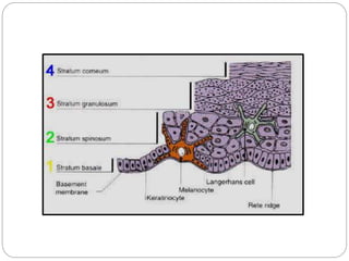
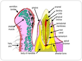
ORAL MUCOUS MEMBRANE
1
Dr. Anila K
Reader
Dept of Oral Pathology
NDC, Raichur
DEFINITION
2
 MUCOUS MEMBRANE: The moist lining of the
gastrointestinal tract, nasal passage & other body
cavities that communicate with the exteriors.
 It is defined as a moist lining of oral cavity that
communicates with the exterior.
 Called as oral mucous membrane or oral mucosa.
 Mucous: Latin word
Mean slimy or viscous
3
4
 The oral mucosa consists of two layers:
an epithelium (stratified squamous
epithelium) & an underlying layer of
connective tissue, which is the lamina
propria.
 Beneath selected areas of the oral
mucosa is a loose connective tissue, the
submucosa.
Functions of oral mucosa
Protection
Sensation
Permeability & absorption
Secretion
 Thermal regulation
Functions
 Protection: Barrier for mechanical trauma and
microbiological insults. Protects the deeper tissues and
organs from oral environment.
 Sensation: In mouth , the receptors respond to
temperature (heat and cold), touch, pain, taste buds,
thirst; reflexes such as swallowing, etching, gagging
and salivating.
PERMEABILITY & ABSORPTION:
 OMM does not have absorptive capacity.
Differences in permeability exist regionally
depending on epithelial thickness &
keratinization.
 It explains why nitroglycerin is advised
sublingually in angina.
 Secretion: Salivary secretion which contributes to
the maintenance of a moist surface. Major and
minor salivary glands helps in secretion of saliva.
 Sebaceous glands frequently present in OMM, but
secretions are insignificant. (FORDYCE
GRANULES, Buccal mucosa).
 Thermal regulation: Important in dogs not in
CLASSIFICATION OF ORAL
MUCOSA
based on function
9
 Masticatory
mucosa, keratinized stratified
squamous epithelium, found on
the hard palate and attached gingiva.
Masticatory mucosa
It covers - 25%
epithelium – keratinized.
lamina propria contains
collagenous fibres are
tightly bound to each other
and to underlying
structure.
fibres are more thicker
than lining mucosa.
e,g. gingiva & hard palate.
Smooth &shiny
Covers 65% of oral cavity.
Epithelium – non-keratinized stratified
squamous epithelium.
Lamina propria contains the typical
collagenous, elastic & reticular fibres.
These fibres are not thick & tightly
bound.
E.g. vermillion border, ventral surface
of tongue, floor of mouth, cheek.
Lining mucosa
Buccal mucosa refers to the inside lining
of the cheeks and is part of the lining
mucosa.
Labial mucosa refers to the inside lining
of the lips and is part of the lining
mucosa.
 Specialized mucosa, specifically in the regions
of the taste buds on lingual papillae on the dorsal
surface of the tongue that contains nerve
endings for general sensory reception and taste
perception
 It covers only – 15%
ACCORDING TO
KERATINIZATION:
 Keratinized epithelium---------
Orthokeratinized
Parakeratinized
 Nonkeratinized epithelium
HISTOLOGIC FEATURES
 Oral mucosa is made of- Superficial epithelium
Supporting connective
tissue
 Epithelium - May be keratinized or nonkeratinized.
Both made up of 90% keratinocytes
& 10%
non-keratinocytes
 The interface between epithelium and
connective tissue is not flat, rather irregular.
 Epithelial projections are called rete ridges,
rete pegs or epithelial ridges.
 Connective tissues projections are called
connective tissue papillae.
 E and C is separated by basement
membrane of 1-2microns.
Advantages of irregular epithelial-connective tissue
interface
 Increases the surface area of contact
 Better adhesion
 Transport of nutrients
 Dispersion of forces over greater area of
connective tissue.
 Number and configuration of rete ridges vary in
different regions of oral mucosa.
 Connective tissue beneath the epithelium is
divided into two layers:
1. Lamina propria (Corium)- Supports the
epithelium
2. Submucosa- Attaches oral mucosa to
underlying
structure
Structure of oral epithelium
 Microscopically oral epithelium shows different
layers which vary in keratinized and non-
keratinized.
 Majority of the cells have capacity to produce
keratin therefore called – keratinocytes.
 Unique features :
1. Keratin tonofilaments as a component of
cytoskeleton
Keratinized Epithelium
 Light microscopic
structure: Various cell
layers are :
 Stratum basal
 Stratum spinosum
 Stratum granulosum
 Stratum corneum
Stratum basal
Single layer of columnar cells rest on the
basement membrane.
Site of cell division (stratum germinativum)
Consist of Progenitor & maturing cell
population which appear same microscopically.
Basophilic cytoplasm, centrally placed
hyperchromatic nucleus occupying 1/3rd of
cytoplasm.
Arranged perpendicular to basement
membrane.
 Stratum spinosum:
12 to 22 layers of polyhedral cells above basal
layer.
Decreased basophilia in cytoplasm
Cells are larger than basal cells, centrally placed
round or ovoid nucleus.
Nuclear cytoplamic ratio is 1:6.
Prickle cell layer.
Shrinkage of cells away from each other during
histologic preparation & remaining in contact only
at desmosomal attachments together are
responsible for prickle appearance.
Stratum granulosum
Layer of flat cells above the prickle cell layer.
Cytoplasm is filled with basophilic granules
called keratohyline granules.
Hence the name granulosum.
Nucleus is flattened with long axis parallel to
outer surface of epithelium.
In parakeratinized epithelium, granules in
granular cell layer is less prominent.
Stratum corneum:
Composed of extremely flat dehydrated cells.
 The cells are very flat, devoid of nuclei, &
full of keratin filaments surrounded by a
matrix.
 Nucleus undergoes degeneration.
 If nucleus is completely absent-
orthokeratinization
 Pyknotic nucleus is retained-
parakeratinization.
 These surface cells are continually being
sloughed & are replaced by the continual
migration of cells from the underlying layers-
desquamation.
 Layers of oral mucosa
40
28.03.2013
dr amrutha
41
42
43
44
45
Ultra structure or electron microscopic
structure
 Stratum Basale:
 Basal cells contain nucleus occupying 1/3rd of the
cells with evenly distributed chromatin and 2-
3nucleoli.
 Involved in protein synthesis.
 Cytoplasm has rich cell organelles like RER,
Mitochondria, golgi complex, few lysosomes etc.
 Attached each other desmosomes.
 Attached to basement membrane by
hemidesmosomes.
Cells possess keratin intermediate filament or
tonofilament of 7.5 to 10 nm diameter.
48
Stratum spinosum:
 Nucleus has evenly distributed chromatin and 2-
3nucleoli.
 Cytoplasm is rich in organelles.
 Concentration of tonofilaments increases and
arranged to form bundles.
 Cells are attached with desmosomes.
 Number and width of desmosomes is more in
keratinized epithelium.
 Upper part of spinous cell layer – new cytoplasmic
organelles called odland bodies are seen.
 Also known as membrane coating granules,
keratinosomes, microphages or cementosomes.
 Odland bodies are ovoid membrane bound
organelles containing series of parallel internal
lamellae alternate electron lucent and electron
dense bands.
 Derived from golgi bodies.
Granular cell layer
 Cell and nucleus are flatter and long axis parallel to
the epithelial surface.
 Decrease in the number of cytoplasmic organelles.
 Amount of tonofilaments is more.
 Odland bodies fuse with the superficial cell
membrane and discharge contents into intercellular
spaces.
 Lipid rich permeability barrier is formed that limits
the movement of substances.
 Cytoplasm shows heavy keratohyline granules.
 Variable in size ranging from 0.1-1.5 microns.
 They are angular or irregular, hence thought to
be synthesized by ribosomes.
 Contain sulphur rich proteins, fillagrin, and
loricrin which provide an embedding matrix for
tonofilaments.
 Help in aggregating tonofilaments.
Stratum corneum
 Cellular organelles are almost completely lost.
 The nucleus may be lost or remain pyknotic.
 Cell membrane is thickened.
 Desmosomes become less distinct.
 Large amount of bundles of keratin tonofilaments
are found to be embedded in a matrix to from
thick keratin.
 Keratin is a tough insoluble protein which is more
or less completely fills the interior of shrunken
cells.
 Superficial cells tend to degenerate resulting in
desquamation of cells.
57
58
 Nonkeratinized epithelium
- The stratum basale,
- The stratum intermedium,
- The stratum superficiale.
STRATUM BASALE
59
 Cells are cuboidal or low columnar.
 Cells form a single layer resting on the
basement membrane at the interface of the
epithelium & lamina propria.
 The basal cells show the most mitotic activity.
Basophilic cytoplasm, centrally placed
hyperchromatic nucleus occupying 1/3rd of
cytoplasm.
Basophilia is due to high RNA content.
Nucleus is arranged perpendicular to
basement membrane.
 Involved in protein synthesis and therefore
cytoplasm contains rich organelles like
RER, mitochondria, golgi complex.
Attached to each other by desmosomes.
Attached to basement membrane by
hemidesmosomes.
Cells possess keratin intermediate filament
or tonofilament but less in number.
STRATUM INTERMEDIUM
62
 Is several cells thick.
 The cells are polyhedral shaped.
 Cytoplasm takes up eosinophilic stain.
 Cells are larger than basal cells and
centrally placed round nucleus.
 Prickly appearance is less distinct.
 Nucleus has evenly distributed chromatin
and 2-3 nucleoli.
 Concentration of tono-filaments increases
but dispersed & separate and hence found
in unbundled form.
 Cells are attached with desmosomes.
 Number and width of desmosomes is less
in nonkeratinized epithelium.
 In nonkeratinized epithelium, odland bodies
appear as spherical membrane bound
organelles.
 Electron dense core in center, from which
delicate radiating strands are observed (no
lamellae).
 These bodies may have similar function
here but contents have different lipid
Cells contain glycogen
Keratohyalin granules are rare.
If present they appear as regular
spherical structure surrounded with
ribosome but not associated with
tonofilaments.
Stratum superficiale
 Composed of few layers of flattened cells.
 Cellular organelles decrease in number but
still present .
 Desmosomes become less distinct.
 Large amount of keratin tonofilaments are
found dispersed in unbundled form.
 Nucleus is still retained
 Superficial cells tend to degenerate
resulting in desquamation of cells.
 Cells form a surface that is flexible &
tolerant to compression & distention
 Also called stratum distendum,
reflecting its mechanical flexibility.
Nonkeratinocytes in Oral Epithelium
 Constitute 10% of epithelial cell population.
 Present in both keratinized & non-keratinized
epithelium.
 As they lack desmosomes (except Merkel cell),
during histologic processing the cytoplasm shrinks
around the nucleus to produce a clear halo.( clear-
lack keratin intermediate filaments) .
 So these cells are called “Clear cells”.
 All cells lack tonofilaments.
Types of non-keratinocytes
 Melanocytes
 Merkel cell
 Langherhans cell
 Lymphocyte
Melanocyte:
 Origin- Neural crest cells
 Migrate to connective tissue in 8th week of IUL &
enter the epithelium at about 11th week of
gestation.
 These cells have long, thin extensions of the
cytoplasmic membrane, called dendrites.
 These are self reproducing population of
dendritic cells which lack desmosomes &
 Contain ovoid melanin containing organelles
(melanosomes) in dendrites of cell.
 Located in basal layer of oral epithelium and
stained by silver stain.
Langerhans cells:
 Previously thought to be ageing melanocytes
 Now consider as macrophages which originate
from bone marrow.
 Migrate to epithelium in 16th to 20th week of IUL.
Capable of limited divisions in epithelium
 These are dendritic cells with no desmosome or
tonofilaments. Present in suprabasal cell layer
78
• Characterized ultra structurally by small rod
or flask shaped, or racquet shaped
longitudinally striated granules called
Birbeck granules.
• Are antigen presenting cells.
• They engulf antigens from the external
environment & the intracellular lysosomes
split the antigens into peptide components.
• These fragments are then transferred to T-
Birbeck granules (Inset)
Merkel cell:
 These are modified keratinocytes.
 Located in basal layer of epithelium.
 Previously thought to arise from neural crest cells
 Recent evidences indicate epithelial origin (may
arise from division of a epithelial cell i.e.
keratinocyte).
 It has few tonofilaments & also contacts adjacent
cells through desmosomes. So Merkel cell does
not always resemble other clear cell in histologic
 Nucleus is deeply invaginated
 Ultra structurally fine filaments are present,
numerous mitochondria & 80-180 nm electron
dense granules at basal aspect of cell may be
observed.
 These granules may liberate a transmitter
substance across the junction between merkel
cell & nerve fibres.
 It suggests that merkel cell are sensory &
Inflammatory cells (lymphocyte):
 In clinically normal mucosa, a number of
inflammatory cells are often seen.
 These cells do not reproduce in epithelium.
 The cell most commonly seen in epithelium is
lymphocyte although PMNLs & mast cells are
also present.
 Posses no desmosome, no tonofilaments,
with large circular nucleus & scanty
cytoplasm.
 Lining mucosa is non- keratinized mucosa and
includes
 Labial mucosa
 Buccal mucosa
 Alveolar mucosa
 Vestibular mucosa
 Floor of the mouth
 Ventral surface of tongue
 Soft palate.
LINING MUCOSA
Labial and Buccal mucosa
 Epithelium is thick, non keratinized stratified
squamous, of about 0.5mm thickness.
 Rete-ridges are short and irregular.
 Lamina propria is thick.
 Dense fibrous connective tissue.
 Rich vascular supply and anastomosing
capillaries.
 Submucosa has dense connective tissue with fat,
minor salivary gland and sometimes sebaceous
glands.
 Mucosa is firmly attached to underlying muscle by
collagen and elastin.
Vestibular and Alveolar mucosa
 Vestibular mucosa lines the v shaped
vestibules.
 Alveolar mucosa appears reddish, lines the
alveolar bone.
 Loosely attached to underlying structures.
 Thin non-keratinized epithelium with
relatively flat epithelial connective tissue
junction.
 Small rete-ridges and sometimes
connective tissue papillae may be absent.
Ventral surface of tongue and floor of the
mouth
 Epithelium of floor of the mouth is very thin
(0.1mm), non keratinized type.
 Short, cylindrical papillae are present which are
widely separated.
 Extensive vascular supply with short anatomizing
capillary loops helps in rapid absorption of
medicines.
 In submucosa loose fibrous connective tissue
with fat and minor salivary gland is present.
 Ventral surface of tongue has thin and irregular
submucosa, may contain fat, muscle and small
vessels.
SOFT PALATE
98
 Is covered by lining mucosa, which is
more pink than the hard palate.
 Non-keratinized stratified squamous
epithelium, shows presence of taste buds.
 Lamina propria is highly vascular.
 Epithelial connective tissue interface is
irregular with short rete-ridges.
 A layer of elastic fibers separates the
lamina propria from the underlying
submucosa.
 Submucosa has minor salivary glands.
VERMILION BORDER
 It is the junction between the skin & mucous
membrane.
 It is the transitional zone between the skin
covering the external surface of lip and labial
mucosa lining inner aspect.
 Skin is composed of keratinized stratified
squamous epithelium with all appendages like
hair follicles, sweat glands and sebaceaous
100
 The labial mucosa is lined by non-
keratinized stratified squamous epithelium.
 Connective tissue shows minor salivary
glands.
 Transitional zone- thin lining with mild
keratinization.
 Central most region shows orbicularis oris
muscles.
 Ectopic sebaceous glands are seen in the
vermillion border at the corners of the
mouth or more laterally in the cheeks
101
 Long connective tissue papilae.
 Connective tissue characteristically devoid of
glands which cause mucosa to dry up
Hard palate
 Forms the roof of oral cavity.
 Has bony support.
 Macroscopically divided into different zones
1. Gingival zone: peripheral portion of hard palate
adjacent to teeth.
2. Mid palatine raphae: a narrow zone in the
midline of the hard palate extending antero-
posteriorly. Appears depressed compared to
 Incisive papilla: it is an oval prominence seen
at the extreme anterior region of palate
immediately behind the maxillary central
incisors. It covers the opening of incisive canal.
 Anterolateral region (fatty area-adipose):
between raphae and gingiva. Containing much
of fat tissue in submucosa.
 Posterolateral region (glandular region):
between raphae and gingiva. Containing much
DIAGRAM OF REGIONS OF
PALATE.
106
Palatal rugae
Transverse
ridges
•Role in
suckling in
infants.
•Helps in
backward
movement of
food during
mastication.
Microscopically
 Covered by thick orthrokeratinized stratified
squamous epithelium.
 Average thickness is 0.25mm.
 Lamina propria has long papillae with narrow,
blunt, dome shaped tips.
 Thick collagen bundles specially under the
rugae area.
 Moderate vascularity with short capillary loops.
110
 In the anterior lateral regions of the hard palate,
the submucosa contains fatty tissue.
 The lateral regions of the posterior parts contain
the palatine glands, which extend posteriorly into
the soft palate.
 These glands are purely mucous glands
containing only mucous acini.
 The median raphae has no submucosa & there
is only dense fibrous attachment to the
underlying bone.
Gingiva
 Gingiva is that part of masticatory mucosa
that covers the alveolar process of jaws and
surrounds the cervical portion of teeth.
Macroscopically
 Free gingiva is part of gingiva that forms
collar of tissue around the tooth and not
attached to tooth.
 Also called as marginal gingiva.
Gingiva
 Interdental gingiva also called as papillary
gingiva is part of gingiva that fills the
interproximal spaces.
 The interdental COL is valley buccolingually,
the depression of interdental papilla
between the facial and lingual papillae that
conforms the shape of contact area.
 Attached gingiva is the band of keratinized
mucosa that is firmly bound to the underlying
bone. It is present between the free gingival
margin and more movable alveolar mucosa
 Width is around 2-5mm.
 Gingival sulcus is the space or potential
space between the tooth surface and the
narrow unattached cervical margin of gingiva
that is free gingiva.
 It is lined with sulcular epithelium and extends
from free gingival margin to the junctional
epithelium. Average depth is 0.69 mm, range
is 0.5 – 1mm.
 Free gingival groove separates the attached
gingiva from marginal gingiva. It is present in
about 1/3rd of the adults.
 Colour of gingiva generally is coral pink, scalloped
shape surrounding the tooth in a collar like
fashion.
 Attached gingiva is stippled as a result of traction
of fiber bundles which are tightly bound to the
underlying periosteum.
 This disappears in inflammation (oedema).
Microscopically
 Epithelium: more commonly thick parakeratinized
stratified squamous epithelium( 0.25mm thick).
 Oral region of Gingiva (outer portion), sulcular
epithelium, junctional epithelium.
 Oral region of Gingiva (outer portion):
Parakerationized stratified squamous epithelium.
Degree of gingival keratinisation diminishes with
age and the onset of menopause.
 Sulcular epithelium:
Thin non-keratinized
 Junctional epithelium:
Junctional epithelium
 Junction between tooth and gingiva –
dentogingival junction
 Part of the gingiva that serve to attach
gingiva to the tooth- junctional epithelium
 Epithelium has layer of fatten squamous
cells parallel to the tooth surface.
 It is an immature epithelium because it lacks
terminal differentiation.
 The cells have high amount of rER, golgi
body, and few tonofilaments.
 Lesser desmosomal density (4 times less
than gingival epithelium)
 Epithelium is attached to the tooth by
hemidesmosome followed by lamina
lucida and lamina dense.
 Epithelium connective tissue junction is
flat.
 Connective tissue is made up of
inflammatory cells and deep connective
tissue of periodontal ligament.
Tongue
 Dorsal surface of tongue is covered by a
functionally masticatory mucosa but which is
also highly extensible.
 Has different types of lingual papillae, some
of which have mechanical function and other
gustatory function by virtue of presence of
taste buds.
Tongue
 It is divided into two parts by a V shaped
groove known as sulcus terminalis.
 Anterior 2/3rds or papillary portion or body of
the tongue contains lingual papillae.
 Posterior one third is lymphatic portion or
base of the tongue contains lingual tonsil.
 The circumvallate papillae are large papillae
surrounded by a deep sulcus or circular groove into
which the ducts of minor salivary gland (serous
gland of von Ebner) open.
 They are 8-12 in number, adjacent and anterior to
sulcus terminalis. Concerned with bitter taste.
 Foliate papillae are leaf like that may be present on
the lateral margins of the posterior part of the
tongue.
 More in other mammals than in humans.
 Papillae consists of 4-11 parallel ridges which
alternate with deep grooves.
DIAGRAM OF TONGUE, WITH
SPECIALIZED MUCOSA SHOWN
ON DORSUM OF TONGUE
125
 Fungiform papillae are mushroome like
papillae that are scattered in the anterior
2/3rd of tongue between the numerous
filliform papillae at the tip of tongue and at
the lateral borders.
 Smooth round and red.
 Concerned with sweet taste at the tip and
salty taste at border.
 Filliform papillae are hair like that cover the
entire 2/3rd of tongue and form a tough
abrasive surface.
 Cone shaped structures without taste bud.
Microscopic features
 Epithelium of circumvallate papillae on superior
surface is keratinized but on lateral borders
nonkeratinized with taste buds.
 Lamina propria has cylindrical connective tissue
core of 2mm diameter.
 Papillary projections into keratinized epithelium of
120 s in length.
 Submucosa is not distinct.
 Direct contact with perimysium of lingual
musculature is present.
 Serous glands of von Ebner is the main source of
salivary lipase, present in lamina propria.
 Foliate papillae are covered by non
keratinized epithelium with taste buds in its
lateral walls.
 Lamina propria is cylindrical connective
tissue core, papillary projections are
superficial.
 Fungiform papillae have non keratinized
epithelium with taste buds on superior
surface.
 Highly vascular cylindrical connective tissue
core so appear red.
 Filliform papillae have keratinized epithelium
with no taste buds.
Oral-Mucous-Membrane.pptx

Oral-Mucous-Membrane.pptx

  • 1.
    ORAL MUCOUS MEMBRANE 1 Dr.Anila K Reader Dept of Oral Pathology NDC, Raichur
  • 2.
    DEFINITION 2  MUCOUS MEMBRANE:The moist lining of the gastrointestinal tract, nasal passage & other body cavities that communicate with the exteriors.  It is defined as a moist lining of oral cavity that communicates with the exterior.  Called as oral mucous membrane or oral mucosa.  Mucous: Latin word Mean slimy or viscous
  • 3.
  • 4.
    4  The oralmucosa consists of two layers: an epithelium (stratified squamous epithelium) & an underlying layer of connective tissue, which is the lamina propria.  Beneath selected areas of the oral mucosa is a loose connective tissue, the submucosa.
  • 5.
    Functions of oralmucosa Protection Sensation Permeability & absorption Secretion  Thermal regulation
  • 6.
    Functions  Protection: Barrierfor mechanical trauma and microbiological insults. Protects the deeper tissues and organs from oral environment.  Sensation: In mouth , the receptors respond to temperature (heat and cold), touch, pain, taste buds, thirst; reflexes such as swallowing, etching, gagging and salivating.
  • 7.
    PERMEABILITY & ABSORPTION: OMM does not have absorptive capacity. Differences in permeability exist regionally depending on epithelial thickness & keratinization.  It explains why nitroglycerin is advised sublingually in angina.
  • 8.
     Secretion: Salivarysecretion which contributes to the maintenance of a moist surface. Major and minor salivary glands helps in secretion of saliva.  Sebaceous glands frequently present in OMM, but secretions are insignificant. (FORDYCE GRANULES, Buccal mucosa).  Thermal regulation: Important in dogs not in
  • 9.
  • 10.
     Masticatory mucosa, keratinizedstratified squamous epithelium, found on the hard palate and attached gingiva.
  • 11.
    Masticatory mucosa It covers- 25% epithelium – keratinized. lamina propria contains collagenous fibres are tightly bound to each other and to underlying structure. fibres are more thicker than lining mucosa. e,g. gingiva & hard palate.
  • 12.
    Smooth &shiny Covers 65%of oral cavity. Epithelium – non-keratinized stratified squamous epithelium. Lamina propria contains the typical collagenous, elastic & reticular fibres. These fibres are not thick & tightly bound. E.g. vermillion border, ventral surface of tongue, floor of mouth, cheek. Lining mucosa
  • 13.
    Buccal mucosa refersto the inside lining of the cheeks and is part of the lining mucosa. Labial mucosa refers to the inside lining of the lips and is part of the lining mucosa.
  • 15.
     Specialized mucosa,specifically in the regions of the taste buds on lingual papillae on the dorsal surface of the tongue that contains nerve endings for general sensory reception and taste perception  It covers only – 15%
  • 17.
    ACCORDING TO KERATINIZATION:  Keratinizedepithelium--------- Orthokeratinized Parakeratinized  Nonkeratinized epithelium
  • 18.
    HISTOLOGIC FEATURES  Oralmucosa is made of- Superficial epithelium Supporting connective tissue  Epithelium - May be keratinized or nonkeratinized. Both made up of 90% keratinocytes & 10% non-keratinocytes
  • 19.
     The interfacebetween epithelium and connective tissue is not flat, rather irregular.  Epithelial projections are called rete ridges, rete pegs or epithelial ridges.  Connective tissues projections are called connective tissue papillae.  E and C is separated by basement membrane of 1-2microns.
  • 21.
    Advantages of irregularepithelial-connective tissue interface  Increases the surface area of contact  Better adhesion  Transport of nutrients  Dispersion of forces over greater area of connective tissue.  Number and configuration of rete ridges vary in different regions of oral mucosa.
  • 22.
     Connective tissuebeneath the epithelium is divided into two layers: 1. Lamina propria (Corium)- Supports the epithelium 2. Submucosa- Attaches oral mucosa to underlying structure
  • 24.
    Structure of oralepithelium  Microscopically oral epithelium shows different layers which vary in keratinized and non- keratinized.  Majority of the cells have capacity to produce keratin therefore called – keratinocytes.  Unique features : 1. Keratin tonofilaments as a component of cytoskeleton
  • 25.
    Keratinized Epithelium  Lightmicroscopic structure: Various cell layers are :  Stratum basal  Stratum spinosum  Stratum granulosum  Stratum corneum
  • 27.
    Stratum basal Single layerof columnar cells rest on the basement membrane. Site of cell division (stratum germinativum) Consist of Progenitor & maturing cell population which appear same microscopically.
  • 28.
    Basophilic cytoplasm, centrallyplaced hyperchromatic nucleus occupying 1/3rd of cytoplasm. Arranged perpendicular to basement membrane.
  • 32.
     Stratum spinosum: 12to 22 layers of polyhedral cells above basal layer. Decreased basophilia in cytoplasm Cells are larger than basal cells, centrally placed round or ovoid nucleus.
  • 33.
    Nuclear cytoplamic ratiois 1:6. Prickle cell layer. Shrinkage of cells away from each other during histologic preparation & remaining in contact only at desmosomal attachments together are responsible for prickle appearance.
  • 35.
    Stratum granulosum Layer offlat cells above the prickle cell layer. Cytoplasm is filled with basophilic granules called keratohyline granules. Hence the name granulosum. Nucleus is flattened with long axis parallel to outer surface of epithelium. In parakeratinized epithelium, granules in granular cell layer is less prominent.
  • 37.
    Stratum corneum: Composed ofextremely flat dehydrated cells.  The cells are very flat, devoid of nuclei, & full of keratin filaments surrounded by a matrix.
  • 38.
     Nucleus undergoesdegeneration.  If nucleus is completely absent- orthokeratinization  Pyknotic nucleus is retained- parakeratinization.  These surface cells are continually being sloughed & are replaced by the continual migration of cells from the underlying layers- desquamation.
  • 40.
     Layers oforal mucosa 40
  • 41.
  • 42.
  • 43.
  • 44.
  • 45.
  • 46.
    Ultra structure orelectron microscopic structure  Stratum Basale:  Basal cells contain nucleus occupying 1/3rd of the cells with evenly distributed chromatin and 2- 3nucleoli.  Involved in protein synthesis.  Cytoplasm has rich cell organelles like RER, Mitochondria, golgi complex, few lysosomes etc.
  • 47.
     Attached eachother desmosomes.  Attached to basement membrane by hemidesmosomes. Cells possess keratin intermediate filament or tonofilament of 7.5 to 10 nm diameter.
  • 48.
  • 50.
    Stratum spinosum:  Nucleushas evenly distributed chromatin and 2- 3nucleoli.  Cytoplasm is rich in organelles.  Concentration of tonofilaments increases and arranged to form bundles.  Cells are attached with desmosomes.  Number and width of desmosomes is more in keratinized epithelium.
  • 51.
     Upper partof spinous cell layer – new cytoplasmic organelles called odland bodies are seen.  Also known as membrane coating granules, keratinosomes, microphages or cementosomes.  Odland bodies are ovoid membrane bound organelles containing series of parallel internal lamellae alternate electron lucent and electron dense bands.  Derived from golgi bodies.
  • 53.
    Granular cell layer Cell and nucleus are flatter and long axis parallel to the epithelial surface.  Decrease in the number of cytoplasmic organelles.  Amount of tonofilaments is more.  Odland bodies fuse with the superficial cell membrane and discharge contents into intercellular spaces.  Lipid rich permeability barrier is formed that limits the movement of substances.
  • 54.
     Cytoplasm showsheavy keratohyline granules.  Variable in size ranging from 0.1-1.5 microns.  They are angular or irregular, hence thought to be synthesized by ribosomes.  Contain sulphur rich proteins, fillagrin, and loricrin which provide an embedding matrix for tonofilaments.  Help in aggregating tonofilaments.
  • 56.
    Stratum corneum  Cellularorganelles are almost completely lost.  The nucleus may be lost or remain pyknotic.  Cell membrane is thickened.  Desmosomes become less distinct.  Large amount of bundles of keratin tonofilaments are found to be embedded in a matrix to from thick keratin.  Keratin is a tough insoluble protein which is more or less completely fills the interior of shrunken cells.  Superficial cells tend to degenerate resulting in desquamation of cells.
  • 57.
  • 58.
    58  Nonkeratinized epithelium -The stratum basale, - The stratum intermedium, - The stratum superficiale.
  • 59.
    STRATUM BASALE 59  Cellsare cuboidal or low columnar.  Cells form a single layer resting on the basement membrane at the interface of the epithelium & lamina propria.  The basal cells show the most mitotic activity.
  • 60.
    Basophilic cytoplasm, centrallyplaced hyperchromatic nucleus occupying 1/3rd of cytoplasm. Basophilia is due to high RNA content. Nucleus is arranged perpendicular to basement membrane.
  • 61.
     Involved inprotein synthesis and therefore cytoplasm contains rich organelles like RER, mitochondria, golgi complex. Attached to each other by desmosomes. Attached to basement membrane by hemidesmosomes. Cells possess keratin intermediate filament or tonofilament but less in number.
  • 62.
    STRATUM INTERMEDIUM 62  Isseveral cells thick.  The cells are polyhedral shaped.  Cytoplasm takes up eosinophilic stain.  Cells are larger than basal cells and centrally placed round nucleus.  Prickly appearance is less distinct.
  • 63.
     Nucleus hasevenly distributed chromatin and 2-3 nucleoli.  Concentration of tono-filaments increases but dispersed & separate and hence found in unbundled form.  Cells are attached with desmosomes.  Number and width of desmosomes is less in nonkeratinized epithelium.
  • 64.
     In nonkeratinizedepithelium, odland bodies appear as spherical membrane bound organelles.  Electron dense core in center, from which delicate radiating strands are observed (no lamellae).  These bodies may have similar function here but contents have different lipid
  • 67.
    Cells contain glycogen Keratohyalingranules are rare. If present they appear as regular spherical structure surrounded with ribosome but not associated with tonofilaments.
  • 68.
    Stratum superficiale  Composedof few layers of flattened cells.  Cellular organelles decrease in number but still present .  Desmosomes become less distinct.  Large amount of keratin tonofilaments are found dispersed in unbundled form.  Nucleus is still retained  Superficial cells tend to degenerate resulting in desquamation of cells.
  • 69.
     Cells forma surface that is flexible & tolerant to compression & distention  Also called stratum distendum, reflecting its mechanical flexibility.
  • 71.
    Nonkeratinocytes in OralEpithelium  Constitute 10% of epithelial cell population.  Present in both keratinized & non-keratinized epithelium.  As they lack desmosomes (except Merkel cell), during histologic processing the cytoplasm shrinks around the nucleus to produce a clear halo.( clear- lack keratin intermediate filaments) .  So these cells are called “Clear cells”.  All cells lack tonofilaments.
  • 72.
    Types of non-keratinocytes Melanocytes  Merkel cell  Langherhans cell  Lymphocyte
  • 73.
    Melanocyte:  Origin- Neuralcrest cells  Migrate to connective tissue in 8th week of IUL & enter the epithelium at about 11th week of gestation.  These cells have long, thin extensions of the cytoplasmic membrane, called dendrites.  These are self reproducing population of dendritic cells which lack desmosomes &
  • 74.
     Contain ovoidmelanin containing organelles (melanosomes) in dendrites of cell.  Located in basal layer of oral epithelium and stained by silver stain.
  • 77.
    Langerhans cells:  Previouslythought to be ageing melanocytes  Now consider as macrophages which originate from bone marrow.  Migrate to epithelium in 16th to 20th week of IUL. Capable of limited divisions in epithelium  These are dendritic cells with no desmosome or tonofilaments. Present in suprabasal cell layer
  • 78.
    78 • Characterized ultrastructurally by small rod or flask shaped, or racquet shaped longitudinally striated granules called Birbeck granules. • Are antigen presenting cells. • They engulf antigens from the external environment & the intracellular lysosomes split the antigens into peptide components. • These fragments are then transferred to T-
  • 79.
  • 81.
    Merkel cell:  Theseare modified keratinocytes.  Located in basal layer of epithelium.  Previously thought to arise from neural crest cells  Recent evidences indicate epithelial origin (may arise from division of a epithelial cell i.e. keratinocyte).  It has few tonofilaments & also contacts adjacent cells through desmosomes. So Merkel cell does not always resemble other clear cell in histologic
  • 82.
     Nucleus isdeeply invaginated  Ultra structurally fine filaments are present, numerous mitochondria & 80-180 nm electron dense granules at basal aspect of cell may be observed.  These granules may liberate a transmitter substance across the junction between merkel cell & nerve fibres.  It suggests that merkel cell are sensory &
  • 84.
    Inflammatory cells (lymphocyte): In clinically normal mucosa, a number of inflammatory cells are often seen.  These cells do not reproduce in epithelium.  The cell most commonly seen in epithelium is lymphocyte although PMNLs & mast cells are also present.  Posses no desmosome, no tonofilaments, with large circular nucleus & scanty cytoplasm.
  • 88.
     Lining mucosais non- keratinized mucosa and includes  Labial mucosa  Buccal mucosa  Alveolar mucosa  Vestibular mucosa  Floor of the mouth  Ventral surface of tongue  Soft palate. LINING MUCOSA
  • 89.
  • 90.
     Epithelium isthick, non keratinized stratified squamous, of about 0.5mm thickness.  Rete-ridges are short and irregular.  Lamina propria is thick.  Dense fibrous connective tissue.  Rich vascular supply and anastomosing capillaries.  Submucosa has dense connective tissue with fat, minor salivary gland and sometimes sebaceous glands.  Mucosa is firmly attached to underlying muscle by collagen and elastin.
  • 92.
    Vestibular and Alveolarmucosa  Vestibular mucosa lines the v shaped vestibules.  Alveolar mucosa appears reddish, lines the alveolar bone.  Loosely attached to underlying structures.  Thin non-keratinized epithelium with relatively flat epithelial connective tissue junction.  Small rete-ridges and sometimes connective tissue papillae may be absent.
  • 95.
    Ventral surface oftongue and floor of the mouth  Epithelium of floor of the mouth is very thin (0.1mm), non keratinized type.  Short, cylindrical papillae are present which are widely separated.  Extensive vascular supply with short anatomizing capillary loops helps in rapid absorption of medicines.  In submucosa loose fibrous connective tissue with fat and minor salivary gland is present.  Ventral surface of tongue has thin and irregular submucosa, may contain fat, muscle and small vessels.
  • 98.
    SOFT PALATE 98  Iscovered by lining mucosa, which is more pink than the hard palate.  Non-keratinized stratified squamous epithelium, shows presence of taste buds.  Lamina propria is highly vascular.  Epithelial connective tissue interface is irregular with short rete-ridges.  A layer of elastic fibers separates the lamina propria from the underlying submucosa.  Submucosa has minor salivary glands.
  • 100.
    VERMILION BORDER  Itis the junction between the skin & mucous membrane.  It is the transitional zone between the skin covering the external surface of lip and labial mucosa lining inner aspect.  Skin is composed of keratinized stratified squamous epithelium with all appendages like hair follicles, sweat glands and sebaceaous 100
  • 101.
     The labialmucosa is lined by non- keratinized stratified squamous epithelium.  Connective tissue shows minor salivary glands.  Transitional zone- thin lining with mild keratinization.  Central most region shows orbicularis oris muscles.  Ectopic sebaceous glands are seen in the vermillion border at the corners of the mouth or more laterally in the cheeks 101
  • 103.
     Long connectivetissue papilae.  Connective tissue characteristically devoid of glands which cause mucosa to dry up
  • 104.
    Hard palate  Formsthe roof of oral cavity.  Has bony support.  Macroscopically divided into different zones 1. Gingival zone: peripheral portion of hard palate adjacent to teeth. 2. Mid palatine raphae: a narrow zone in the midline of the hard palate extending antero- posteriorly. Appears depressed compared to
  • 105.
     Incisive papilla:it is an oval prominence seen at the extreme anterior region of palate immediately behind the maxillary central incisors. It covers the opening of incisive canal.  Anterolateral region (fatty area-adipose): between raphae and gingiva. Containing much of fat tissue in submucosa.  Posterolateral region (glandular region): between raphae and gingiva. Containing much
  • 106.
    DIAGRAM OF REGIONSOF PALATE. 106
  • 107.
    Palatal rugae Transverse ridges •Role in sucklingin infants. •Helps in backward movement of food during mastication.
  • 108.
    Microscopically  Covered bythick orthrokeratinized stratified squamous epithelium.  Average thickness is 0.25mm.  Lamina propria has long papillae with narrow, blunt, dome shaped tips.  Thick collagen bundles specially under the rugae area.  Moderate vascularity with short capillary loops.
  • 110.
    110  In theanterior lateral regions of the hard palate, the submucosa contains fatty tissue.  The lateral regions of the posterior parts contain the palatine glands, which extend posteriorly into the soft palate.  These glands are purely mucous glands containing only mucous acini.  The median raphae has no submucosa & there is only dense fibrous attachment to the underlying bone.
  • 111.
    Gingiva  Gingiva isthat part of masticatory mucosa that covers the alveolar process of jaws and surrounds the cervical portion of teeth. Macroscopically  Free gingiva is part of gingiva that forms collar of tissue around the tooth and not attached to tooth.  Also called as marginal gingiva.
  • 112.
    Gingiva  Interdental gingivaalso called as papillary gingiva is part of gingiva that fills the interproximal spaces.  The interdental COL is valley buccolingually, the depression of interdental papilla between the facial and lingual papillae that conforms the shape of contact area.
  • 113.
     Attached gingivais the band of keratinized mucosa that is firmly bound to the underlying bone. It is present between the free gingival margin and more movable alveolar mucosa  Width is around 2-5mm.  Gingival sulcus is the space or potential space between the tooth surface and the narrow unattached cervical margin of gingiva that is free gingiva.  It is lined with sulcular epithelium and extends from free gingival margin to the junctional epithelium. Average depth is 0.69 mm, range is 0.5 – 1mm.
  • 116.
     Free gingivalgroove separates the attached gingiva from marginal gingiva. It is present in about 1/3rd of the adults.  Colour of gingiva generally is coral pink, scalloped shape surrounding the tooth in a collar like fashion.  Attached gingiva is stippled as a result of traction of fiber bundles which are tightly bound to the underlying periosteum.  This disappears in inflammation (oedema).
  • 117.
    Microscopically  Epithelium: morecommonly thick parakeratinized stratified squamous epithelium( 0.25mm thick).  Oral region of Gingiva (outer portion), sulcular epithelium, junctional epithelium.  Oral region of Gingiva (outer portion): Parakerationized stratified squamous epithelium. Degree of gingival keratinisation diminishes with age and the onset of menopause.  Sulcular epithelium: Thin non-keratinized  Junctional epithelium:
  • 120.
    Junctional epithelium  Junctionbetween tooth and gingiva – dentogingival junction  Part of the gingiva that serve to attach gingiva to the tooth- junctional epithelium  Epithelium has layer of fatten squamous cells parallel to the tooth surface.  It is an immature epithelium because it lacks terminal differentiation.  The cells have high amount of rER, golgi body, and few tonofilaments.  Lesser desmosomal density (4 times less than gingival epithelium)
  • 121.
     Epithelium isattached to the tooth by hemidesmosome followed by lamina lucida and lamina dense.  Epithelium connective tissue junction is flat.  Connective tissue is made up of inflammatory cells and deep connective tissue of periodontal ligament.
  • 122.
    Tongue  Dorsal surfaceof tongue is covered by a functionally masticatory mucosa but which is also highly extensible.  Has different types of lingual papillae, some of which have mechanical function and other gustatory function by virtue of presence of taste buds.
  • 123.
    Tongue  It isdivided into two parts by a V shaped groove known as sulcus terminalis.  Anterior 2/3rds or papillary portion or body of the tongue contains lingual papillae.  Posterior one third is lymphatic portion or base of the tongue contains lingual tonsil.
  • 124.
     The circumvallatepapillae are large papillae surrounded by a deep sulcus or circular groove into which the ducts of minor salivary gland (serous gland of von Ebner) open.  They are 8-12 in number, adjacent and anterior to sulcus terminalis. Concerned with bitter taste.  Foliate papillae are leaf like that may be present on the lateral margins of the posterior part of the tongue.  More in other mammals than in humans.  Papillae consists of 4-11 parallel ridges which alternate with deep grooves.
  • 125.
    DIAGRAM OF TONGUE,WITH SPECIALIZED MUCOSA SHOWN ON DORSUM OF TONGUE 125
  • 128.
     Fungiform papillaeare mushroome like papillae that are scattered in the anterior 2/3rd of tongue between the numerous filliform papillae at the tip of tongue and at the lateral borders.  Smooth round and red.  Concerned with sweet taste at the tip and salty taste at border.  Filliform papillae are hair like that cover the entire 2/3rd of tongue and form a tough abrasive surface.  Cone shaped structures without taste bud.
  • 129.
    Microscopic features  Epitheliumof circumvallate papillae on superior surface is keratinized but on lateral borders nonkeratinized with taste buds.  Lamina propria has cylindrical connective tissue core of 2mm diameter.  Papillary projections into keratinized epithelium of 120 s in length.  Submucosa is not distinct.  Direct contact with perimysium of lingual musculature is present.  Serous glands of von Ebner is the main source of salivary lipase, present in lamina propria.
  • 131.
     Foliate papillaeare covered by non keratinized epithelium with taste buds in its lateral walls.  Lamina propria is cylindrical connective tissue core, papillary projections are superficial.  Fungiform papillae have non keratinized epithelium with taste buds on superior surface.  Highly vascular cylindrical connective tissue core so appear red.  Filliform papillae have keratinized epithelium with no taste buds.