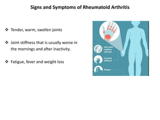
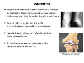
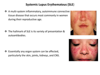
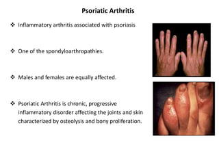
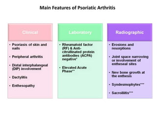
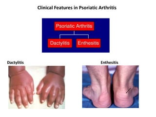
Rheumatological diseases
Rheumatic Diseases
Common Rheumatic Diseases :
 Rheumatoid arthritis (RA)
 Osteoarthritis
 Systemic lupus erythematous (SLE)
 Sjogren syndrome
 Dermatomyositis/polymyositis
 Scleroderma/systemic sclerosis
 Ankylosing spondylitis.
 Psoriatic arthritis
The Most Common Autoimmune Disease
 1% population or 3 per 10,000
 3:1 female to male
 Highest in 3rd and 4th decades
 Two peaks incidence
 Morbid/mortal disease
 Description dates to the 17th century -A New World Disease
Rheumatoid arthritis (RA)
 Rheumatoid Arthritis (RA) is a chronic inflammatory disorder that may affect
many tissues and organs, but mainly attacks the joints producing an
inflammatory synovitis.
 RA mainly affects the joints.
 May also produce inflammation in the lungs, pericardium, pleura, and sclera.
 Rheumatoid Arthritis is a chronic disease who’s pain intensity and deterioration
of joint structures progress over time often leading to deformations and
disability.
Signs and Symptoms of Rheumatoid Arthritis
 Tender, warm, swollen joints
 Joint stiffness that is usually worse in
the mornings and after inactivity.
 Fatigue, fever and weight loss
RA Diagnosis & Tests
 X Rays of hands and feet are generally performed in people with RA.
 Magnetic Resonance Imaging (MRI)
 Ultrasounds
 Blood Tests: Rheumatoid Factor
Treatment
 Surgery
• Removal of inflamed synovium
• Arthroplasty
 Physical therapy
 Cortisone Therapy
 Anti-inflammatory Agents
• Aspirin
• Ibuprofen
• Naproxen
 Acetaminophen
• Tylenol
Osteoarthritis
 Most common rheumatic disease and is characterized
by progressive loss of cartilage and reactive changes
at the margins of the joint and in the subchondral bone
 Primarily affects weight-bearing joints
such as the knees, hips and lumbosacral spine
 In early disease, pain occurs only after joint use
and is relieved by rest
 As the disease progresses, pain occurs with
minimal motion or even at rest
Classification of OA
Primary OA Secondary OA
Etiology is unknown Etiology is known
• More common than secondary OA Due to a predisposing cause such as:
1) Injury to the joint
• Common-in elders where there is no 2) Previous infection
previous pathology 3) RA
4) CDH
• Its mainly due to wear and tear changes 5) Deformity
occurring in old ages mainly in weight 6)Obesity
bearing joints
Clinical features of OA
 Pain
 Stiffness
 Muscle spasm
 Restricted movement
 Deformity
 Muscle weakness or wasting
 Joint enlargement and instability
 Crepitus
 Joint Effusion
Possible Anatomic Sites Of Pain Generation in OA
Special Investigations
 Blood tests: Normal
 Radiological features:
1) Cartilage loss
2) Subchondral sclerosis
3) Cysts
4) Osteophytes
Treatment
 Education
 Physiotherapy
– Exercise program
– Pain relief modalities
 Aids and appliances
 Medical Treatment
 Surgical Treatment
Systemic Lupus Erythematous (SLE)
 A multi-system inflammatory, autoimmune connective
tissue disease that occurs most commonly in women
during their reproductive age.
 The hallmark of SLE is its variety of presentation &
autoantibodies.
 Essentially any organ system can be affected,
particularly the skin, joints, kidneys, and CNS.
Epidemiology of SLE
 More common in urban than in rural areas
 4 -5 cases per 10,000
 Female : male= 9:1 (adult)
 Onset age= 65% between 15-25 ys(late onset ?40-55)
 Identical twin:30%
 First degree relative:5%
 Child of SLE mother has risk of SLE
(with positive anti –Ro/SSA antibody) ≈2%
Pathophysiology of SLE
 Systemic lupus erythematous (SLE) is characterized by a global loss of self-
tolerance with activation of auto reactive T and B cells leading to production of
pathogenic autoantibodies and tissue injury.
 Autoimmune reactions directed against constituents of cell nucleus, DNA
Clinical Features and Clinical Manifestations
 Clinical Features:
• Musculoskeletal disease
• Mucocutaneous involvement
• Renal Disease
• Central nervous system disease
• Cardiopulmonary disease
• Hematologic abnormalities
• Gastrointestinal involvement
 Clinical Manifestation:
• Severe fatigue
• Fever
• Weight loss
• Anorexia
• Lymphadenopathy
Treatment of SLE
 Arthritis, Arthralgias, Myalgias: Glomerulonephritis
NSAIDS, Anti-Malarials (eg. Plaquenil), Steroids
steroids-injections, oral methotrexate Pulse cytotoxic
Mycophenolate mofetil
 Photosensitivity, Dermatitis
Avoid Sun exposure CNS disease
Topical steroids, Plaquenil Anti-coagulants for thrombosis
Steroids and cytotoxic for
vasculitis
 Weight loss and fatigue Infarction
Steroids Steroids
Cytotoxic
 Abortion, Fetal loss Prostacyclin
ASA, Immunosuppression
 Thrombosis
Anti-coagulants
Sjogren syndrome
 A chronic, slowly progressive autoimmune
disease characterized by lymphocytic
infiltration of the exocrine glands resulting in
xerostomia and dry eyes
 1/3 have systemic manifestations
 Few develop lymphoma
 Female-to-male ratio, 9:1
2 Forms: Sjogren syndrome
 Primary Sjogren's syndrome
The disease presents alone
 Secondary Sjogren's :
Associated with other autoimmune diseases
• RA
• SLE
• Scleroderma
• Mixed CT disease
• Primary biliary cirrhosis
• Vasculitis
• Chronic active hepatitis
Sjogren syndrome: Etiopathology
 Etiology -not well understood
 Findings suggest an on-going interaction between
The innate and acquired immune systems
 Lymphocytic (T,B) infiltration of exocrine glands
+
 B lymphocyte hyper-reactivity
 Inherited susceptibility+ exo /endogenous antigens
 Trigger a self-perpetuating inflammatory response
Sjogren’s: Clinical Manifestation
 Medications that cause similar symptoms
• Antidepressants
• Anticholinergic
• Beta blockers
• Diuretics
• Antihistamines
• Women on HRT
• Anxiety
 Glandular: Extra glandular:
• Xerophthalmia • Arthralgias/arthritis
• Xerostomia • Raynaud's phenomenon
• Bilateral parotid swelling • Lymphadenopathy
Investigations
 Routine:
• Mild normochromic, normocytic anaemia
• ESR rise- in 70%
 Mouth:
• Sialometry
• Sialography
• Imaging: Ultrasound, MRI or MR sialography of salivary glands.
• Salivary gland Biopsy
 Eye:
• Schirmer test
Sjogren’s: Treatment
 Artificial tears, Rx -corneal ulcerations
 Avoid drugs that secretions (Diuretics, anti HTs,anticholinergic &
antidepressants)
 Xerostomia: Best replacement - water
 Vaginal dryness: Propionic acid gels
 Secretagogues: Oral Pilocarpine / Cevimeline
 Arthralgias : HCQ
 RTA: Oral Soda bicarb
 Systemic vasculitis: Steroids,
 immunosuppressives, M Abs
 High-grade lymphomas: Chemo (CHOP) + M Abs
Dermatomyositis
 Dermatomyositis is a disease of the connective-tissue which is defined by
swelling of the skin and muscles.
 Dermatomyositis affects adults and children however it is more common in
females than males.
 It mostly affects the skin and muscles, but it may also affect the lungs and
oesophagus
Dermatomyositis: Symptoms
 Skin rash
 Symmetric proximal muscle weakness
 Muscle pain
 Temporary paralysis
 Difficulty in swallowing
 Scaly skin eruption
 Flat, erythematous lesion on the shoulders and chest
 Reddish-purple eruption on the upper eyelid
 Erythema
 Psoriasiform scalp changes
 Gastrointestinal infection
 Lung problems
Diagnostic Criteria
 Bohan and Peter Criteria: Symmetric proximal muscle weakness
 Typical rash
 Elevated serum muscle enzymes
 Myopathic changes on EMG
 Characteristic muscle biopsy abnormalities and absence of histopathologic signs
of other myopathies
Treatment
 Improve muscle strength and avoid development of extra muscular
complications
 Glucocorticoids are the cornerstone of initial therapy
 Typically initiate prednisone at 1 mg/kg to a maximum dose of 80 mg
 Initial treatment with high doses for the first several months to establish
disease control
 Slow taper to the lowest effective dose for total duration of 9-12 months
 First line agents include Azathioprine or methotrexate
 More than 80% of patients will improve with glucocorticoids alone
Sclerosis
 Multisystem disorder
 Unknown Etiology
 Thickening of skin caused by accumulation of
connective tissue (collagen types I and III)
 Involvement of visceral organs
Sclerosis: Etiology
 Unknown
 Environmental Exposures
• Silica exposure in men conferred increased risk
• Silicone breast implants: no definite risk identified
• Aniline laced Contaminated rapseed oil in Spain
• Vinyl chloride exposure increased risk of SSc like disorder: Eosinophilic
Fasciitis
• Bleomycin
• L-tryptophan: Eosinophilia Myalgia syndrome
 Genetic Factor
Familial Clustering: 1.5-2.5% of those with 1st degree relative
Sclerosis: Pathogenesis
Forms of Systemic Sclerosis
 Limited Scleroderma
• Crest Syndrome
• Calcinosis
• Raynaud’s
• Esophageal Dysmotility
• Sclerodactyly
• Telangiectasisa
 Diffuse Scleroderma
• Organ Involved
• Skin
• Musculoskeletal
• Pulmonary
• Renal
Forms of Systemic Sclerosis
 Limited Scleroderma
• Crest Syndrome
• Calcinosis
• Raynaud’s
• Esophageal Dysmotility
• Sclerodactyly
• Telangiectasisa
 Diffuse Scleroderma
• Organ Involved
• Skin
• Musculoskeletal
• Pulmonary
• Renal
Treatment: Sclerosis
 Corticosteroids
 Immunosuppressive Therapy
 Antispasmodics (muscle relaxants)
 Nutritional therapy
 High-protein diet with supplementary vitamins
Ankylosing Spondylitis
 A systemic rheumatic disease and is one of
the seronegative spondyloarthropathies
 The typical patient is young, aged 18-30
 Men are affected more than women by
a ratio about of 3:1
Signs and Symptoms
 Mild to severe back and buttock pain that is
often worse in the early morning hours
 Continued inflammation of the:
• Ligaments
• Tendons
• Joint capsules
• joints of the spine
 Common symptom is generalized fatigue.
Diagnosis
 A blood test for the HLA-B27 gene
 X-ray
 Tomography and magnetic resonance imaging of the sacroiliac joints
 Schober's test
Treatment
 Anti-inflammatory drugs
• NSAIDs such as Aspirin, Ibuprofen, Phenylbutazone, Indomethacin, Naproxen
and COX-2 inhibitors.
 DMARDs
• Such as Cyclosporine, Methotrexate, Sulfasalazine, and Corticosteroids
 TNFα blockers (antagonists)
• Etanercept, infliximab and Adalimumab
 Surgical Management
• Osteotomy for marked deformities of the hip/spine.
• Occasionally, hip or knee Arthroplasty is used
Psoriatic Arthritis
 Inflammatory arthritis associated with psoriasis
 One of the spondyloarthropathies.
 Males and females are equally affected.
 Psoriatic Arthritis is chronic, progressive
inflammatory disorder affecting the joints and skin
characterized by osteolysis and bony proliferation.
Signs and Symptoms
 Morning stiffness lasting >30 min in 50% of patients
 Patients may present with less joint tenderness than is usually seen in RA
 Dactylitis may be noted in >40% of patients2,4
 Ridging, pitting of nails, onycholysis – up 90% of patients vs nail changes in only
40% of psoriasis cases2,3
Main Features of Psoriatic Arthritis
Clinical Features in Psoriatic Arthritis
Dactylitis Enthesitis
Treatment
 NSAIDS
 DMARDS
• MTX
• Leflunomide
• Sulfasalazine
• Cyclosporine
• TNF α inhibitor
 Coordinate b/w Rheumatology and Dermatology

Various rheumatological diseases

  • 1.
  • 2.
    Rheumatic Diseases Common RheumaticDiseases :  Rheumatoid arthritis (RA)  Osteoarthritis  Systemic lupus erythematous (SLE)  Sjogren syndrome  Dermatomyositis/polymyositis  Scleroderma/systemic sclerosis  Ankylosing spondylitis.  Psoriatic arthritis
  • 3.
    The Most CommonAutoimmune Disease  1% population or 3 per 10,000  3:1 female to male  Highest in 3rd and 4th decades  Two peaks incidence  Morbid/mortal disease  Description dates to the 17th century -A New World Disease
  • 4.
    Rheumatoid arthritis (RA) Rheumatoid Arthritis (RA) is a chronic inflammatory disorder that may affect many tissues and organs, but mainly attacks the joints producing an inflammatory synovitis.  RA mainly affects the joints.  May also produce inflammation in the lungs, pericardium, pleura, and sclera.  Rheumatoid Arthritis is a chronic disease who’s pain intensity and deterioration of joint structures progress over time often leading to deformations and disability.
  • 5.
    Signs and Symptomsof Rheumatoid Arthritis  Tender, warm, swollen joints  Joint stiffness that is usually worse in the mornings and after inactivity.  Fatigue, fever and weight loss
  • 6.
    RA Diagnosis &Tests  X Rays of hands and feet are generally performed in people with RA.  Magnetic Resonance Imaging (MRI)  Ultrasounds  Blood Tests: Rheumatoid Factor
  • 7.
    Treatment  Surgery • Removalof inflamed synovium • Arthroplasty  Physical therapy  Cortisone Therapy  Anti-inflammatory Agents • Aspirin • Ibuprofen • Naproxen  Acetaminophen • Tylenol
  • 8.
    Osteoarthritis  Most commonrheumatic disease and is characterized by progressive loss of cartilage and reactive changes at the margins of the joint and in the subchondral bone  Primarily affects weight-bearing joints such as the knees, hips and lumbosacral spine  In early disease, pain occurs only after joint use and is relieved by rest  As the disease progresses, pain occurs with minimal motion or even at rest
  • 9.
    Classification of OA PrimaryOA Secondary OA Etiology is unknown Etiology is known • More common than secondary OA Due to a predisposing cause such as: 1) Injury to the joint • Common-in elders where there is no 2) Previous infection previous pathology 3) RA 4) CDH • Its mainly due to wear and tear changes 5) Deformity occurring in old ages mainly in weight 6)Obesity bearing joints
  • 10.
    Clinical features ofOA  Pain  Stiffness  Muscle spasm  Restricted movement  Deformity  Muscle weakness or wasting  Joint enlargement and instability  Crepitus  Joint Effusion
  • 11.
    Possible Anatomic SitesOf Pain Generation in OA
  • 12.
    Special Investigations  Bloodtests: Normal  Radiological features: 1) Cartilage loss 2) Subchondral sclerosis 3) Cysts 4) Osteophytes
  • 13.
    Treatment  Education  Physiotherapy –Exercise program – Pain relief modalities  Aids and appliances  Medical Treatment  Surgical Treatment
  • 14.
    Systemic Lupus Erythematous(SLE)  A multi-system inflammatory, autoimmune connective tissue disease that occurs most commonly in women during their reproductive age.  The hallmark of SLE is its variety of presentation & autoantibodies.  Essentially any organ system can be affected, particularly the skin, joints, kidneys, and CNS.
  • 15.
    Epidemiology of SLE More common in urban than in rural areas  4 -5 cases per 10,000  Female : male= 9:1 (adult)  Onset age= 65% between 15-25 ys(late onset ?40-55)  Identical twin:30%  First degree relative:5%  Child of SLE mother has risk of SLE (with positive anti –Ro/SSA antibody) ≈2%
  • 16.
    Pathophysiology of SLE Systemic lupus erythematous (SLE) is characterized by a global loss of self- tolerance with activation of auto reactive T and B cells leading to production of pathogenic autoantibodies and tissue injury.  Autoimmune reactions directed against constituents of cell nucleus, DNA
  • 17.
    Clinical Features andClinical Manifestations  Clinical Features: • Musculoskeletal disease • Mucocutaneous involvement • Renal Disease • Central nervous system disease • Cardiopulmonary disease • Hematologic abnormalities • Gastrointestinal involvement  Clinical Manifestation: • Severe fatigue • Fever • Weight loss • Anorexia • Lymphadenopathy
  • 18.
    Treatment of SLE Arthritis, Arthralgias, Myalgias: Glomerulonephritis NSAIDS, Anti-Malarials (eg. Plaquenil), Steroids steroids-injections, oral methotrexate Pulse cytotoxic Mycophenolate mofetil  Photosensitivity, Dermatitis Avoid Sun exposure CNS disease Topical steroids, Plaquenil Anti-coagulants for thrombosis Steroids and cytotoxic for vasculitis  Weight loss and fatigue Infarction Steroids Steroids Cytotoxic  Abortion, Fetal loss Prostacyclin ASA, Immunosuppression  Thrombosis Anti-coagulants
  • 19.
    Sjogren syndrome  Achronic, slowly progressive autoimmune disease characterized by lymphocytic infiltration of the exocrine glands resulting in xerostomia and dry eyes  1/3 have systemic manifestations  Few develop lymphoma  Female-to-male ratio, 9:1
  • 20.
    2 Forms: Sjogrensyndrome  Primary Sjogren's syndrome The disease presents alone  Secondary Sjogren's : Associated with other autoimmune diseases • RA • SLE • Scleroderma • Mixed CT disease • Primary biliary cirrhosis • Vasculitis • Chronic active hepatitis
  • 21.
    Sjogren syndrome: Etiopathology Etiology -not well understood  Findings suggest an on-going interaction between The innate and acquired immune systems  Lymphocytic (T,B) infiltration of exocrine glands +  B lymphocyte hyper-reactivity  Inherited susceptibility+ exo /endogenous antigens  Trigger a self-perpetuating inflammatory response
  • 22.
    Sjogren’s: Clinical Manifestation Medications that cause similar symptoms • Antidepressants • Anticholinergic • Beta blockers • Diuretics • Antihistamines • Women on HRT • Anxiety  Glandular: Extra glandular: • Xerophthalmia • Arthralgias/arthritis • Xerostomia • Raynaud's phenomenon • Bilateral parotid swelling • Lymphadenopathy
  • 23.
    Investigations  Routine: • Mildnormochromic, normocytic anaemia • ESR rise- in 70%  Mouth: • Sialometry • Sialography • Imaging: Ultrasound, MRI or MR sialography of salivary glands. • Salivary gland Biopsy  Eye: • Schirmer test
  • 24.
    Sjogren’s: Treatment  Artificialtears, Rx -corneal ulcerations  Avoid drugs that secretions (Diuretics, anti HTs,anticholinergic & antidepressants)  Xerostomia: Best replacement - water  Vaginal dryness: Propionic acid gels  Secretagogues: Oral Pilocarpine / Cevimeline  Arthralgias : HCQ  RTA: Oral Soda bicarb  Systemic vasculitis: Steroids,  immunosuppressives, M Abs  High-grade lymphomas: Chemo (CHOP) + M Abs
  • 25.
    Dermatomyositis  Dermatomyositis isa disease of the connective-tissue which is defined by swelling of the skin and muscles.  Dermatomyositis affects adults and children however it is more common in females than males.  It mostly affects the skin and muscles, but it may also affect the lungs and oesophagus
  • 26.
    Dermatomyositis: Symptoms  Skinrash  Symmetric proximal muscle weakness  Muscle pain  Temporary paralysis  Difficulty in swallowing  Scaly skin eruption  Flat, erythematous lesion on the shoulders and chest  Reddish-purple eruption on the upper eyelid  Erythema  Psoriasiform scalp changes  Gastrointestinal infection  Lung problems
  • 27.
    Diagnostic Criteria  Bohanand Peter Criteria: Symmetric proximal muscle weakness  Typical rash  Elevated serum muscle enzymes  Myopathic changes on EMG  Characteristic muscle biopsy abnormalities and absence of histopathologic signs of other myopathies
  • 28.
    Treatment  Improve musclestrength and avoid development of extra muscular complications  Glucocorticoids are the cornerstone of initial therapy  Typically initiate prednisone at 1 mg/kg to a maximum dose of 80 mg  Initial treatment with high doses for the first several months to establish disease control  Slow taper to the lowest effective dose for total duration of 9-12 months  First line agents include Azathioprine or methotrexate  More than 80% of patients will improve with glucocorticoids alone
  • 29.
    Sclerosis  Multisystem disorder Unknown Etiology  Thickening of skin caused by accumulation of connective tissue (collagen types I and III)  Involvement of visceral organs
  • 30.
    Sclerosis: Etiology  Unknown Environmental Exposures • Silica exposure in men conferred increased risk • Silicone breast implants: no definite risk identified • Aniline laced Contaminated rapseed oil in Spain • Vinyl chloride exposure increased risk of SSc like disorder: Eosinophilic Fasciitis • Bleomycin • L-tryptophan: Eosinophilia Myalgia syndrome  Genetic Factor Familial Clustering: 1.5-2.5% of those with 1st degree relative
  • 31.
  • 32.
    Forms of SystemicSclerosis  Limited Scleroderma • Crest Syndrome • Calcinosis • Raynaud’s • Esophageal Dysmotility • Sclerodactyly • Telangiectasisa  Diffuse Scleroderma • Organ Involved • Skin • Musculoskeletal • Pulmonary • Renal
  • 33.
    Forms of SystemicSclerosis  Limited Scleroderma • Crest Syndrome • Calcinosis • Raynaud’s • Esophageal Dysmotility • Sclerodactyly • Telangiectasisa  Diffuse Scleroderma • Organ Involved • Skin • Musculoskeletal • Pulmonary • Renal
  • 34.
    Treatment: Sclerosis  Corticosteroids Immunosuppressive Therapy  Antispasmodics (muscle relaxants)  Nutritional therapy  High-protein diet with supplementary vitamins
  • 35.
    Ankylosing Spondylitis  Asystemic rheumatic disease and is one of the seronegative spondyloarthropathies  The typical patient is young, aged 18-30  Men are affected more than women by a ratio about of 3:1
  • 36.
    Signs and Symptoms Mild to severe back and buttock pain that is often worse in the early morning hours  Continued inflammation of the: • Ligaments • Tendons • Joint capsules • joints of the spine  Common symptom is generalized fatigue.
  • 37.
    Diagnosis  A bloodtest for the HLA-B27 gene  X-ray  Tomography and magnetic resonance imaging of the sacroiliac joints  Schober's test
  • 38.
    Treatment  Anti-inflammatory drugs •NSAIDs such as Aspirin, Ibuprofen, Phenylbutazone, Indomethacin, Naproxen and COX-2 inhibitors.  DMARDs • Such as Cyclosporine, Methotrexate, Sulfasalazine, and Corticosteroids  TNFα blockers (antagonists) • Etanercept, infliximab and Adalimumab  Surgical Management • Osteotomy for marked deformities of the hip/spine. • Occasionally, hip or knee Arthroplasty is used
  • 39.
    Psoriatic Arthritis  Inflammatoryarthritis associated with psoriasis  One of the spondyloarthropathies.  Males and females are equally affected.  Psoriatic Arthritis is chronic, progressive inflammatory disorder affecting the joints and skin characterized by osteolysis and bony proliferation.
  • 40.
    Signs and Symptoms Morning stiffness lasting >30 min in 50% of patients  Patients may present with less joint tenderness than is usually seen in RA  Dactylitis may be noted in >40% of patients2,4  Ridging, pitting of nails, onycholysis – up 90% of patients vs nail changes in only 40% of psoriasis cases2,3
  • 41.
    Main Features ofPsoriatic Arthritis
  • 42.
    Clinical Features inPsoriatic Arthritis Dactylitis Enthesitis
  • 43.
    Treatment  NSAIDS  DMARDS •MTX • Leflunomide • Sulfasalazine • Cyclosporine • TNF α inhibitor  Coordinate b/w Rheumatology and Dermatology