INDICATIONS FOR BONE
MARROW EXAMINATION
By
Sivaranjini. N
Guide: Dr. S.P. Hiryur
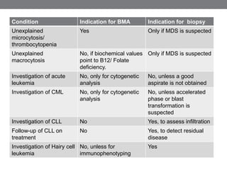
Bone marrow examination is an important diagnostic tool to
evaluate various disorders including both neoplastic and non-
neoplastic hematological diseases.
The bone marrow evaluation may either confirm clinically
suspected disease or may provide a previously unsuspected
diagnosis.
A request for bone marrow examination
should be regarded as a request for a
consultation on the patient in question and
not simply as a request to carry out a
technical procedure.
-B J Bain
• Bone marrow examination should be preceded by
evaluation of the medical history and clinical features,
examination of a blood film and assessment of results of a
full blood count, other laboratory tests, and radiological
investigations.
• This is essential to ensure that all appropriate tests are
performed on the material obtained and to permit an
adequate evaluation.
Tests performed on a bone marrow aspirate
Tests applied routinely
• Cytology on a Romanowsky stained film
• A Perls’ stain for haemosiderin
Tests applied selectively
• Immunophenotyping
• Cytogenetic analysis
• Molecular genetic analysis
• Culture for Mycobacteria, Leishmania, Histoplasma, or
other microorganisms
• Ultrastructural examination
Tests Appropriate anticoagulant
Cytogenetic analysis Preservative free heparin
Immunophenotyping Heparin/ EDTA
Molecular genetic analysis EDTA
Ultrastructural examination Glutaraldehyde
Indications for a trephine biopsy
• Inadequate or failed aspirate.
• Need for accurate assessment of cellularity.
• Suspected focal lesion (for example, suspected
granulomatous disease or lymphoma).
• Suspected bone marrow fibrosis.
• Need to study bone marrow architecture.
• Need to study bone structure or bone marrow blood
vessels.
• Patients who have a hypocellular bone marrow or bone
marrow fibrosis are likely to need a trephine biopsy for
adequate assessment. In such patients, an aspirate will
probably be inadequate or even impossible.
• Only a trephine biopsy shows the architecture of the bone
marrow and permits the detection of an abnormal
distribution of cells, bone marrow granulomas, and focal
lymphoid infiltrates.
Definite indications for performing a trephine
biopsy
• Investigation of suspected Hodgkin’s disease and non-
Hodgkin’s lymphoma
• Staging of non-Hodgkin’s lymphoma
• Diagnosis and follow up of hairy cell leukaemia
• Evaluation and follow up of chronic lymphocytic leukaemia
• Investigation of suspected myeloproliferative disorders
(polycythaemia rubra vera, essential thrombocythaemia,
idiopathic myelofibrosis, and systemic mastocytosis)
• Diagnosis of aplastic anaemia, hypoplastic myelodysplastic
syndromes, and hypoplastic acute myeloid leukaemia
• Investigation of an unexplained leucoerythroblastic blood film
• Investigation of a fever of unknown origin
• Investigation of patients in whom multiple myeloma is
suspected and investigation of selected patients with serum
paraproteins without other evidence of multiple myeloma
• Diagnosis of suspected metastatic carcinoma
• Diagnosis, staging, and follow up of small cell tumours of
• childhood
• Investigation of suspected bone disease
• Evaluation of any patient in whom an adequate bone
marrow aspirate cannot be obtained
• Possible indications
• Investigation of suspected acute myeloid leukaemia
• Investigation of suspected myelodysplastic syndrome
• Staging of Hodgkin’s disease
• Evaluation of chronic myeloid (granulocytic) leukaemia
• Investigation of suspected primary amyloidosis
Cases where trephine biopsy is not indicated
• In suspected chromosomal disorders in neonates when
rapid confirmation is required: Cytogenetic analysis (may
produce results in 1 day cf. several days if culture,
peripheral blood lymphocytes are used)
• Confirmation of normal bone marrow if bone marrow is
being aspirated for allogeneic transplantation
• Investigations of suspected storage disease
Indications for a bone marrow aspiration with
or without a trephine biopsy and relevance
of other techniques applicable to the
aspirate
• Unexpected microcytosis/ macrocytosis/
thrombocytopenia : Biopsy is indicated in cases where
MDS is suspected.
• It is acceptable to omit bone marrow examination in a
case of macrocytic anemia if the peripheral blood features
are totally typical and if assays of vitamin B12 and folic
acid are suggestive of a deficiency state.
• Unexplained Anemia: Biopsy is usually indicated.
Cytogenetic analysis for MDS and ultrastructural
examination for Congenital Dyserythropoeitic Anaemia if
suspected.
Investigation of pancytopenia/ suspected
aplastic anaemia
• Biopsy is indicated.
• Cytogenetic analysis if MDS is suspected;
• Appropriate culture if mycobacterial infection or
leishmaniasis is suspected
• Bone marrow is a useful source of DNA if investigation for
Pearson’s syndrome is required;
• Cytogenetic analysis if a haemophagocytic syndrome is
suspected (Because Epstein-Barr virus related
haemophagocytic syndrome may be associated with a
clonal proliferation of neoplastic T cells.)
• Investigation of fever of unknown origin: Biopsy is
indicated. Cultures for mycobacteria and also, if there is a
possibility of previous exposure, for leishmania and
histoplasma.
• Investigation of a leucoerythroblastic blood film and
suspected bone marrow infiltration: Biopsy is indicated.
Cytogenetic analysis if a haematological neoplasm is
suspected; if an abnormal infiltrate is found,
immunophenotyping and cytogenetic analysis may be
useful; cytogenetic analysis is indicated if a small cell
tumour of childhood is suspected
• Investigation of suspected MDS or myelodysplastic/
myeloproliferative disorder: Biopsy is indicated.
Cytogenetic analysis; investigation of colony forming units
if juvenile myelomonocytic leukaemia is suspected
• Investigation of suspected myeloproliferative disorder
(polycythaemia rubra vera, essential thrombocythaemia,
idiopathic myelofibrosis, or systemic mastocytosis):
Biopsy is indicated. Cytogenetic analysis; investigation of
colony forming units
• Diagnosis and follow up of hairy cell leukaemia: Biopsy is
indicated. Immunophenotyping, unless there are sufficient
circulating cells for it to be performed on peripheral blood
cells; tartrate resistant acid phosphatase stain if detailed
immunophenotyping is not available
Investigation of acute leukemia
• It is possible to establish a diagnosis of acute leukaemia
from peripheral blood examination, bone marrow
aspiration should nevertheless be carried out.
• The likelihood of successful cytogenetic analysis is higher
if bone marrow cells are used and because a baseline is
needed for comparison with bone marrow aspirates
performed during treatment.
• In addition, bone marrow aspiration permits the
assessment of trilineage dysplasia, which may be of
prognostic relevance.
Investigation of suspected chronic myeloid
leukaemia
• The diagnosis can be made without difficulty from the
peripheral blood features and the immunophenotype
.
• However, cytogenetic analysis is more often successful
when performed on the bone marrow and aspiration is
therefore indicated.
• If the accelerated phase of the disease or blast
transformation is suspected, bone marrow aspiration and
trephine biopsy are both important.
Investigation of chronic lymphocytic leukaemia
• The diagnosis can be made without difficulty from the peripheral
blood features and the immunophenotype.
• Patients with early stage disease do not require active treatment and
bone marrow biopsy is not essential for management.
• A bone marrow trephine biopsy is indicated in patients in whom
treatment is necessary, either those with more advanced disease or
younger patients in whom intensive treatment is planned.
• A trephine biopsy is essential for follow up of intensive treatment
because it may show residual focal disease when a bone marrow
aspirate is normal.
• The trephine biopsy permits an accurate assessment of the extent of
infiltration and gives information of prognostic importance.
Investigation of Non-Hodgkin’s lymphoma
• Diagnosis is usually based on a lymph node biopsy.
• In patients with bone marrow involvement, diagnosis can be
reliably based on cytology, immunophenotyping, and the
pattern of bone marrow infiltration.
• When there are circulating lymphoma cells,
immunophenotyping can be performed on the peripheral blood
and the bone marrow aspirate is of little importance.
• The trephine biopsy is much more important because it permits
an assessment of the pattern and extent of infiltration, which is
of both diagnostic and prognostic relevance, and may
demonstrate lymphoma when no abnormal cells have been
detected in the blood or the bone marrow aspirate.
• A bone marrow biopsy performed in patients with low grade
lymphoma sometimes shows unexpected high grade
transformation, which necessitates a different therapeutic
approach.
Investigation of Hodgkin’s disease
• The biopsy will usually have been done for the investigation of
pancytopenia or of systemic symptoms such as fever.
• Diagnosis by bone marrow biopsy is relatively frequent in HIV
positive patients, who often present with stage IVB disease.
• Trephine biopsy is not mandatory in the staging of Hodgkin’s
disease diagnosed at another site.
• Patients with clinical stage IA disease very rarely have bone
marrow infiltration and it has been considered justifiable to
avoid trephine biopsy in such patients.
• If bone marrow examination is required in Hodgkin’s disease, a
trephine biopsy is essential because, even when the marrow is
involved, it is rare for neoplastic cells to be detected in an
aspirate.
Investigation of multiple myeloma
• Bone marrow aspiration and trephine biopsy should be
regarded as complementary investigations in suspected
multiple myeloma
• A trephine biopsy is usually also performed. Because infiltration
is often focal, it is sometimes essential for a diagnosis.
• A trephine biopsy may demonstrate much more extensive
infiltration than is suspected from the bone marrow aspirate.
• A trephine biopsy is much more useful than an aspirate in
assessing suspected light chain associated amyloidosis.
• Cytogenetic analysis may be useful because demonstration of
poor prognosis abnormalities may influence management;
immunophenotyping is only needed if cytology of the aspirate is
not diagnostic and if it is not certain whether or not a
monoclonal plasma cell population is present
Investigation of MGUS
• In suspected cases of MGUS, it is reasonable not to
perform a bone marrow examination if a low concentration
paraprotein, has been detected almost incidentally in a
patient who does not have anaemia, bone pain,
hypercalcaemia, or other relevant clinical features.
• However, if investigation is undertaken then both a bone
marrow aspirate and a trephine biopsy should be
performed.
Investigation of Metastatic tumours
• A bone marrow trephine biopsy is better for suspected
metastatic disease because the rate of detection of tumour
cells is higher than with an aspirate.
• Because of the possibility of assessing tissue structure and
applying histochemical stains, it may be possible to predict the
tissue of origin of metastatic tumour through a trephine biopsy.
• Bone marrow trephine biopsy is essential in the initial staging
of certain paediatric tumours, such as neuroblastoma,
rhabdomyosarcoma, PNET, and Ewing’s tumour.
• The detection of bone marrow infiltration in the initial staging
bone marrow biopsy means that trephine biopsy is also
required during follow up
Investigation of Granulomatous Disease
• A trephine biopsy is always indicated.
• The detection rate is much higher because the increased
reticulin deposition associated with granuloma formation
often means that there is a dry tap or only relatively
normal bone marrow is aspirated.
Indications for a trephine biopsy
• Inadequate or failed aspirate.
• Need for accurate assessment of cellularity.
• Suspected focal lesion (for example, suspected
granulomatous disease or lymphoma).
• Suspected bone marrow fibrosis.
• Need to study bone marrow architecture.
• Need to study bone structure or bone marrow blood
vessels.
Cases where trephine biopsy is not indicated
• In suspected chromosomal disorders in neonates when
rapid confirmation is required: Cytogenetic analysis (may
produce results in 1 day cf. several days if culture,
peripheral blood lymphocytes are used)
• Confirmation of normal bone marrow if bone marrow is
being aspirated for allogeneic transplantation
• Investigations of suspected storage disease
Condition Indication for BMA Indication for biopsy
Unexplained
microcytosis/
thrombocytopenia
Yes Only if MDS is suspected
Unexplained
macrocytosis
No, if biochemical values
point to B12/ Folate
deficiency.
Only if MDS is suspected
Investigation of acute
leukemia
No, only for cytogenetic
analysis
No, unless a good
aspirate is not obtained
Investigation of CML No, only for cytogenetic
analysis
No, unless accelerated
phase or blast
transformation is
suspected
Investigation of CLL No Yes, to assess infiltration
Follow-up of CLL on
treatment
No Yes, to detect residual
disease
Investigation of Hairy cell
leukemia
No, unless for
immunophenotyping
Yes
Condition Indication for BMA Indication for biopsy
Investigation of Non-
Hodgkin’s lymphoma
No, unless for
immunophenotyping
Yes
Investigation of
Hodgkin’s lymphoma
No Rarely
Investigation of Multiple
Myeloma
References
• Lee S, Erber W, Portwit A, Tomonaga M, Peterson L.
ICSH guidelines for the standardization of bone
marrow specimens and reports. International Journal of
Laboratory Hematology. 2008;30(5):349-364.
• Bain B. Bone marrow aspiration. Journal of Clinical
Pathology. 2001; 54(9):657-663.
• Bain B. Bone marrow trephine biopsy. Journal of
Clinical Pathology. 2001; 54(10):737-742.

Indications for bone marrow examination

  • 1.
    INDICATIONS FOR BONE MARROWEXAMINATION By Sivaranjini. N Guide: Dr. S.P. Hiryur
  • 2.
    Bone marrow examinationis an important diagnostic tool to evaluate various disorders including both neoplastic and non- neoplastic hematological diseases. The bone marrow evaluation may either confirm clinically suspected disease or may provide a previously unsuspected diagnosis.
  • 3.
    A request forbone marrow examination should be regarded as a request for a consultation on the patient in question and not simply as a request to carry out a technical procedure. -B J Bain
  • 4.
    • Bone marrowexamination should be preceded by evaluation of the medical history and clinical features, examination of a blood film and assessment of results of a full blood count, other laboratory tests, and radiological investigations. • This is essential to ensure that all appropriate tests are performed on the material obtained and to permit an adequate evaluation.
  • 6.
    Tests performed ona bone marrow aspirate Tests applied routinely • Cytology on a Romanowsky stained film • A Perls’ stain for haemosiderin Tests applied selectively • Immunophenotyping • Cytogenetic analysis • Molecular genetic analysis • Culture for Mycobacteria, Leishmania, Histoplasma, or other microorganisms • Ultrastructural examination
  • 7.
    Tests Appropriate anticoagulant Cytogeneticanalysis Preservative free heparin Immunophenotyping Heparin/ EDTA Molecular genetic analysis EDTA Ultrastructural examination Glutaraldehyde
  • 8.
    Indications for atrephine biopsy • Inadequate or failed aspirate. • Need for accurate assessment of cellularity. • Suspected focal lesion (for example, suspected granulomatous disease or lymphoma). • Suspected bone marrow fibrosis. • Need to study bone marrow architecture. • Need to study bone structure or bone marrow blood vessels.
  • 9.
    • Patients whohave a hypocellular bone marrow or bone marrow fibrosis are likely to need a trephine biopsy for adequate assessment. In such patients, an aspirate will probably be inadequate or even impossible. • Only a trephine biopsy shows the architecture of the bone marrow and permits the detection of an abnormal distribution of cells, bone marrow granulomas, and focal lymphoid infiltrates.
  • 10.
    Definite indications forperforming a trephine biopsy • Investigation of suspected Hodgkin’s disease and non- Hodgkin’s lymphoma • Staging of non-Hodgkin’s lymphoma • Diagnosis and follow up of hairy cell leukaemia • Evaluation and follow up of chronic lymphocytic leukaemia • Investigation of suspected myeloproliferative disorders (polycythaemia rubra vera, essential thrombocythaemia, idiopathic myelofibrosis, and systemic mastocytosis) • Diagnosis of aplastic anaemia, hypoplastic myelodysplastic syndromes, and hypoplastic acute myeloid leukaemia • Investigation of an unexplained leucoerythroblastic blood film • Investigation of a fever of unknown origin • Investigation of patients in whom multiple myeloma is suspected and investigation of selected patients with serum paraproteins without other evidence of multiple myeloma
  • 11.
    • Diagnosis ofsuspected metastatic carcinoma • Diagnosis, staging, and follow up of small cell tumours of • childhood • Investigation of suspected bone disease • Evaluation of any patient in whom an adequate bone marrow aspirate cannot be obtained
  • 12.
    • Possible indications •Investigation of suspected acute myeloid leukaemia • Investigation of suspected myelodysplastic syndrome • Staging of Hodgkin’s disease • Evaluation of chronic myeloid (granulocytic) leukaemia • Investigation of suspected primary amyloidosis
  • 13.
    Cases where trephinebiopsy is not indicated • In suspected chromosomal disorders in neonates when rapid confirmation is required: Cytogenetic analysis (may produce results in 1 day cf. several days if culture, peripheral blood lymphocytes are used) • Confirmation of normal bone marrow if bone marrow is being aspirated for allogeneic transplantation • Investigations of suspected storage disease
  • 14.
    Indications for abone marrow aspiration with or without a trephine biopsy and relevance of other techniques applicable to the aspirate
  • 15.
    • Unexpected microcytosis/macrocytosis/ thrombocytopenia : Biopsy is indicated in cases where MDS is suspected. • It is acceptable to omit bone marrow examination in a case of macrocytic anemia if the peripheral blood features are totally typical and if assays of vitamin B12 and folic acid are suggestive of a deficiency state. • Unexplained Anemia: Biopsy is usually indicated. Cytogenetic analysis for MDS and ultrastructural examination for Congenital Dyserythropoeitic Anaemia if suspected.
  • 16.
    Investigation of pancytopenia/suspected aplastic anaemia • Biopsy is indicated. • Cytogenetic analysis if MDS is suspected; • Appropriate culture if mycobacterial infection or leishmaniasis is suspected • Bone marrow is a useful source of DNA if investigation for Pearson’s syndrome is required; • Cytogenetic analysis if a haemophagocytic syndrome is suspected (Because Epstein-Barr virus related haemophagocytic syndrome may be associated with a clonal proliferation of neoplastic T cells.)
  • 17.
    • Investigation offever of unknown origin: Biopsy is indicated. Cultures for mycobacteria and also, if there is a possibility of previous exposure, for leishmania and histoplasma. • Investigation of a leucoerythroblastic blood film and suspected bone marrow infiltration: Biopsy is indicated. Cytogenetic analysis if a haematological neoplasm is suspected; if an abnormal infiltrate is found, immunophenotyping and cytogenetic analysis may be useful; cytogenetic analysis is indicated if a small cell tumour of childhood is suspected
  • 18.
    • Investigation ofsuspected MDS or myelodysplastic/ myeloproliferative disorder: Biopsy is indicated. Cytogenetic analysis; investigation of colony forming units if juvenile myelomonocytic leukaemia is suspected • Investigation of suspected myeloproliferative disorder (polycythaemia rubra vera, essential thrombocythaemia, idiopathic myelofibrosis, or systemic mastocytosis): Biopsy is indicated. Cytogenetic analysis; investigation of colony forming units • Diagnosis and follow up of hairy cell leukaemia: Biopsy is indicated. Immunophenotyping, unless there are sufficient circulating cells for it to be performed on peripheral blood cells; tartrate resistant acid phosphatase stain if detailed immunophenotyping is not available
  • 19.
    Investigation of acuteleukemia • It is possible to establish a diagnosis of acute leukaemia from peripheral blood examination, bone marrow aspiration should nevertheless be carried out. • The likelihood of successful cytogenetic analysis is higher if bone marrow cells are used and because a baseline is needed for comparison with bone marrow aspirates performed during treatment. • In addition, bone marrow aspiration permits the assessment of trilineage dysplasia, which may be of prognostic relevance.
  • 20.
    Investigation of suspectedchronic myeloid leukaemia • The diagnosis can be made without difficulty from the peripheral blood features and the immunophenotype . • However, cytogenetic analysis is more often successful when performed on the bone marrow and aspiration is therefore indicated. • If the accelerated phase of the disease or blast transformation is suspected, bone marrow aspiration and trephine biopsy are both important.
  • 21.
    Investigation of chroniclymphocytic leukaemia • The diagnosis can be made without difficulty from the peripheral blood features and the immunophenotype. • Patients with early stage disease do not require active treatment and bone marrow biopsy is not essential for management. • A bone marrow trephine biopsy is indicated in patients in whom treatment is necessary, either those with more advanced disease or younger patients in whom intensive treatment is planned. • A trephine biopsy is essential for follow up of intensive treatment because it may show residual focal disease when a bone marrow aspirate is normal. • The trephine biopsy permits an accurate assessment of the extent of infiltration and gives information of prognostic importance.
  • 22.
    Investigation of Non-Hodgkin’slymphoma • Diagnosis is usually based on a lymph node biopsy. • In patients with bone marrow involvement, diagnosis can be reliably based on cytology, immunophenotyping, and the pattern of bone marrow infiltration. • When there are circulating lymphoma cells, immunophenotyping can be performed on the peripheral blood and the bone marrow aspirate is of little importance. • The trephine biopsy is much more important because it permits an assessment of the pattern and extent of infiltration, which is of both diagnostic and prognostic relevance, and may demonstrate lymphoma when no abnormal cells have been detected in the blood or the bone marrow aspirate. • A bone marrow biopsy performed in patients with low grade lymphoma sometimes shows unexpected high grade transformation, which necessitates a different therapeutic approach.
  • 23.
    Investigation of Hodgkin’sdisease • The biopsy will usually have been done for the investigation of pancytopenia or of systemic symptoms such as fever. • Diagnosis by bone marrow biopsy is relatively frequent in HIV positive patients, who often present with stage IVB disease. • Trephine biopsy is not mandatory in the staging of Hodgkin’s disease diagnosed at another site. • Patients with clinical stage IA disease very rarely have bone marrow infiltration and it has been considered justifiable to avoid trephine biopsy in such patients. • If bone marrow examination is required in Hodgkin’s disease, a trephine biopsy is essential because, even when the marrow is involved, it is rare for neoplastic cells to be detected in an aspirate.
  • 24.
    Investigation of multiplemyeloma • Bone marrow aspiration and trephine biopsy should be regarded as complementary investigations in suspected multiple myeloma • A trephine biopsy is usually also performed. Because infiltration is often focal, it is sometimes essential for a diagnosis. • A trephine biopsy may demonstrate much more extensive infiltration than is suspected from the bone marrow aspirate. • A trephine biopsy is much more useful than an aspirate in assessing suspected light chain associated amyloidosis. • Cytogenetic analysis may be useful because demonstration of poor prognosis abnormalities may influence management; immunophenotyping is only needed if cytology of the aspirate is not diagnostic and if it is not certain whether or not a monoclonal plasma cell population is present
  • 25.
    Investigation of MGUS •In suspected cases of MGUS, it is reasonable not to perform a bone marrow examination if a low concentration paraprotein, has been detected almost incidentally in a patient who does not have anaemia, bone pain, hypercalcaemia, or other relevant clinical features. • However, if investigation is undertaken then both a bone marrow aspirate and a trephine biopsy should be performed.
  • 26.
    Investigation of Metastatictumours • A bone marrow trephine biopsy is better for suspected metastatic disease because the rate of detection of tumour cells is higher than with an aspirate. • Because of the possibility of assessing tissue structure and applying histochemical stains, it may be possible to predict the tissue of origin of metastatic tumour through a trephine biopsy. • Bone marrow trephine biopsy is essential in the initial staging of certain paediatric tumours, such as neuroblastoma, rhabdomyosarcoma, PNET, and Ewing’s tumour. • The detection of bone marrow infiltration in the initial staging bone marrow biopsy means that trephine biopsy is also required during follow up
  • 27.
    Investigation of GranulomatousDisease • A trephine biopsy is always indicated. • The detection rate is much higher because the increased reticulin deposition associated with granuloma formation often means that there is a dry tap or only relatively normal bone marrow is aspirated.
  • 28.
    Indications for atrephine biopsy • Inadequate or failed aspirate. • Need for accurate assessment of cellularity. • Suspected focal lesion (for example, suspected granulomatous disease or lymphoma). • Suspected bone marrow fibrosis. • Need to study bone marrow architecture. • Need to study bone structure or bone marrow blood vessels.
  • 29.
    Cases where trephinebiopsy is not indicated • In suspected chromosomal disorders in neonates when rapid confirmation is required: Cytogenetic analysis (may produce results in 1 day cf. several days if culture, peripheral blood lymphocytes are used) • Confirmation of normal bone marrow if bone marrow is being aspirated for allogeneic transplantation • Investigations of suspected storage disease
  • 30.
    Condition Indication forBMA Indication for biopsy Unexplained microcytosis/ thrombocytopenia Yes Only if MDS is suspected Unexplained macrocytosis No, if biochemical values point to B12/ Folate deficiency. Only if MDS is suspected Investigation of acute leukemia No, only for cytogenetic analysis No, unless a good aspirate is not obtained Investigation of CML No, only for cytogenetic analysis No, unless accelerated phase or blast transformation is suspected Investigation of CLL No Yes, to assess infiltration Follow-up of CLL on treatment No Yes, to detect residual disease Investigation of Hairy cell leukemia No, unless for immunophenotyping Yes
  • 31.
    Condition Indication forBMA Indication for biopsy Investigation of Non- Hodgkin’s lymphoma No, unless for immunophenotyping Yes Investigation of Hodgkin’s lymphoma No Rarely Investigation of Multiple Myeloma
  • 33.
    References • Lee S,Erber W, Portwit A, Tomonaga M, Peterson L. ICSH guidelines for the standardization of bone marrow specimens and reports. International Journal of Laboratory Hematology. 2008;30(5):349-364. • Bain B. Bone marrow aspiration. Journal of Clinical Pathology. 2001; 54(9):657-663. • Bain B. Bone marrow trephine biopsy. Journal of Clinical Pathology. 2001; 54(10):737-742.

Editor's Notes

  • #22 A bone marrow aspirate is therefore not essential in patients with early stage disease, particularly in elderly patients in whom treatment may never become necessary.