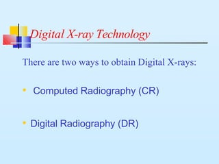
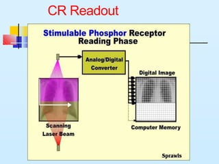
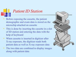
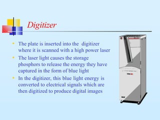
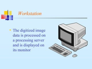
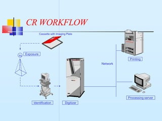
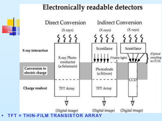
COMPUTED
RADIOGRAPHY &
DIGITAL
RADIOGRAPHY
Dr Amit
Digital X-ray Technology
There are two ways to obtain Digital X-rays:
• Computed Radiography (CR)
• Digital Radiography (DR)
What is CR?
• Computed Radiography (CR) is a process of
capturing radiographic data from a
conventional X-ray machine and processing
the data digitally to produce crisp and high
quality radiographic images
What is CR?…
• For exposure, an Imaging Plate (IP) is placed in a
cassette instead of a piece of film. The IP captures
and "stores" the X-rays
• The image is "developed" in a CR reader instead
of a film processor. The CR reader extracts the
information stored in the plate and produces a
digital image
• Images produced have much better contrast than a
Conventional X-ray film-screen system
Basic Modules of CR
Digitizer
Preview & ID Station
Processing Server
ID Tablet
Laser Camera
Cassette with
Imaging Plate
Imaging Plate (IP)
• The Imaging Plate looks like the intensifying
screens found in Conventional film-screen
cassettes
• They are made of photostimulable
phosphor.
Imaging Plate (IP)
• Instead of emitting light immediately when exposed
to X-rays, the photostimulable phosphor has the
special property of storing the X-ray energy in a
latent form and releasing the same when stimulated
by a laser energy in the CR Reader / Digitizer
Imaging Plate (IP)…
• Storage phosphors are unique because they
respond to a very wide range of X-ray exposures
• This latitude gives the flexibility in selecting
X-ray technique and takes care of under or over
exposure
• Regardless of the exposure, the image can be
displayed correctly
• As a consequence, retakes due to inappropriate
exposures are drastically reduced.
Storage phosphor principle
Absorption Emission
laser stimulation
x-rays
electron
trap
electron
trap
Storage phosphor principle…
• The imaging plate is coated with
photostimulable phosphor, also called storage
phosphor
• The phosphor material is generally a kind of
Barium fluorohalide
• The Imaging Plate contains not only the
phosphor layer, but also a protective coat, a
conductive layer, support and laminate layers
Storage phosphor principle…
• Incident X-rays excite electrons into a higher energy
level (electron traps)
• A latent image is created in the form of “stored energy”
• Stimulation with a scanning laser beam releases
electrons
• Typical wavelength of the stimulating laser is 633 nm
• Falling back, electrons emit luminescent light
• Typical wavelength of the emitted light is 390 nm
CR Readout
Storage phosphor principle…
• The emitted light intensity is proportional to
the original incident X-ray intensity
• The emitted light is captured with an optical
array and a photomultiplier and is digitized
• The residual image is erased from the plate by
an intense light source, which returns all
electrons to their original stat, making the plate
ready to be reused for new exposures
• The storage phosphor plate fits inside a standard
size cassette and is exposed to X-rays exactly like
film
• The X-ray energy is stored on the plate in the form
of latent energy
How is a Storage Phosphor plate
exposed?
Patient ID Station
• Before exposing the cassette, the patient
demographic and exam data is stored on the
microchip attached on cassette
• This is done by inserting the cassette in a slot
of ID station and entering the data with the
help of keyboard
• When cassette is inserted in digitizer after
X-ray exposure, the digitizer reads both
patient data as well as X-ray exposure data
• The two data are combined to display images
along with patient data
Digitizer
• The plate is inserted into the digitizer
where it is scanned with a high power laser
• The laser light causes the storage
phosphors to release the energy they have
captured in the form of blue light
• In the digitizer, this blue light energy is
converted to electrical signals which are
then digitized to produce digital images
What happens to a Storage Phosphor
Plate after it is scanned?
• After exposure and scanning, the phosphor
plate is "erased" by exposing to a bright light
exposure within the digitizer
• The previous image stored in the phosphors is
removed and the plate is ready to be exposed
again
Workstation
• The digitized image
data is processed on
a processing server
and is displayed on
its monitor
How many times can we use a
Storage Phosphor Plate?
• The life of a phosphor plate depends on
how carefully it is handled. Physical
damage to the plate will limit its useful
life
• If properly cared for, a plate will produce
thousands of images
• Imaging Plates are known to last more
than 50000 Exposure Cycles
How is the workflow different with
CR ?
• Instead of taking the film cassette to a dark
room for processing, the technologist takes
the cassette with imaging plate to the CR
reader for digital processing of the image
• Instead of manually taking the films to the
reporting radiologists , the soft copy
images reach the workstation almost
immediately
How is the workflow different with
CR ?…
• The time required to acquire a Digital
image is much less compared to
conventional darkroom process
• The film is the first product in Conventional
where as the film is the last product in CR
CR WORKFLOW
Rx
Network
Digitizer
Printing
Identification
Processing server
Exposure
Cassette with Imaging Plate
Digital Radiography
This technique is performed by digital X-ray
machines with flat panel detectors
Digital Radiography
Digital Radiography uses two types of
detectors:
• Direct
• Indirect
Direct detectors
• Direct detectors automatically convert X- rays into electronic
signals.
■
■
X.rays interact with semiconductor material
■ Amorphous selenium
X.rays converted directly into electrical charge
■ No intermediate steps
Indirect detectors
• Indirect detectors are so named because this technique still uses a
scintillator to converts x-rays to light before conversion to an
electrical charge for subsequent readout.
• X-ray photons encounter a cesium iodide (CsI) scintillator and
are converted to light. The light then reaches a low-noise
photodiode array and is converted into an electrical charge. Each
photodiode represents a single pixel, and each produces an
electrical charge that is read out digitally before finally being sent
to the image processor
■ TFT = THIN-FILM TRANSISTOR ARRAY
Limitations of Conventional X-ray process
(X-ray film / Screen/Darkroom)
• Film has a limited exposure latitude i.e less
detail contrast
• Time consuming & cumbersome
• Intolerant to exposure errors
• Repeat X-rays ( More radiation exposure )
• Film wastage
Limitations of Conventional X-ray process
(X-ray film / Screen/Darkroom)…
• Cannot be duplicated without loss of quality
• Film storage is a problem
• Scatter radiation reduces contrast and increases
patient dose
• Quality control is an issue
What are the advantages of
Digital X-rays?
• Post processing (soft tissue and bony details can
be viewed at same time )
• Reduction in hazardous X-ray dose to patients
• More info on one image
• Constant image quality
• Possibility of viewing X-ray images wherever
needed
What are the advantages of
Digital X-rays?…
• Digital images are of extremely high
quality
• Digital images have a future scope of
better image management
• Facility of giving multiple images of
investigative studies on a single high
definition laser film
Thank
you

CR DR system.pptx

  • 1.
  • 2.
    Digital X-ray Technology Thereare two ways to obtain Digital X-rays: • Computed Radiography (CR) • Digital Radiography (DR)
  • 3.
    What is CR? •Computed Radiography (CR) is a process of capturing radiographic data from a conventional X-ray machine and processing the data digitally to produce crisp and high quality radiographic images
  • 4.
    What is CR?… •For exposure, an Imaging Plate (IP) is placed in a cassette instead of a piece of film. The IP captures and "stores" the X-rays • The image is "developed" in a CR reader instead of a film processor. The CR reader extracts the information stored in the plate and produces a digital image • Images produced have much better contrast than a Conventional X-ray film-screen system
  • 5.
    Basic Modules ofCR Digitizer Preview & ID Station Processing Server ID Tablet Laser Camera Cassette with Imaging Plate
  • 6.
    Imaging Plate (IP) •The Imaging Plate looks like the intensifying screens found in Conventional film-screen cassettes • They are made of photostimulable phosphor.
  • 7.
    Imaging Plate (IP) •Instead of emitting light immediately when exposed to X-rays, the photostimulable phosphor has the special property of storing the X-ray energy in a latent form and releasing the same when stimulated by a laser energy in the CR Reader / Digitizer
  • 8.
    Imaging Plate (IP)… •Storage phosphors are unique because they respond to a very wide range of X-ray exposures • This latitude gives the flexibility in selecting X-ray technique and takes care of under or over exposure • Regardless of the exposure, the image can be displayed correctly • As a consequence, retakes due to inappropriate exposures are drastically reduced.
  • 9.
    Storage phosphor principle AbsorptionEmission laser stimulation x-rays electron trap electron trap
  • 10.
    Storage phosphor principle… •The imaging plate is coated with photostimulable phosphor, also called storage phosphor • The phosphor material is generally a kind of Barium fluorohalide • The Imaging Plate contains not only the phosphor layer, but also a protective coat, a conductive layer, support and laminate layers
  • 11.
    Storage phosphor principle… •Incident X-rays excite electrons into a higher energy level (electron traps) • A latent image is created in the form of “stored energy” • Stimulation with a scanning laser beam releases electrons • Typical wavelength of the stimulating laser is 633 nm • Falling back, electrons emit luminescent light • Typical wavelength of the emitted light is 390 nm
  • 12.
  • 13.
    Storage phosphor principle… •The emitted light intensity is proportional to the original incident X-ray intensity • The emitted light is captured with an optical array and a photomultiplier and is digitized • The residual image is erased from the plate by an intense light source, which returns all electrons to their original stat, making the plate ready to be reused for new exposures
  • 14.
    • The storagephosphor plate fits inside a standard size cassette and is exposed to X-rays exactly like film • The X-ray energy is stored on the plate in the form of latent energy How is a Storage Phosphor plate exposed?
  • 15.
    Patient ID Station •Before exposing the cassette, the patient demographic and exam data is stored on the microchip attached on cassette • This is done by inserting the cassette in a slot of ID station and entering the data with the help of keyboard • When cassette is inserted in digitizer after X-ray exposure, the digitizer reads both patient data as well as X-ray exposure data • The two data are combined to display images along with patient data
  • 16.
    Digitizer • The plateis inserted into the digitizer where it is scanned with a high power laser • The laser light causes the storage phosphors to release the energy they have captured in the form of blue light • In the digitizer, this blue light energy is converted to electrical signals which are then digitized to produce digital images
  • 17.
    What happens toa Storage Phosphor Plate after it is scanned? • After exposure and scanning, the phosphor plate is "erased" by exposing to a bright light exposure within the digitizer • The previous image stored in the phosphors is removed and the plate is ready to be exposed again
  • 18.
    Workstation • The digitizedimage data is processed on a processing server and is displayed on its monitor
  • 19.
    How many timescan we use a Storage Phosphor Plate? • The life of a phosphor plate depends on how carefully it is handled. Physical damage to the plate will limit its useful life • If properly cared for, a plate will produce thousands of images • Imaging Plates are known to last more than 50000 Exposure Cycles
  • 20.
    How is theworkflow different with CR ? • Instead of taking the film cassette to a dark room for processing, the technologist takes the cassette with imaging plate to the CR reader for digital processing of the image • Instead of manually taking the films to the reporting radiologists , the soft copy images reach the workstation almost immediately
  • 21.
    How is theworkflow different with CR ?… • The time required to acquire a Digital image is much less compared to conventional darkroom process • The film is the first product in Conventional where as the film is the last product in CR
  • 22.
  • 23.
    Digital Radiography This techniqueis performed by digital X-ray machines with flat panel detectors
  • 24.
    Digital Radiography Digital Radiographyuses two types of detectors: • Direct • Indirect
  • 25.
    Direct detectors • Directdetectors automatically convert X- rays into electronic signals. ■ ■ X.rays interact with semiconductor material ■ Amorphous selenium X.rays converted directly into electrical charge ■ No intermediate steps
  • 26.
    Indirect detectors • Indirectdetectors are so named because this technique still uses a scintillator to converts x-rays to light before conversion to an electrical charge for subsequent readout. • X-ray photons encounter a cesium iodide (CsI) scintillator and are converted to light. The light then reaches a low-noise photodiode array and is converted into an electrical charge. Each photodiode represents a single pixel, and each produces an electrical charge that is read out digitally before finally being sent to the image processor
  • 27.
    ■ TFT =THIN-FILM TRANSISTOR ARRAY
  • 28.
    Limitations of ConventionalX-ray process (X-ray film / Screen/Darkroom) • Film has a limited exposure latitude i.e less detail contrast • Time consuming & cumbersome • Intolerant to exposure errors • Repeat X-rays ( More radiation exposure ) • Film wastage
  • 29.
    Limitations of ConventionalX-ray process (X-ray film / Screen/Darkroom)… • Cannot be duplicated without loss of quality • Film storage is a problem • Scatter radiation reduces contrast and increases patient dose • Quality control is an issue
  • 30.
    What are theadvantages of Digital X-rays? • Post processing (soft tissue and bony details can be viewed at same time ) • Reduction in hazardous X-ray dose to patients • More info on one image • Constant image quality • Possibility of viewing X-ray images wherever needed
  • 31.
    What are theadvantages of Digital X-rays?… • Digital images are of extremely high quality • Digital images have a future scope of better image management • Facility of giving multiple images of investigative studies on a single high definition laser film
  • 32.