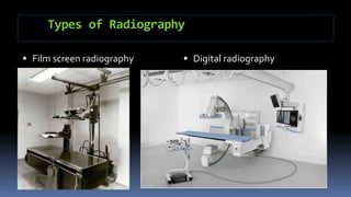
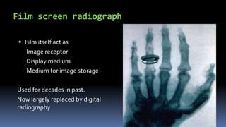
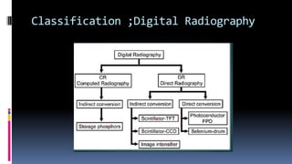
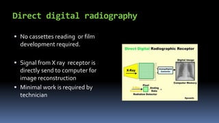
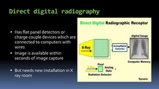
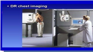
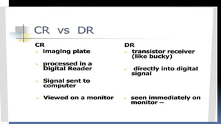
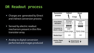
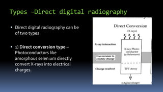
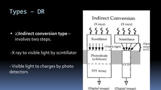
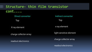
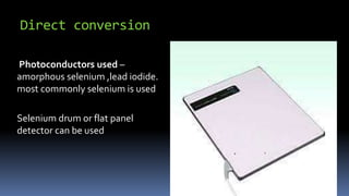
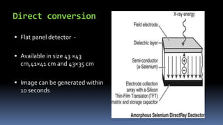
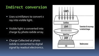
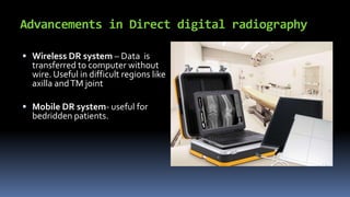
Presentation
Presented by
Dr Tarun Kumar
Topics covered/ Table of contents
1)Types of radiography
2) Film screen radiography ,digital radiography
3) Computed and direct digital radiography
4) Process of imaging in direct digital radiography
5) Advantages of direct digital radiography
6) Advancement in direct digital radiography
Types of Radiography
 Film screen radiography  Digital radiography
Film screen radiograph
 Film itself act as
Image receptor
Display medium
Medium for image storage
Used for decades in past.
Now largely replaced by digital
radiography
Film screen radiography cont...
 Uses intensifying screens
 Film is placed between two
intensifying screens
 Screens emit light when x-rays
strike them causing change in
film
 Film is processed chemically
Limitations of film screen radiography
 No Manipulation – Cant be manipulated i.e. magnification,
cropping ,windowing, etc are not possible
 Storage – Image quality deteriorates over time
- Requires large space and more management for
storage
 Transmission – loss of quality during transmission.
- Loss of quality during duplication
Digital radiography
When digital detectors are used
to capture information then its
called digital radiography
Films and Screens are replaced by
digital detectors.
• Image reception ,display and
storage are done separately.
Advantages of digital radiography
Manipulation – can be magnified ,compressed ,cropped
,contrast enhanced.
Storage – stored in digital files requiring much less space
- no reduction in quality on long time storage
Communication – Can me made into multiple copies without
loss of. quality
- Can be easily send anywhere through PACS
Two Types of digital radiography
 Computed radiography system i.e. CR
 Direct digital radiography system i.e. DR
Classification ;Digital Radiography
Computed radiography system CR
 Operational in Radiology
department of PMCH
 CR contains photo illuminable
Phosphor plates acting as
detecting and storage layer in
place of conventional films.
 Coating of europium activated
barium fluorohalide is used in
these phosphor plates.
Process of imaging in computed radiography
 On exposure to X ray energy is
absorbed and stored temporarily
by crystals in the plate
 Readout process – plate is scanned
with high energy laser beam and
energy stored in .crystals is set free
in form of light photons.
 Light thus emitted is collected and
converted into digital images.
Computed Radiography cont...
 Imaging plate can be reused after exposure to white light
 It can be easily integrated into existing conventional setup and
produce digital images that can be stored and communicated
digitally.
Imaging plates are expensive and can easily get damaged by
manual handling.
Direct digital radiography
 No cassettes reading or film
development required.
 Signal from X ray receptor is
directly send to computer for
image reconstruction
 Minimal work is required by
technician
Direct digital radiography
 Has flat panel detectors or
charge couple devices which are
connected to computers with
wires
 Image is available within
seconds of image capture
 But needs new installation in X
ray room
DR Readout process
 Charges are generated by Direct
and indirect conversion process
 Sensed by electric readout
mechanism present in thin film
transistor array
 Analog to digital conversion
performed and images produced
Types –Direct digital radiography
 Direct digital radiography can be
of two types
 1) Direct conversion type –
Photoconductors like
amorphous selenium directly
convert X-rays into electrical
charges.
Types – DR
 2)Indirect conversion type –
involves two steps.
-X ray to visible light by scintillator
-Visible light to charges by photo
detectors
Thin film transistors
 Used in both direct and indirect conversion
 Structure – Deposited in multiple layer on glass substrate.
lowest layer has readout electronics
higher layer has charge collectors
X ray element and light sensitive element are
deposited on top layer
Structure- thin film transistor
cont....
Direct converter Indirect converter
Top
X ray element
charge collector array
readout electronics
Top
x ray element
light sensitive element
charge collector array
readout electronics
Thin film transistor cont..
Encased in protective layer for insulation
connected to computers through wire for image reconstruction
Direct conversion
Photoconductors used –
amorphous selenium ,lead iodide.
most commonly selenium is used
Selenium drum or flat panel
detector can be used
Direct conversion
 Flat panel detector -
 Available in size 43 ×43
cm,41×41 cm and 43×35 cm
 Image can be generated within
10 seconds
Indirect conversion
 Uses scintillators to convert x
ray into visible light.
 Visible light is converted into
charge by photo iodide array.
 Charge Collected at photo
iodide is converted to digital
signal by readout electronics.
Types ; scintillator
 Unstructured scintillator
visible light emitted can spread to
adjacent structure
 Structured scintillator
Consists of caesium iodide crystals
on detectors which are parallel
needles channel most signal
directly to photo iodide layer.
Image processing
 Raw data is processed by computers and converted into
image
 Processing includes removing technical artefacts and
optimising contrast.
 Removing unwanted signals called noise
 Windowing and reversing of contrast is done for better view
Advantages of DR over CR
 Quick image development, within 10 sec
 Very less work done by technician so less manpower is
required
Less radiation dose than film screen or conventional
radiography.
Image transmission in DR
 As image is generated within seconds it can be duplicated and
sent to various departments simultaneously.
 Helps in Inter department coordination for better patient
management
Advancements in Direct digital radiography
 Wireless DR system – Data is
transferred to computer without
wire. Useful in difficult regions like
axilla andTM joint
 Mobile DR system- useful for
bedridden patients.
Advancement in DR system
 Automatic image stitching – image reconstruction after
multiple sequential exposure. Useful for imaging large
anatomical regions like whole spine in a single film.
 Computer aided diagnosis- suspicious looking areas are
marked by computer for review by radiologist.
Direct digital radiography(1) (1)

Direct digital radiography(1) (1)

  • 1.
  • 2.
    Topics covered/ Tableof contents 1)Types of radiography 2) Film screen radiography ,digital radiography 3) Computed and direct digital radiography 4) Process of imaging in direct digital radiography 5) Advantages of direct digital radiography 6) Advancement in direct digital radiography
  • 3.
    Types of Radiography Film screen radiography  Digital radiography
  • 4.
    Film screen radiograph Film itself act as Image receptor Display medium Medium for image storage Used for decades in past. Now largely replaced by digital radiography
  • 5.
    Film screen radiographycont...  Uses intensifying screens  Film is placed between two intensifying screens  Screens emit light when x-rays strike them causing change in film  Film is processed chemically
  • 6.
    Limitations of filmscreen radiography  No Manipulation – Cant be manipulated i.e. magnification, cropping ,windowing, etc are not possible  Storage – Image quality deteriorates over time - Requires large space and more management for storage  Transmission – loss of quality during transmission. - Loss of quality during duplication
  • 7.
    Digital radiography When digitaldetectors are used to capture information then its called digital radiography Films and Screens are replaced by digital detectors. • Image reception ,display and storage are done separately.
  • 8.
    Advantages of digitalradiography Manipulation – can be magnified ,compressed ,cropped ,contrast enhanced. Storage – stored in digital files requiring much less space - no reduction in quality on long time storage Communication – Can me made into multiple copies without loss of. quality - Can be easily send anywhere through PACS
  • 9.
    Two Types ofdigital radiography  Computed radiography system i.e. CR  Direct digital radiography system i.e. DR
  • 10.
  • 11.
    Computed radiography systemCR  Operational in Radiology department of PMCH  CR contains photo illuminable Phosphor plates acting as detecting and storage layer in place of conventional films.  Coating of europium activated barium fluorohalide is used in these phosphor plates.
  • 12.
    Process of imagingin computed radiography  On exposure to X ray energy is absorbed and stored temporarily by crystals in the plate  Readout process – plate is scanned with high energy laser beam and energy stored in .crystals is set free in form of light photons.  Light thus emitted is collected and converted into digital images.
  • 13.
    Computed Radiography cont... Imaging plate can be reused after exposure to white light  It can be easily integrated into existing conventional setup and produce digital images that can be stored and communicated digitally. Imaging plates are expensive and can easily get damaged by manual handling.
  • 14.
    Direct digital radiography No cassettes reading or film development required.  Signal from X ray receptor is directly send to computer for image reconstruction  Minimal work is required by technician
  • 15.
    Direct digital radiography Has flat panel detectors or charge couple devices which are connected to computers with wires  Image is available within seconds of image capture  But needs new installation in X ray room
  • 18.
    DR Readout process Charges are generated by Direct and indirect conversion process  Sensed by electric readout mechanism present in thin film transistor array  Analog to digital conversion performed and images produced
  • 19.
    Types –Direct digitalradiography  Direct digital radiography can be of two types  1) Direct conversion type – Photoconductors like amorphous selenium directly convert X-rays into electrical charges.
  • 20.
    Types – DR 2)Indirect conversion type – involves two steps. -X ray to visible light by scintillator -Visible light to charges by photo detectors
  • 21.
    Thin film transistors Used in both direct and indirect conversion  Structure – Deposited in multiple layer on glass substrate. lowest layer has readout electronics higher layer has charge collectors X ray element and light sensitive element are deposited on top layer
  • 22.
    Structure- thin filmtransistor cont.... Direct converter Indirect converter Top X ray element charge collector array readout electronics Top x ray element light sensitive element charge collector array readout electronics
  • 23.
    Thin film transistorcont.. Encased in protective layer for insulation connected to computers through wire for image reconstruction
  • 24.
    Direct conversion Photoconductors used– amorphous selenium ,lead iodide. most commonly selenium is used Selenium drum or flat panel detector can be used
  • 25.
    Direct conversion  Flatpanel detector -  Available in size 43 ×43 cm,41×41 cm and 43×35 cm  Image can be generated within 10 seconds
  • 26.
    Indirect conversion  Usesscintillators to convert x ray into visible light.  Visible light is converted into charge by photo iodide array.  Charge Collected at photo iodide is converted to digital signal by readout electronics.
  • 27.
    Types ; scintillator Unstructured scintillator visible light emitted can spread to adjacent structure  Structured scintillator Consists of caesium iodide crystals on detectors which are parallel needles channel most signal directly to photo iodide layer.
  • 28.
    Image processing  Rawdata is processed by computers and converted into image  Processing includes removing technical artefacts and optimising contrast.  Removing unwanted signals called noise  Windowing and reversing of contrast is done for better view
  • 29.
    Advantages of DRover CR  Quick image development, within 10 sec  Very less work done by technician so less manpower is required Less radiation dose than film screen or conventional radiography.
  • 30.
    Image transmission inDR  As image is generated within seconds it can be duplicated and sent to various departments simultaneously.  Helps in Inter department coordination for better patient management
  • 31.
    Advancements in Directdigital radiography  Wireless DR system – Data is transferred to computer without wire. Useful in difficult regions like axilla andTM joint  Mobile DR system- useful for bedridden patients.
  • 32.
    Advancement in DRsystem  Automatic image stitching – image reconstruction after multiple sequential exposure. Useful for imaging large anatomical regions like whole spine in a single film.  Computer aided diagnosis- suspicious looking areas are marked by computer for review by radiologist.