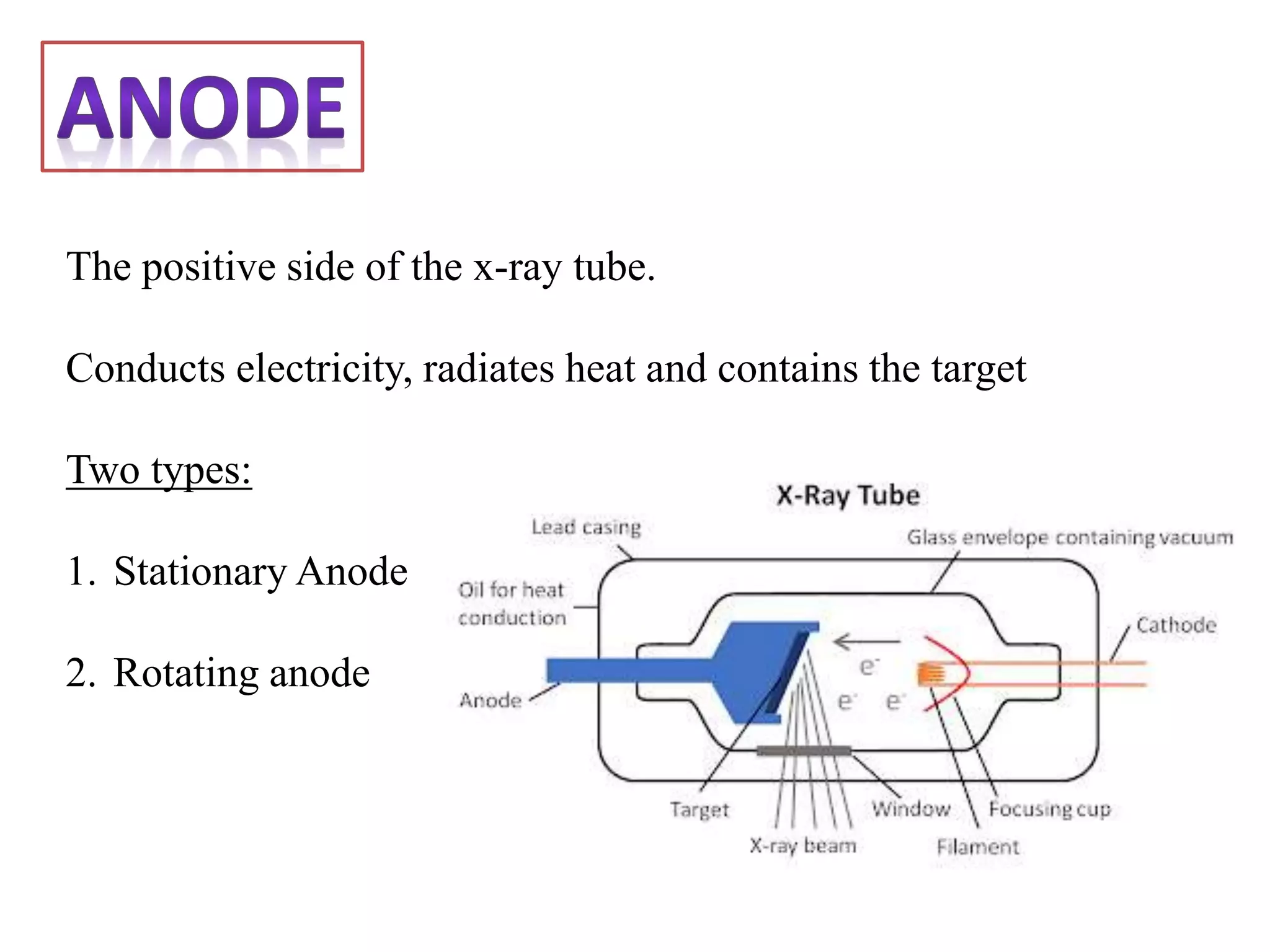
SlideShare a Scribd company logo
Supervising Faculty: Dr. B. P. Baruah, Professor, Dept. of Radiodiagnosis
Mentor: Dr. Amit Gupta, Senior Resident, Dept. of Radiodiagnosis
Presenter: Dr. Tarun Goyal, Junior Resident (PG 1), Dept. of Radiodiagnosis
School Of Medical Sciences & Research, Sharda University, G.Noida
An x-ray tube functions as a specific energy converter, receiving
the electrical energy and converting it into two other forms of
energy:
X RADIATION & HEAT
Heat is considered the undesirable product of this conversion
process; therefore x-radiation is created by taking the energy
from the electrons and converting it into photons.
This very specific energy conversion takes place in the x-ray tube.
It is a component of x-ray imaging system, it is contained in a
protective housing and therefore is inaccessible.
Contains two Principal elements:
• CATHODE: is the source of electrons
• ANODE: acts as the target for electrons and releases x-rays
The two electrodes of
the x ray are sealed in
a vacuum.
Other components of the x ray tubes are:
1. Expansion bellows (provide space for oil to expand)
2. Tube envelope (evacuated); preferably made of
Pyrex glass
3. Tube housing
4. Cooling dielectric oil
5. Rotor
6. Induction stator
7. Tube window
Negative terminal of the x ray tube.
Cathode or filament; both terms are used interchangeably.
Act as source of electrons.
Cathode has two other elements
• Connecting wires: which supply both VOLTAGE and AMPERAGE
• Metallic focusing cup
The X-ray tube current, measured in milliamperes, refers to the
number of electrons flowing per second from the filament to the
target
FILAMENT… WHAT DOES IT MADE UP OF AND WHY?
Made up of TUNGSTEN wires of about 0.2mm in diameter,
which is coiled to form a vertical spiral about 0.2 cm in
diameter and 1cm or less in length.
Now why tungsten?? Despite it is not efficient
emitter as other metals?
Because it can be easily drawn into thin wires that are quite
strong and has a high melting point (3370’C) and has little
tendency to vaporize…
Thermionic emission
After heating of filament, some
electrons acquire energy to move
small distances, normally, within
the metal and cannot escape it.
This escape of electrons is known
as THERMIONIC EMISSION.
The electron cloud
surrounding the
filament produced due
to this effect is termed
as “EDISON EFFECT”
Surrounds the cathode with the same negative potential as cathode.
Its electrical forces cause electron stream to converge onto target anode in required size
and shape instead of spreading out.
Mostly made up of NICKEL
Types
1. Single filament: use for single exposure x
rays.
2. Double filament: consists of spiral wires in
each filament.
3. Triple focus: used in highly specialized x rays
4. Stereoscopic Angiographic tube: useful in
angiography
Line Focus Principle
It will help in heat dissipation
and yet achieve small focal
spot size to produce good
radiographic details….
The positive side of the x-ray tube.
Conducts electricity, radiates heat and contains the target
Two types:
1. Stationary Anode
2. Rotating anode
Stationary Anode
consists of small tungsten plate, 2 to 3 mm thick, embedded in
large copper mass..
Tungsten is used due to its atomic
number(74), hence produce x rays
more efficiently, also has high
melting point 0f 3370’C.
Also it is good for heat dissipation.
Tungsten cannot bear repeated
exposures, hence covered with
copper..
Copper anode act to increase the
total thermal capacity of anode
and speed the rating of cooling
Usually used in dental x-ray machine
ROTATING ANODE
The principle is used to produce x-ray tubes
capable of withstanding the heat generated
by large exposures.
Rotates at the speed of 3600 rpm which is
practically not achievable due to mechanical
factors such as slipping between the rotor
and bearing. Hence 3000 rpm is assumed.
The purpose of rotating anode is to spread the heat produced during an
exposure over a large area of the anode.
It consists of a disc which is made up of tungsten or tungsten and it’s alloy
with beveled edge at 6 to 20 degree.
This beveled edge is used to take advantage of line focus principle.
FUNCTIONS OF DIFFERENT PARTS OF TUBES..
1. Stator Coils: lies at neck of the tube outside the envelope which produce the
magnetic field to provide power to effect rotation of anode.
2. Rotor: the magnetic field induces current in the copper rotor of induction
motor and provides power to rotate the anode assembly.
3. Lubricants: as there is a lot of friction, bearings need to be lubricated. Dry
lubricant (graphite) would wear as powder and will destroy the vacuum;
hence metallic lubricants are used especially silver.
4. Anode stem: made of molybdenum and has a role in heat dissipation as it
restricts the expansion of the anode. Molybdenum is chosen due to it its high
melting point (2600’C) and a poor conductor of heat. Length of the anode
stem proportional to inertia of tungsten disc and hence proportional to load
on the bearings.
5. Metal bellows: allows the oil to expand without increasing pressure on tube.
Heat Dissipation
In Stationary Anode: takes place
by absorption and conductivity
by copper anode.
In Rotating Anode: it occurs by
the radiating through the vacuum
to the tube wall and then into
the surrounding oil and tube
housing.
Drawbacks with the Rotating Anode
Thermal stress: life of anode is minimized due to roughening and pitting of the anode
surface exposed to electron beam.
Output of x-rays decreased due to more scattering of the x-rays and increased
absorption of x rays by target itself.
Now how to overcome this stress?
90% TUNGSTEN + 10% RHENIUM
Resistant to surface roughening and has
high thermal capacity
GRID CONTROLLED X-RAY TUBES
A grid controlled x-ray tube contains its own switch which allows
the x-ray tube to be turned on and off rapidly, as in required with
cinefluorograhy
Cinefluorography is the process of making X-ray motion films by
photographing the image from a fluorescent screen, rather than
by capturing the X-rays directly on the film emulsion.
Focusing cup acts as third electrode which can be electrically
negative relative to filament.
The voltage applied between the focusing cup and filament act
like a switch to turn the tube current on and off.
Angled Tube Head
Due to the anode heel effect,
the x-ray beam is not uniform in
the direction parallel to the
anode-cathode axis of the x-ray
tube. This is used in
mammography by aligning the
cathode over the chest wall end
(thicker area to penetrate,
higher energy beam) and the
anode over the nipple end
(thinner area to penetrate,
lower energy beam).
Expectation of the Target material
• Need good differentiation of low contrast structures
• Need very high spatial resolution for micro-calcifications
The Target in the Mammographic X ray tube
Target Construction
Need material that produces characteristic x-rays with energies of
17-20 keV (20-30 keV for larger breasts) to produce the best
contrast.
The commonly used material is Molybdenum (characteristic x-rays
at 17.5 and 19.6 keV).
TUBE SHIELDING AND HIGH VOLTAGE CABLES
Tube housing is lined with lead to absorb primary and secondary x-rays.
Provide shielding for the high voltages required to produce x-ray
High voltage cables contain a grounding sheath of wires to provide proper grounding of tube
to earth and thick mineral oil is present between tube and grounded cables to prevent short
circuiting.
Housing is sealed as to exclude the air and vacuum is created
X ray tube is within tube housing and oil inside the housing surrounding the tube.
Function of oil:
1. Good electrical insulating and thermal cooling properties.
2. Carry heat away from the tube
Tube Rating
Tube ratings are the defined input parameters (kVp, mA, exposure) that can be
safely used during its operation without causing damage to the x ray tube itself
unique to each individual x-ray tube model.
Increasing the kVp, mA, or exposure time increases the thermal energy produced
per examination
By creating tube ratings the operator can ensure that the parameters set are
appropriate for the examination whilst minimizing the risk of damage to the x-ray
tube.
A graph that indicates the maximum exposure values that may be made without
damage to the tube.
Tube Rating Charts
The X axis and Y axis show scales of the two radiographic
parameters of mA and kV. For a given mA, any combination of kVp
and time that lies below the mA curve is safe.
Each chart contains a family
of curves representing the
various tube currents in mA.
Heat loss by radiation is
proportional to the fourth
power of the temperature
Anode Cooling Chart
Provides the thermal capacity of
the anode and its heat
dissipation characteristics.
Thermal energy is measured in
British Thermal Units (BTU’s)
where x—ray thermal energy is
measured in Heat Units (HU).
Thus: 1 kVp, 1 mA, 1s = 1 HU.
Has a metal casting instead of glass envelope.
Has three ceramic insulators; two of which provide
insulation for two high-voltage cables and one
supports anode stem.
Anode as heavy as 2000gm can be used which is
of 700gm in conventional x ray tubes.
Aluminum oxide is used as ceramic insulators.
Three most imp. Advantages of these tubes:
1. Less off focus radiation
2. Longer tube life with high tube currents
3. Higher tube loading.
Introduced by PHILIPS MEDICAL SYSTEM as ‘ CERAMIC SUPER ROLATIX TUBE.
SOME IMPORTANT NOTE WORTHY POINTS….
Unit of electric current is AMPERE; i.e rate of flow when 1 coulomb of electricity flows
through a conductor in 1 second. i.e 6.25*1018 electrons from cathode to anode.
Electron current across x ray tube is in one direction only…
Tungsten which gets evaporated more from cathode deposits as thin layer on glass wall;
aging tub acquire bronze color known as sunburn.
The diameter of the tungsten disc determines the total length of the target track.
Space Charge: electrons emitted from the tungsten filament forms a small cloud in front
of the filament which is negatively charged.
Space charge Effect: tendency of the space charge to restrict the emission of other
electrons from the filament
What is it?
A flat, light-tight container in which x-ray films are placed for
exposure to ionizing radiation and usually backed by lead to
eliminate the effects of back scatter radiation.
A light –tight metal container which is designed to hold the X-ray
film and intensifying screens in close contact.
Used in conventional and Computed radiography (CR) for the screen
film system and imaging plate respectively.
Parts Of Cassette
FRONT PART :
faces the x-ray tube and consists
of a sturdy metal frame into
which is fixed a sheet of either
light metal such as aluminum, or
plastic material; the critical point
being that it must be
transparent to x-rays. The frame
constitutes a shallow container
into which can be placed thin
intensifying screens and a film.
front is made of low atomic
number material (e.g. plastic or
carbon)
Constructed from a strong metal
customary to spray the internal surface with lead paint, to absorb secondary
radiation, preventing it from being scattered back onto the film.
THE BACK OF CASSETTE
contains a felt pad; function of the
felt is to maintain the screen, the
film and its fellow screen in
uniform, firm contact.
front and back of the cassette are
held tightly together, either by
spring clips on the edge opposite to
the hinge or by means of pivoted
resilient metal bars on the back of
the cassette
CASSETTE - CROSS SECTION
HINGE
LEAD FOIL
INTENSIFYING
SCREENS
FILM
FOAM PRESSURE PAD
1. To contain a film
2. To exclude light
3. To maintain the film in close, uniform contact with both screens
during the exposure
4. To protect the intensifying screens from physical damage.
Cassettes are used in
association with
intensifying screens and
screen films
• Should be light
• Ease of operation
• Size: Slightly larger than x-ray beam and area to radiographed.
• Robust structure
• Flexibility:
i. Non flexible - so as not to allow the film to bend.
ii. Flexible - for panoramic machines.
Single Screen Cassettes:
• Have a single intensifying screen
• Designed to be used with single sided emulsion film.
• Principal application in mammography
Double screen cassettes:
• Have screen on both sides
• Used with double sided emulsion film
• Used in general radiography
Curved cassettes:
• Internally constructed as conventional cassette with double
sided intensifying screen
• Only shape is curved to provide good contact between object
and cassette.
Gridded cassette:
• Have a secondary radiation grid between the front
intensifying screen and the front of cassette.
• Used for radiography where conventional Bucky system is
unavailable e.g. in ward radiography.
Multi section cassettes:
Designed to hold 3-7 films, with their respective intensifying screens
and spacers of about 5 or 10 mm in thickness.
Advantage of using spacers:
Films which are separated by 5mm spacers will produce images of
body sections 5mm apart and like wise with 10mm spacers.
Vacuum cassettes
•Made from flexible vinyl material
•Have a valve attached at one end
•Inside is removable plastic folder containing single intensifying screen
•For use a single sided emulsion film is inserted inside the folder .
•The entire folder is placed inside the cassette and sealed.
Formatter cassettes
Consist of frame designed to hold two single sided emulsion
films by their edges
Do not contain intensifying screens
USES:
Used for imaging from CRT and TV monitors in ultrasound,
nuclear medicine CT,DSA and MRI
Angiography cassettes
• Can hold up to 30 sheets of films, size 35*35 cm each
positioned with metal spacers
Photo fluorography cassette
• Photo fluorography or the recording of images from output
phosphor of an image intensifier tube is usually carried out on
70 mm or 105 mm roll film or on 100 mm sheet film
Computed radiography (CR) cassettes
• Use PSP plates in place of film and screens
• The plates are coated with europium activated Barium Fluoro
Halide
• Information is stored in PSP imaging plates as electrons, in semi
stable higher energy states
• Image information is acquired by scanning the plate by a laser
beam
Care Of Cassette
General care is aimed at the avoidance of rough handling by all who
use them.
Helpful to mark each cassette, with identifying numerals which are
inconspicuous; makes it easy to eliminate, if radiographic faults are
observed.
Screens come with a sticker indicating the film speed and this sticker
is placed on the outside of the cassette.
Flexible cassettes may tear at the edges allowing the entry of light,
and this must be regularly checked.
INTENSIFYING SCREENS.
An intensifying screen is a plastic sheet coated with
fluorescent material called phosphors; which convert
photon energy to light.
LUMINESCENCE is the emission of light from a
substance bombarded by radiation.
Two types: Fluorescence and Phosphorescence.
Fluorescence means that luminescence is excited only
during the period of irradiation and will terminate at
completion of the X-ray exposure. The phosphors in
intensifying screens produce fluorescence.
Phosphorescence is afterglow.
• INTENSIFYING SCREENS CONVERT X-RAYS
INTO VISIBLE LIGHT. THIS LIGHT THEN
EXPOSES THE FILM.
• USING INTENSIFYING SCREENS REDUCES
THE DOSE REQUIRED FOR AN
EXAMINATION, WHICH CAN RESULT IN
SHORTER EXPOSURE TIMES AND HENCE
LESS MOVEMENT UNSHARPNESS.
INTENSIFYING SCREENS.
• FOR EVERY X-RAY PHOTON ABSORBED,
HUNDREDS OF LIGHT PHOTONS ARE
EMITTED BY THE SCREEN BY THE PROCESS
OF FLUORESCENCE
• THE LIGHT FOGS THE FILM
• THIS INTENSIFIES THE EFFECT OF THE
RADIATION
INTENSIFYING SCREENS.
SUPERCOAT PHOSPHOR
SUBSTRATUM
REFLECTIVE OR
ABSORBATIVEBASE
STRUCTURE OF AN INTENSIFYING
SCREEN.
STRUCTURE OF AN INTENSIFYING
SCREEN.
BASE:
• ACTS AS A FLEXIBLE SUPPORT FOR ALL THE
OTHER LAYERS
• MADE OF POLYESTER
STRUCTURE OF AN INTENSIFYING
SCREEN.
REFLECTIVE / ABSORPTIVE LAYER:
• EITHER REFLECTS OR ABSORBS LIGHT THAT IS
EMITTED BY THE SCREEN AND NOT
TRAVELLING TOWARDS THE FILM.
• USUALLY INCORPORATED IN THE UPPER PART
OF THE BASE.
IF THE LAYER IS REFLECTIVE.
• DIRECTS LIGHT BACK TOWARDS THE FILM.
• THE LIGHT THEN CONTRIBUTES TO
EXPOSURE.
• THIS INCREASES THE SPEED OF THE FILM /
SCREEN COMBINATION.
BUT:
• THIS INCREASES THE AMOUNT OF
UNSHARPNESS PRODUCED.
BASE
REFLECTIVE LAYER
LIGHT RELECTED
TOWARDS FILM:-
REDUCES DOSE
REQUIRED BUT
INCREASES
UNSHARPNESS
IF THE LAYER IS REFLECTIVE.
IF THE LAYER IS ABSORPTIVE.
• LIGHT TRAVELLING AWAY FROM THE FILM
IS ABSORBED BY A DYE AND DOES NOT
CONTRIBUTE TO IMAGE FORMATION.
• THIS IMPROVES THE SHARPNESS OF THE
IMAGE, BUT
• REDUCES THE SPEED OF THE SYSTEM
(INCREASES PATIENT DOSE).
BASE
ABSORPTION LAYER
LIGHT ABSORBED – NO
REFLECTION:-
INCREASES DOSE
REQUIRED BUT
REDUCES
UNSHARPNESS
IF THE LAYER IS ABSORPTIVE.
STRUCTURE OF AN INTENSIFYING
SCREEN.
SUBSTRATUM:
• THIN LAYER OF ADHESIVE THAT ATTACHES
THE PHOSPHOR LAYER TO THE BASE.
STRUCTURE OF THE INTENSIFYING
SCREEN.
PHOSPHOR LAYER:
• A SUSPENSION OF PHOSPHOR CRYSTALS
WITHIN A BINDER.
• THIS LAYER EMITS LIGHT WHEN HIT BY X-
RAYS
• THE COLOUR OF THE EMITTED LIGHT IS
DEPENDENT UPON THE PHOSPHOR USED
STRUCTURE OF AN INTENSIFYING
SCREEN.
SUPERCOAT:
• ACTS AS A PROTECTIVE TOP COAT TO THE
INTENSIFYING SCREEN, IN PARTICULAR THE
PHOSPHOR LAYER
• MAKES THE SURFACE OF THE SCREEN EASY
TO CLEAN AND IS A POOR GENERATOR OF
STATIC ELECTRICITY
Characteristics of Intensifying Screens
1. An intensifying screen consists of a base of polyester or
cellulose triacetate similar to radiographic film
2. This base must be radio parent and chemically inert.
3. combine characteristics of toughness and flexibility
4. should neither curl nor discolor with age.
5. The base is first coated with a reflective layer of
titanium dioxide to bounce light back onto the film.
Divergence of the light rays causes unsharpness of the
image.
6. The flexibility of the material is important to allow the
screen to bend without cracking - an intensifying screen
of this type is used in the panoramic cassette.
SCREEN PERFORMANCE
IS A THREE STAGE PROCESS:
• ABSORPTION.
• CONVERSION.
• EMISSION.
ABSORPTION
CONVERSION
EMISSION
SCREEN PERFORMANCE
QUANTUM DETECTION EFFICIENCY:
• THE EFFICIENCY OF A SCREEN IN ABSORBING
X-RAY PHOTONS .
CONVERSION EFFICIENCY:
• THE EFFICIENCY OF THE SCREEN IN
CONVERTING X-RAY PHOTONS INTO LIGHT.
SCREEN PERFORMANCE
WHEN SCREENS ARE USED THERE IS ALWAYS A
DEGREE OF UNSHARPNESS IN THE IMAGE
THIS UNSHARPNESS IS DUE TO:-
1. LIGHT DIVERGENCE
2. CROSSOVER
SCREEN PERFORMANCE
DIVERGENCE
• A SINGLE PHOTON HITS A CRYSTAL WHICH
EMITS LIGHT
• THIS LIGHT DIVERGES (KNOWN AS THE
‘TORCHBEAM ‘ EFFECT)
• THIS DIVERGENCE CAUSES UNSHARPNESS
SCREEN PERFORMANCE
UNSHARPNESS & LIGHT DIVERGENCE.
SCREEN
FILM
EXPOSED AREA
CRYSTAL
X-ray film is a photographic film
with radiosensitive emulsion
coated on a transparent base.
BASE
1.Cellulose nitrate
2.Triacetate
3.Polyester
EMULSION
1.Gelatin
2.Silver halide
3.Anti frothing agents
4.Anti foggants
5.Wetting agents
6.Fungicides
CROSSOVER
• LIGHT FORM ONE SCREEN PASSES THROUGH
THE EMULSION CLOSEST TO IT AND FOGS THE
EMULSION ON THE OPPOSITE SIDE OF THE
FILM
• THIS CAUSES UNSHARPNESS
SCREEN PERFORMANCE
UNSHARPNESS & THE CROSSOVER EFFECT.
SCREEN
SCREEN
FILM
TYPES OF SCREEN.
• SCREENS USED TODAY ARE OF THE RARE
EARTH VARIETY (NAMED AFTER THE TYPES OF
PHOSPHORS USED).
• DEPENDING ON THE PHOSPHOR, RARE EARTH
PHOSPHORS EMIT EITHER BLUE OR GREEN
LIGHT.
• ACTIVATORS ARE ADDED TO THE PHOSPHOR.
TYPES OF SCREEN.
PHOSPHOR ACTIVATOR EMISSION
GADOLINIUM
OXYSULPHIDE
TERBIUM GREEN
YTTRIUM
OXYSULPHIDE
TERBIUM BLUE
LANTHANUM
OXYBROMIDE
THULIUM BLUE
• IT IS IMPORTANT THAT THE SPECTRAL
EMISSION OF THE SCREEN IS MATCHED TO
THE SPECTRAL SENSITIVITY OF THE FILM
• IF THIS DOES NOT HAPPEN, FILM QUALITY
IS VERY MUCH REDUCED
MATCHING FILM SENSITIVITY &
SCREEN EMISSION.
66
MATCHING FILM SENSITIVITY &
SCREEN EMISSION.
300 500 600 700400
100
WAVELENGTH (nm)
FILMSENSITIVITY(%)
SCREEN EMISSION & FILM
SENSITIVITY MATCHED
FILM SENSITIVITY & SCREEN
EMISSION NOT MATCHING.
300 500 600 700400
100
WAVELENGTH (nm)
FILMSENSITIVITY(%)
SCREEN EMISSION &
FILM SENSITIVITY
UNMATCHED
• THE COMBINATION OF A PARTICULAR TYPE
OF FILM AND SCREEN INFLUENCE THE
DOSE OF RADIATION REQUIRED TO
PRODUCE THE IMAGE
• THIS IS REFERRED TO AS THE SPEED OF THE
SYSTEM
FILM / SCREEN SPEED.
• THE FASTER A FILM / SCREEN COMBINATION,
THE LESS RADIATION REQUIRED TO PRODUCE
A DIAGNOSTIC IMAGE.
• THE SLOWER A FILM / SCREEN COMBINATION,
THE MORE RADIATION REQUIRED.
Increasing Film Speed.
1. Thicker phosphor layers.
2. Higher conversion efficiency phosphor.
3. Higher absorption phosphor.
SPEED & UNSHARPNESS.
• AS A GENERAL RULE, A FASTER SYSTEM
REQUIRES LESS EXPOSURE TO RADIATION,
BUT PRODUCES A LESS DETAILED IMAGE.
• A SLOWER SYSTEM REQUIRES MORE
EXPOSURE TO RADIATION, BUT PRODUCES
A MORE DETAILED IMAGE
CASSETTE FRONT
FOAM (RIGID CASSETTES)
SCREEN SUPPORT
PHOSPHOR/COATING
FILM
PHOSPHOR/COATING
SCREEN SUPPORT
FOAM (RIGID CASSETTES)
CASSETE BACK
X-RAYS
References/ Bibliography..
1. CHRISTENSEN’S PHYSICS OF DIAGNOSTIC RADIOLOGY..
2. Farr’s Physics for Medical Imaging, Second edition.
3. www.radiopaedia.com
4. www.radiologycafe.com
5. INTENSIFYING SCREENS, CASSETTES AND SCREEN FILMS
Paper by N. Serman & S. Singer
6. Internet as source of pictures.
X ray tube, cassette and screens

More Related Content

What's hot

Radiographic exposure and image quality
Radiographic exposure and image qualityRadiographic exposure and image quality
Radiographic exposure and image qualityRad Tech
 
Intensifying Screen (x-ray)
Intensifying Screen (x-ray)Intensifying Screen (x-ray)
Intensifying Screen (x-ray)
Celjhon Ariño
 
CR & DR
CR & DRCR & DR
Modern x-ray tube
Modern x-ray tubeModern x-ray tube
Modern x-ray tube
sachidanand giri
 
Mobile & portable radiography
Mobile & portable radiographyMobile & portable radiography
Mobile & portable radiography
Priyanka Parimala
 
X ray tube
X ray tubeX ray tube
Faults in x ray tube and its care
Faults in x ray tube and its careFaults in x ray tube and its care
Faults in x ray tube and its care
SaruGosain
 
Portable n mobile unit
Portable n mobile unitPortable n mobile unit
Portable n mobile unitSudil Paudyal
 
X ray film
X  ray filmX  ray film
X ray film
Jay Patel
 
X-RAY TUBE (ANODE)
X-RAY TUBE (ANODE)X-RAY TUBE (ANODE)
X-RAY TUBE (ANODE)
Nik Noor Ashikin Nik Ab Razak
 
Radiographic film
Radiographic filmRadiographic film
Radiographic filmairwave12
 
Production and control of scatter radiation (beam
Production and control of scatter radiation (beamProduction and control of scatter radiation (beam
Production and control of scatter radiation (beam
SUJAN KARKI
 
factor affecting quality of radiograph
factor affecting quality of radiographfactor affecting quality of radiograph
factor affecting quality of radiograph
Islamfaiq9
 
Grids
GridsGrids
The x ray tube
The x ray tubeThe x ray tube
The x ray tubeairwave12
 
Factors affecting Quality and Quantity of X-ray beam
Factors affecting Quality and Quantity of X-ray beamFactors affecting Quality and Quantity of X-ray beam
Factors affecting Quality and Quantity of X-ray beam
Vinay Desai
 
Computed Radiography and digital radiography
Computed Radiography and  digital radiographyComputed Radiography and  digital radiography
Computed Radiography and digital radiography
Durga Singh
 
Intensifying screen avinesh shrestha
Intensifying screen avinesh shresthaIntensifying screen avinesh shrestha
Intensifying screen avinesh shrestha
Avinesh Shrestha
 
MACRORADIOGRAPHY.pptx
MACRORADIOGRAPHY.pptxMACRORADIOGRAPHY.pptx
MACRORADIOGRAPHY.pptx
ThejaTej6
 
Dark room
Dark roomDark room
Dark room
kunalbedse
 

What's hot (20)

Radiographic exposure and image quality
Radiographic exposure and image qualityRadiographic exposure and image quality
Radiographic exposure and image quality
 
Intensifying Screen (x-ray)
Intensifying Screen (x-ray)Intensifying Screen (x-ray)
Intensifying Screen (x-ray)
 
CR & DR
CR & DRCR & DR
CR & DR
 
Modern x-ray tube
Modern x-ray tubeModern x-ray tube
Modern x-ray tube
 
Mobile & portable radiography
Mobile & portable radiographyMobile & portable radiography
Mobile & portable radiography
 
X ray tube
X ray tubeX ray tube
X ray tube
 
Faults in x ray tube and its care
Faults in x ray tube and its careFaults in x ray tube and its care
Faults in x ray tube and its care
 
Portable n mobile unit
Portable n mobile unitPortable n mobile unit
Portable n mobile unit
 
X ray film
X  ray filmX  ray film
X ray film
 
X-RAY TUBE (ANODE)
X-RAY TUBE (ANODE)X-RAY TUBE (ANODE)
X-RAY TUBE (ANODE)
 
Radiographic film
Radiographic filmRadiographic film
Radiographic film
 
Production and control of scatter radiation (beam
Production and control of scatter radiation (beamProduction and control of scatter radiation (beam
Production and control of scatter radiation (beam
 
factor affecting quality of radiograph
factor affecting quality of radiographfactor affecting quality of radiograph
factor affecting quality of radiograph
 
Grids
GridsGrids
Grids
 
The x ray tube
The x ray tubeThe x ray tube
The x ray tube
 
Factors affecting Quality and Quantity of X-ray beam
Factors affecting Quality and Quantity of X-ray beamFactors affecting Quality and Quantity of X-ray beam
Factors affecting Quality and Quantity of X-ray beam
 
Computed Radiography and digital radiography
Computed Radiography and  digital radiographyComputed Radiography and  digital radiography
Computed Radiography and digital radiography
 
Intensifying screen avinesh shrestha
Intensifying screen avinesh shresthaIntensifying screen avinesh shrestha
Intensifying screen avinesh shrestha
 
MACRORADIOGRAPHY.pptx
MACRORADIOGRAPHY.pptxMACRORADIOGRAPHY.pptx
MACRORADIOGRAPHY.pptx
 
Dark room
Dark roomDark room
Dark room
 

Similar to X ray tube, cassette and screens

Production of x rays
Production of x raysProduction of x rays
Production of x rays
aslam bs
 
X ray tube
X ray tubeX ray tube
X ray tubeRad Tech
 
Study of Xray machines and functions.pdf
Study of Xray machines and functions.pdfStudy of Xray machines and functions.pdf
Study of Xray machines and functions.pdf
gametheory686sharech
 
X-RAY TUBE.pptx
X-RAY TUBE.pptxX-RAY TUBE.pptx
X-RAY TUBE.pptx
sudhanracki
 
Production of x rays
Production of x rays Production of x rays
Production of x rays
DrMeghaBahal
 
medical equipment lec 2 x-rays
medical equipment lec 2 x-raysmedical equipment lec 2 x-rays
medical equipment lec 2 x-rays
cairo university
 
Components of X-Ray Tube
Components of X-Ray TubeComponents of X-Ray Tube
Components of X-Ray Tube
Alisha Karmali
 
Basic principle of x ray
Basic principle of x rayBasic principle of x ray
Basic principle of x ray
prahlad maurya
 
X-ray Tube (Cathode)
X-ray Tube (Cathode)X-ray Tube (Cathode)
X-ray Tube (Cathode)
Nik Noor Ashikin Nik Ab Razak
 
X- Ray physics- X-Ray Tube, Transformer, Generator and Rectifiers by kajalsra...
X- Ray physics- X-Ray Tube, Transformer, Generator and Rectifiers by kajalsra...X- Ray physics- X-Ray Tube, Transformer, Generator and Rectifiers by kajalsra...
X- Ray physics- X-Ray Tube, Transformer, Generator and Rectifiers by kajalsra...
DrKajalLimbad
 
X ray tube
X ray tube X ray tube
X ray tube
DrMansi Mehta
 
Cttubeanddetectors
Cttubeanddetectors Cttubeanddetectors
Ct tube and detectors
Ct tube and detectorsCt tube and detectors
Ct tube and detectors
sandip suman
 
Production of x rays & generators
Production of x rays & generatorsProduction of x rays & generators
Production of x rays & generators
Rakesh Ca
 
Production of x rays chapter -1
Production  of  x   rays  chapter -1Production  of  x   rays  chapter -1
Production of x rays chapter -1
sadhamhussain52
 
X-ray ppt.pptx
X-ray ppt.pptxX-ray ppt.pptx
X-ray ppt.pptx
khaziathar777
 
Ch.2-Anatomy of x-ray machine.ppt
Ch.2-Anatomy of x-ray machine.pptCh.2-Anatomy of x-ray machine.ppt
Ch.2-Anatomy of x-ray machine.ppt
Abubakar Hammadama
 
X-ray Tube & Production.pptx
X-ray Tube & Production.pptxX-ray Tube & Production.pptx
X-ray Tube & Production.pptx
GMC Anantnag
 
X-ray Tube & Production.pptx
X-ray Tube & Production.pptxX-ray Tube & Production.pptx
X-ray Tube & Production.pptx
GMC Anantnag
 

Similar to X ray tube, cassette and screens (20)

Production of x rays
Production of x raysProduction of x rays
Production of x rays
 
Neelam
NeelamNeelam
Neelam
 
X ray tube
X ray tubeX ray tube
X ray tube
 
Study of Xray machines and functions.pdf
Study of Xray machines and functions.pdfStudy of Xray machines and functions.pdf
Study of Xray machines and functions.pdf
 
X-RAY TUBE.pptx
X-RAY TUBE.pptxX-RAY TUBE.pptx
X-RAY TUBE.pptx
 
Production of x rays
Production of x rays Production of x rays
Production of x rays
 
medical equipment lec 2 x-rays
medical equipment lec 2 x-raysmedical equipment lec 2 x-rays
medical equipment lec 2 x-rays
 
Components of X-Ray Tube
Components of X-Ray TubeComponents of X-Ray Tube
Components of X-Ray Tube
 
Basic principle of x ray
Basic principle of x rayBasic principle of x ray
Basic principle of x ray
 
X-ray Tube (Cathode)
X-ray Tube (Cathode)X-ray Tube (Cathode)
X-ray Tube (Cathode)
 
X- Ray physics- X-Ray Tube, Transformer, Generator and Rectifiers by kajalsra...
X- Ray physics- X-Ray Tube, Transformer, Generator and Rectifiers by kajalsra...X- Ray physics- X-Ray Tube, Transformer, Generator and Rectifiers by kajalsra...
X- Ray physics- X-Ray Tube, Transformer, Generator and Rectifiers by kajalsra...
 
X ray tube
X ray tube X ray tube
X ray tube
 
Cttubeanddetectors
Cttubeanddetectors Cttubeanddetectors
Cttubeanddetectors
 
Ct tube and detectors
Ct tube and detectorsCt tube and detectors
Ct tube and detectors
 
Production of x rays & generators
Production of x rays & generatorsProduction of x rays & generators
Production of x rays & generators
 
Production of x rays chapter -1
Production  of  x   rays  chapter -1Production  of  x   rays  chapter -1
Production of x rays chapter -1
 
X-ray ppt.pptx
X-ray ppt.pptxX-ray ppt.pptx
X-ray ppt.pptx
 
Ch.2-Anatomy of x-ray machine.ppt
Ch.2-Anatomy of x-ray machine.pptCh.2-Anatomy of x-ray machine.ppt
Ch.2-Anatomy of x-ray machine.ppt
 
X-ray Tube & Production.pptx
X-ray Tube & Production.pptxX-ray Tube & Production.pptx
X-ray Tube & Production.pptx
 
X-ray Tube & Production.pptx
X-ray Tube & Production.pptxX-ray Tube & Production.pptx
X-ray Tube & Production.pptx
 

More from Tarun Goyal

Foetal git anomalies
Foetal git anomaliesFoetal git anomalies
Foetal git anomalies
Tarun Goyal
 
Gut
GutGut
Radiological anatomy of liver segments
Radiological anatomy of liver segmentsRadiological anatomy of liver segments
Radiological anatomy of liver segments
Tarun Goyal
 
Segmental Anatomy of Liver and its Radiological Correlation
Segmental Anatomy of Liver and its Radiological CorrelationSegmental Anatomy of Liver and its Radiological Correlation
Segmental Anatomy of Liver and its Radiological Correlation
Tarun Goyal
 
Basic principle of ct and ct generations
Basic principle of ct and ct generationsBasic principle of ct and ct generations
Basic principle of ct and ct generations
Tarun Goyal
 
Dose reduction in Conventional Radiography and Fluoroscopy
Dose reduction in Conventional Radiography and FluoroscopyDose reduction in Conventional Radiography and Fluoroscopy
Dose reduction in Conventional Radiography and Fluoroscopy
Tarun Goyal
 

More from Tarun Goyal (6)

Foetal git anomalies
Foetal git anomaliesFoetal git anomalies
Foetal git anomalies
 
Gut
GutGut
Gut
 
Radiological anatomy of liver segments
Radiological anatomy of liver segmentsRadiological anatomy of liver segments
Radiological anatomy of liver segments
 
Segmental Anatomy of Liver and its Radiological Correlation
Segmental Anatomy of Liver and its Radiological CorrelationSegmental Anatomy of Liver and its Radiological Correlation
Segmental Anatomy of Liver and its Radiological Correlation
 
Basic principle of ct and ct generations
Basic principle of ct and ct generationsBasic principle of ct and ct generations
Basic principle of ct and ct generations
 
Dose reduction in Conventional Radiography and Fluoroscopy
Dose reduction in Conventional Radiography and FluoroscopyDose reduction in Conventional Radiography and Fluoroscopy
Dose reduction in Conventional Radiography and Fluoroscopy
 

Recently uploaded

Non-respiratory Functions of the Lungs.pdf
Non-respiratory Functions of the Lungs.pdfNon-respiratory Functions of the Lungs.pdf
Non-respiratory Functions of the Lungs.pdf
MedicoseAcademics
 
24 Upakrama.pptx class ppt useful in all
24 Upakrama.pptx class ppt useful in all24 Upakrama.pptx class ppt useful in all
24 Upakrama.pptx class ppt useful in all
DrSathishMS1
 
ARTHROLOGY PPT NCISM SYLLABUS AYURVEDA STUDENTS
ARTHROLOGY PPT NCISM SYLLABUS AYURVEDA STUDENTSARTHROLOGY PPT NCISM SYLLABUS AYURVEDA STUDENTS
ARTHROLOGY PPT NCISM SYLLABUS AYURVEDA STUDENTS
Dr. Vinay Pareek
 
Prix Galien International 2024 Forum Program
Prix Galien International 2024 Forum ProgramPrix Galien International 2024 Forum Program
Prix Galien International 2024 Forum Program
Levi Shapiro
 
Surat @ℂall @Girls ꧁❤8527049040❤꧂@ℂall @Girls Service Vip Top Model Safe
Surat @ℂall @Girls ꧁❤8527049040❤꧂@ℂall @Girls Service Vip Top Model SafeSurat @ℂall @Girls ꧁❤8527049040❤꧂@ℂall @Girls Service Vip Top Model Safe
Surat @ℂall @Girls ꧁❤8527049040❤꧂@ℂall @Girls Service Vip Top Model Safe
Savita Shen $i11
 
heat stroke and heat exhaustion in children
heat stroke and heat exhaustion in childrenheat stroke and heat exhaustion in children
heat stroke and heat exhaustion in children
SumeraAhmad5
 
Triangles of Neck and Clinical Correlation by Dr. RIG.pptx
Triangles of Neck and Clinical Correlation by Dr. RIG.pptxTriangles of Neck and Clinical Correlation by Dr. RIG.pptx
Triangles of Neck and Clinical Correlation by Dr. RIG.pptx
Dr. Rabia Inam Gandapore
 
Maxilla, Mandible & Hyoid Bone & Clinical Correlations by Dr. RIG.pptx
Maxilla, Mandible & Hyoid Bone & Clinical Correlations by Dr. RIG.pptxMaxilla, Mandible & Hyoid Bone & Clinical Correlations by Dr. RIG.pptx
Maxilla, Mandible & Hyoid Bone & Clinical Correlations by Dr. RIG.pptx
Dr. Rabia Inam Gandapore
 
Are There Any Natural Remedies To Treat Syphilis.pdf
Are There Any Natural Remedies To Treat Syphilis.pdfAre There Any Natural Remedies To Treat Syphilis.pdf
Are There Any Natural Remedies To Treat Syphilis.pdf
Little Cross Family Clinic
 
TEST BANK for Operations Management, 14th Edition by William J. Stevenson, Ve...
TEST BANK for Operations Management, 14th Edition by William J. Stevenson, Ve...TEST BANK for Operations Management, 14th Edition by William J. Stevenson, Ve...
TEST BANK for Operations Management, 14th Edition by William J. Stevenson, Ve...
kevinkariuki227
 
Ophthalmology Clinical Tests for OSCE exam
Ophthalmology Clinical Tests for OSCE examOphthalmology Clinical Tests for OSCE exam
Ophthalmology Clinical Tests for OSCE exam
KafrELShiekh University
 
Couples presenting to the infertility clinic- Do they really have infertility...
Couples presenting to the infertility clinic- Do they really have infertility...Couples presenting to the infertility clinic- Do they really have infertility...
Couples presenting to the infertility clinic- Do they really have infertility...
Sujoy Dasgupta
 
THOA 2.ppt Human Organ Transplantation Act
THOA 2.ppt Human Organ Transplantation ActTHOA 2.ppt Human Organ Transplantation Act
THOA 2.ppt Human Organ Transplantation Act
DrSathishMS1
 
Physiology of Special Chemical Sensation of Taste
Physiology of Special Chemical Sensation of TastePhysiology of Special Chemical Sensation of Taste
Physiology of Special Chemical Sensation of Taste
MedicoseAcademics
 
Factory Supply Best Quality Pmk Oil CAS 28578–16–7 PMK Powder in Stock
Factory Supply Best Quality Pmk Oil CAS 28578–16–7 PMK Powder in StockFactory Supply Best Quality Pmk Oil CAS 28578–16–7 PMK Powder in Stock
Factory Supply Best Quality Pmk Oil CAS 28578–16–7 PMK Powder in Stock
rebeccabio
 
basicmodesofventilation2022-220313203758.pdf
basicmodesofventilation2022-220313203758.pdfbasicmodesofventilation2022-220313203758.pdf
basicmodesofventilation2022-220313203758.pdf
aljamhori teaching hospital
 
Phone Us ❤85270-49040❤ #ℂall #gIRLS In Surat By Surat @ℂall @Girls Hotel With...
Phone Us ❤85270-49040❤ #ℂall #gIRLS In Surat By Surat @ℂall @Girls Hotel With...Phone Us ❤85270-49040❤ #ℂall #gIRLS In Surat By Surat @ℂall @Girls Hotel With...
Phone Us ❤85270-49040❤ #ℂall #gIRLS In Surat By Surat @ℂall @Girls Hotel With...
Savita Shen $i11
 
Pulmonary Thromboembolism - etilogy, types, medical- Surgical and nursing man...
Pulmonary Thromboembolism - etilogy, types, medical- Surgical and nursing man...Pulmonary Thromboembolism - etilogy, types, medical- Surgical and nursing man...
Pulmonary Thromboembolism - etilogy, types, medical- Surgical and nursing man...
VarunMahajani
 
Superficial & Deep Fascia of the NECK.pptx
Superficial & Deep Fascia of the NECK.pptxSuperficial & Deep Fascia of the NECK.pptx
Superficial & Deep Fascia of the NECK.pptx
Dr. Rabia Inam Gandapore
 
micro teaching on communication m.sc nursing.pdf
micro teaching on communication m.sc nursing.pdfmicro teaching on communication m.sc nursing.pdf
micro teaching on communication m.sc nursing.pdf
Anurag Sharma
 

Recently uploaded (20)

Non-respiratory Functions of the Lungs.pdf
Non-respiratory Functions of the Lungs.pdfNon-respiratory Functions of the Lungs.pdf
Non-respiratory Functions of the Lungs.pdf
 
24 Upakrama.pptx class ppt useful in all
24 Upakrama.pptx class ppt useful in all24 Upakrama.pptx class ppt useful in all
24 Upakrama.pptx class ppt useful in all
 
ARTHROLOGY PPT NCISM SYLLABUS AYURVEDA STUDENTS
ARTHROLOGY PPT NCISM SYLLABUS AYURVEDA STUDENTSARTHROLOGY PPT NCISM SYLLABUS AYURVEDA STUDENTS
ARTHROLOGY PPT NCISM SYLLABUS AYURVEDA STUDENTS
 
Prix Galien International 2024 Forum Program
Prix Galien International 2024 Forum ProgramPrix Galien International 2024 Forum Program
Prix Galien International 2024 Forum Program
 
Surat @ℂall @Girls ꧁❤8527049040❤꧂@ℂall @Girls Service Vip Top Model Safe
Surat @ℂall @Girls ꧁❤8527049040❤꧂@ℂall @Girls Service Vip Top Model SafeSurat @ℂall @Girls ꧁❤8527049040❤꧂@ℂall @Girls Service Vip Top Model Safe
Surat @ℂall @Girls ꧁❤8527049040❤꧂@ℂall @Girls Service Vip Top Model Safe
 
heat stroke and heat exhaustion in children
heat stroke and heat exhaustion in childrenheat stroke and heat exhaustion in children
heat stroke and heat exhaustion in children
 
Triangles of Neck and Clinical Correlation by Dr. RIG.pptx
Triangles of Neck and Clinical Correlation by Dr. RIG.pptxTriangles of Neck and Clinical Correlation by Dr. RIG.pptx
Triangles of Neck and Clinical Correlation by Dr. RIG.pptx
 
Maxilla, Mandible & Hyoid Bone & Clinical Correlations by Dr. RIG.pptx
Maxilla, Mandible & Hyoid Bone & Clinical Correlations by Dr. RIG.pptxMaxilla, Mandible & Hyoid Bone & Clinical Correlations by Dr. RIG.pptx
Maxilla, Mandible & Hyoid Bone & Clinical Correlations by Dr. RIG.pptx
 
Are There Any Natural Remedies To Treat Syphilis.pdf
Are There Any Natural Remedies To Treat Syphilis.pdfAre There Any Natural Remedies To Treat Syphilis.pdf
Are There Any Natural Remedies To Treat Syphilis.pdf
 
TEST BANK for Operations Management, 14th Edition by William J. Stevenson, Ve...
TEST BANK for Operations Management, 14th Edition by William J. Stevenson, Ve...TEST BANK for Operations Management, 14th Edition by William J. Stevenson, Ve...
TEST BANK for Operations Management, 14th Edition by William J. Stevenson, Ve...
 
Ophthalmology Clinical Tests for OSCE exam
Ophthalmology Clinical Tests for OSCE examOphthalmology Clinical Tests for OSCE exam
Ophthalmology Clinical Tests for OSCE exam
 
Couples presenting to the infertility clinic- Do they really have infertility...
Couples presenting to the infertility clinic- Do they really have infertility...Couples presenting to the infertility clinic- Do they really have infertility...
Couples presenting to the infertility clinic- Do they really have infertility...
 
THOA 2.ppt Human Organ Transplantation Act
THOA 2.ppt Human Organ Transplantation ActTHOA 2.ppt Human Organ Transplantation Act
THOA 2.ppt Human Organ Transplantation Act
 
Physiology of Special Chemical Sensation of Taste
Physiology of Special Chemical Sensation of TastePhysiology of Special Chemical Sensation of Taste
Physiology of Special Chemical Sensation of Taste
 
Factory Supply Best Quality Pmk Oil CAS 28578–16–7 PMK Powder in Stock
Factory Supply Best Quality Pmk Oil CAS 28578–16–7 PMK Powder in StockFactory Supply Best Quality Pmk Oil CAS 28578–16–7 PMK Powder in Stock
Factory Supply Best Quality Pmk Oil CAS 28578–16–7 PMK Powder in Stock
 
basicmodesofventilation2022-220313203758.pdf
basicmodesofventilation2022-220313203758.pdfbasicmodesofventilation2022-220313203758.pdf
basicmodesofventilation2022-220313203758.pdf
 
Phone Us ❤85270-49040❤ #ℂall #gIRLS In Surat By Surat @ℂall @Girls Hotel With...
Phone Us ❤85270-49040❤ #ℂall #gIRLS In Surat By Surat @ℂall @Girls Hotel With...Phone Us ❤85270-49040❤ #ℂall #gIRLS In Surat By Surat @ℂall @Girls Hotel With...
Phone Us ❤85270-49040❤ #ℂall #gIRLS In Surat By Surat @ℂall @Girls Hotel With...
 
Pulmonary Thromboembolism - etilogy, types, medical- Surgical and nursing man...
Pulmonary Thromboembolism - etilogy, types, medical- Surgical and nursing man...Pulmonary Thromboembolism - etilogy, types, medical- Surgical and nursing man...
Pulmonary Thromboembolism - etilogy, types, medical- Surgical and nursing man...
 
Superficial & Deep Fascia of the NECK.pptx
Superficial & Deep Fascia of the NECK.pptxSuperficial & Deep Fascia of the NECK.pptx
Superficial & Deep Fascia of the NECK.pptx
 
micro teaching on communication m.sc nursing.pdf
micro teaching on communication m.sc nursing.pdfmicro teaching on communication m.sc nursing.pdf
micro teaching on communication m.sc nursing.pdf
 

X ray tube, cassette and screens

  • 1. Supervising Faculty: Dr. B. P. Baruah, Professor, Dept. of Radiodiagnosis Mentor: Dr. Amit Gupta, Senior Resident, Dept. of Radiodiagnosis Presenter: Dr. Tarun Goyal, Junior Resident (PG 1), Dept. of Radiodiagnosis School Of Medical Sciences & Research, Sharda University, G.Noida
  • 2.
  • 3. An x-ray tube functions as a specific energy converter, receiving the electrical energy and converting it into two other forms of energy: X RADIATION & HEAT Heat is considered the undesirable product of this conversion process; therefore x-radiation is created by taking the energy from the electrons and converting it into photons. This very specific energy conversion takes place in the x-ray tube. It is a component of x-ray imaging system, it is contained in a protective housing and therefore is inaccessible.
  • 4. Contains two Principal elements: • CATHODE: is the source of electrons • ANODE: acts as the target for electrons and releases x-rays The two electrodes of the x ray are sealed in a vacuum.
  • 5. Other components of the x ray tubes are: 1. Expansion bellows (provide space for oil to expand) 2. Tube envelope (evacuated); preferably made of Pyrex glass 3. Tube housing 4. Cooling dielectric oil 5. Rotor 6. Induction stator 7. Tube window
  • 6. Negative terminal of the x ray tube. Cathode or filament; both terms are used interchangeably. Act as source of electrons. Cathode has two other elements • Connecting wires: which supply both VOLTAGE and AMPERAGE • Metallic focusing cup The X-ray tube current, measured in milliamperes, refers to the number of electrons flowing per second from the filament to the target
  • 7. FILAMENT… WHAT DOES IT MADE UP OF AND WHY? Made up of TUNGSTEN wires of about 0.2mm in diameter, which is coiled to form a vertical spiral about 0.2 cm in diameter and 1cm or less in length. Now why tungsten?? Despite it is not efficient emitter as other metals? Because it can be easily drawn into thin wires that are quite strong and has a high melting point (3370’C) and has little tendency to vaporize…
  • 8. Thermionic emission After heating of filament, some electrons acquire energy to move small distances, normally, within the metal and cannot escape it. This escape of electrons is known as THERMIONIC EMISSION. The electron cloud surrounding the filament produced due to this effect is termed as “EDISON EFFECT”
  • 9. Surrounds the cathode with the same negative potential as cathode. Its electrical forces cause electron stream to converge onto target anode in required size and shape instead of spreading out. Mostly made up of NICKEL Types 1. Single filament: use for single exposure x rays. 2. Double filament: consists of spiral wires in each filament. 3. Triple focus: used in highly specialized x rays 4. Stereoscopic Angiographic tube: useful in angiography
  • 10. Line Focus Principle It will help in heat dissipation and yet achieve small focal spot size to produce good radiographic details….
  • 11. The positive side of the x-ray tube. Conducts electricity, radiates heat and contains the target Two types: 1. Stationary Anode 2. Rotating anode
  • 12. Stationary Anode consists of small tungsten plate, 2 to 3 mm thick, embedded in large copper mass.. Tungsten is used due to its atomic number(74), hence produce x rays more efficiently, also has high melting point 0f 3370’C. Also it is good for heat dissipation. Tungsten cannot bear repeated exposures, hence covered with copper.. Copper anode act to increase the total thermal capacity of anode and speed the rating of cooling Usually used in dental x-ray machine
  • 13. ROTATING ANODE The principle is used to produce x-ray tubes capable of withstanding the heat generated by large exposures. Rotates at the speed of 3600 rpm which is practically not achievable due to mechanical factors such as slipping between the rotor and bearing. Hence 3000 rpm is assumed. The purpose of rotating anode is to spread the heat produced during an exposure over a large area of the anode. It consists of a disc which is made up of tungsten or tungsten and it’s alloy with beveled edge at 6 to 20 degree. This beveled edge is used to take advantage of line focus principle.
  • 14. FUNCTIONS OF DIFFERENT PARTS OF TUBES.. 1. Stator Coils: lies at neck of the tube outside the envelope which produce the magnetic field to provide power to effect rotation of anode. 2. Rotor: the magnetic field induces current in the copper rotor of induction motor and provides power to rotate the anode assembly. 3. Lubricants: as there is a lot of friction, bearings need to be lubricated. Dry lubricant (graphite) would wear as powder and will destroy the vacuum; hence metallic lubricants are used especially silver. 4. Anode stem: made of molybdenum and has a role in heat dissipation as it restricts the expansion of the anode. Molybdenum is chosen due to it its high melting point (2600’C) and a poor conductor of heat. Length of the anode stem proportional to inertia of tungsten disc and hence proportional to load on the bearings. 5. Metal bellows: allows the oil to expand without increasing pressure on tube.
  • 15.
  • 16. Heat Dissipation In Stationary Anode: takes place by absorption and conductivity by copper anode. In Rotating Anode: it occurs by the radiating through the vacuum to the tube wall and then into the surrounding oil and tube housing.
  • 17. Drawbacks with the Rotating Anode Thermal stress: life of anode is minimized due to roughening and pitting of the anode surface exposed to electron beam. Output of x-rays decreased due to more scattering of the x-rays and increased absorption of x rays by target itself. Now how to overcome this stress? 90% TUNGSTEN + 10% RHENIUM Resistant to surface roughening and has high thermal capacity
  • 18. GRID CONTROLLED X-RAY TUBES A grid controlled x-ray tube contains its own switch which allows the x-ray tube to be turned on and off rapidly, as in required with cinefluorograhy Cinefluorography is the process of making X-ray motion films by photographing the image from a fluorescent screen, rather than by capturing the X-rays directly on the film emulsion. Focusing cup acts as third electrode which can be electrically negative relative to filament. The voltage applied between the focusing cup and filament act like a switch to turn the tube current on and off.
  • 19. Angled Tube Head Due to the anode heel effect, the x-ray beam is not uniform in the direction parallel to the anode-cathode axis of the x-ray tube. This is used in mammography by aligning the cathode over the chest wall end (thicker area to penetrate, higher energy beam) and the anode over the nipple end (thinner area to penetrate, lower energy beam).
  • 20. Expectation of the Target material • Need good differentiation of low contrast structures • Need very high spatial resolution for micro-calcifications The Target in the Mammographic X ray tube Target Construction Need material that produces characteristic x-rays with energies of 17-20 keV (20-30 keV for larger breasts) to produce the best contrast. The commonly used material is Molybdenum (characteristic x-rays at 17.5 and 19.6 keV).
  • 21. TUBE SHIELDING AND HIGH VOLTAGE CABLES Tube housing is lined with lead to absorb primary and secondary x-rays. Provide shielding for the high voltages required to produce x-ray High voltage cables contain a grounding sheath of wires to provide proper grounding of tube to earth and thick mineral oil is present between tube and grounded cables to prevent short circuiting. Housing is sealed as to exclude the air and vacuum is created X ray tube is within tube housing and oil inside the housing surrounding the tube. Function of oil: 1. Good electrical insulating and thermal cooling properties. 2. Carry heat away from the tube
  • 22. Tube Rating Tube ratings are the defined input parameters (kVp, mA, exposure) that can be safely used during its operation without causing damage to the x ray tube itself unique to each individual x-ray tube model. Increasing the kVp, mA, or exposure time increases the thermal energy produced per examination By creating tube ratings the operator can ensure that the parameters set are appropriate for the examination whilst minimizing the risk of damage to the x-ray tube. A graph that indicates the maximum exposure values that may be made without damage to the tube.
  • 23. Tube Rating Charts The X axis and Y axis show scales of the two radiographic parameters of mA and kV. For a given mA, any combination of kVp and time that lies below the mA curve is safe. Each chart contains a family of curves representing the various tube currents in mA. Heat loss by radiation is proportional to the fourth power of the temperature
  • 24. Anode Cooling Chart Provides the thermal capacity of the anode and its heat dissipation characteristics. Thermal energy is measured in British Thermal Units (BTU’s) where x—ray thermal energy is measured in Heat Units (HU). Thus: 1 kVp, 1 mA, 1s = 1 HU.
  • 25. Has a metal casting instead of glass envelope. Has three ceramic insulators; two of which provide insulation for two high-voltage cables and one supports anode stem. Anode as heavy as 2000gm can be used which is of 700gm in conventional x ray tubes. Aluminum oxide is used as ceramic insulators. Three most imp. Advantages of these tubes: 1. Less off focus radiation 2. Longer tube life with high tube currents 3. Higher tube loading. Introduced by PHILIPS MEDICAL SYSTEM as ‘ CERAMIC SUPER ROLATIX TUBE.
  • 26. SOME IMPORTANT NOTE WORTHY POINTS…. Unit of electric current is AMPERE; i.e rate of flow when 1 coulomb of electricity flows through a conductor in 1 second. i.e 6.25*1018 electrons from cathode to anode. Electron current across x ray tube is in one direction only… Tungsten which gets evaporated more from cathode deposits as thin layer on glass wall; aging tub acquire bronze color known as sunburn. The diameter of the tungsten disc determines the total length of the target track. Space Charge: electrons emitted from the tungsten filament forms a small cloud in front of the filament which is negatively charged. Space charge Effect: tendency of the space charge to restrict the emission of other electrons from the filament
  • 27. What is it? A flat, light-tight container in which x-ray films are placed for exposure to ionizing radiation and usually backed by lead to eliminate the effects of back scatter radiation. A light –tight metal container which is designed to hold the X-ray film and intensifying screens in close contact. Used in conventional and Computed radiography (CR) for the screen film system and imaging plate respectively.
  • 28. Parts Of Cassette FRONT PART : faces the x-ray tube and consists of a sturdy metal frame into which is fixed a sheet of either light metal such as aluminum, or plastic material; the critical point being that it must be transparent to x-rays. The frame constitutes a shallow container into which can be placed thin intensifying screens and a film. front is made of low atomic number material (e.g. plastic or carbon)
  • 29. Constructed from a strong metal customary to spray the internal surface with lead paint, to absorb secondary radiation, preventing it from being scattered back onto the film. THE BACK OF CASSETTE contains a felt pad; function of the felt is to maintain the screen, the film and its fellow screen in uniform, firm contact. front and back of the cassette are held tightly together, either by spring clips on the edge opposite to the hinge or by means of pivoted resilient metal bars on the back of the cassette
  • 30. CASSETTE - CROSS SECTION HINGE LEAD FOIL INTENSIFYING SCREENS FILM FOAM PRESSURE PAD
  • 31. 1. To contain a film 2. To exclude light 3. To maintain the film in close, uniform contact with both screens during the exposure 4. To protect the intensifying screens from physical damage. Cassettes are used in association with intensifying screens and screen films
  • 32. • Should be light • Ease of operation • Size: Slightly larger than x-ray beam and area to radiographed. • Robust structure • Flexibility: i. Non flexible - so as not to allow the film to bend. ii. Flexible - for panoramic machines.
  • 33. Single Screen Cassettes: • Have a single intensifying screen • Designed to be used with single sided emulsion film. • Principal application in mammography Double screen cassettes: • Have screen on both sides • Used with double sided emulsion film • Used in general radiography
  • 34. Curved cassettes: • Internally constructed as conventional cassette with double sided intensifying screen • Only shape is curved to provide good contact between object and cassette. Gridded cassette: • Have a secondary radiation grid between the front intensifying screen and the front of cassette. • Used for radiography where conventional Bucky system is unavailable e.g. in ward radiography. Multi section cassettes: Designed to hold 3-7 films, with their respective intensifying screens and spacers of about 5 or 10 mm in thickness. Advantage of using spacers: Films which are separated by 5mm spacers will produce images of body sections 5mm apart and like wise with 10mm spacers.
  • 35. Vacuum cassettes •Made from flexible vinyl material •Have a valve attached at one end •Inside is removable plastic folder containing single intensifying screen •For use a single sided emulsion film is inserted inside the folder . •The entire folder is placed inside the cassette and sealed. Formatter cassettes Consist of frame designed to hold two single sided emulsion films by their edges Do not contain intensifying screens USES: Used for imaging from CRT and TV monitors in ultrasound, nuclear medicine CT,DSA and MRI
  • 36. Angiography cassettes • Can hold up to 30 sheets of films, size 35*35 cm each positioned with metal spacers Photo fluorography cassette • Photo fluorography or the recording of images from output phosphor of an image intensifier tube is usually carried out on 70 mm or 105 mm roll film or on 100 mm sheet film Computed radiography (CR) cassettes • Use PSP plates in place of film and screens • The plates are coated with europium activated Barium Fluoro Halide • Information is stored in PSP imaging plates as electrons, in semi stable higher energy states • Image information is acquired by scanning the plate by a laser beam
  • 37. Care Of Cassette General care is aimed at the avoidance of rough handling by all who use them. Helpful to mark each cassette, with identifying numerals which are inconspicuous; makes it easy to eliminate, if radiographic faults are observed. Screens come with a sticker indicating the film speed and this sticker is placed on the outside of the cassette. Flexible cassettes may tear at the edges allowing the entry of light, and this must be regularly checked.
  • 38.
  • 39. INTENSIFYING SCREENS. An intensifying screen is a plastic sheet coated with fluorescent material called phosphors; which convert photon energy to light. LUMINESCENCE is the emission of light from a substance bombarded by radiation. Two types: Fluorescence and Phosphorescence. Fluorescence means that luminescence is excited only during the period of irradiation and will terminate at completion of the X-ray exposure. The phosphors in intensifying screens produce fluorescence. Phosphorescence is afterglow.
  • 40. • INTENSIFYING SCREENS CONVERT X-RAYS INTO VISIBLE LIGHT. THIS LIGHT THEN EXPOSES THE FILM. • USING INTENSIFYING SCREENS REDUCES THE DOSE REQUIRED FOR AN EXAMINATION, WHICH CAN RESULT IN SHORTER EXPOSURE TIMES AND HENCE LESS MOVEMENT UNSHARPNESS. INTENSIFYING SCREENS.
  • 41. • FOR EVERY X-RAY PHOTON ABSORBED, HUNDREDS OF LIGHT PHOTONS ARE EMITTED BY THE SCREEN BY THE PROCESS OF FLUORESCENCE • THE LIGHT FOGS THE FILM • THIS INTENSIFIES THE EFFECT OF THE RADIATION INTENSIFYING SCREENS.
  • 42.
  • 44. STRUCTURE OF AN INTENSIFYING SCREEN. BASE: • ACTS AS A FLEXIBLE SUPPORT FOR ALL THE OTHER LAYERS • MADE OF POLYESTER
  • 45. STRUCTURE OF AN INTENSIFYING SCREEN. REFLECTIVE / ABSORPTIVE LAYER: • EITHER REFLECTS OR ABSORBS LIGHT THAT IS EMITTED BY THE SCREEN AND NOT TRAVELLING TOWARDS THE FILM. • USUALLY INCORPORATED IN THE UPPER PART OF THE BASE.
  • 46. IF THE LAYER IS REFLECTIVE. • DIRECTS LIGHT BACK TOWARDS THE FILM. • THE LIGHT THEN CONTRIBUTES TO EXPOSURE. • THIS INCREASES THE SPEED OF THE FILM / SCREEN COMBINATION. BUT: • THIS INCREASES THE AMOUNT OF UNSHARPNESS PRODUCED.
  • 47. BASE REFLECTIVE LAYER LIGHT RELECTED TOWARDS FILM:- REDUCES DOSE REQUIRED BUT INCREASES UNSHARPNESS IF THE LAYER IS REFLECTIVE.
  • 48. IF THE LAYER IS ABSORPTIVE. • LIGHT TRAVELLING AWAY FROM THE FILM IS ABSORBED BY A DYE AND DOES NOT CONTRIBUTE TO IMAGE FORMATION. • THIS IMPROVES THE SHARPNESS OF THE IMAGE, BUT • REDUCES THE SPEED OF THE SYSTEM (INCREASES PATIENT DOSE).
  • 49. BASE ABSORPTION LAYER LIGHT ABSORBED – NO REFLECTION:- INCREASES DOSE REQUIRED BUT REDUCES UNSHARPNESS IF THE LAYER IS ABSORPTIVE.
  • 50. STRUCTURE OF AN INTENSIFYING SCREEN. SUBSTRATUM: • THIN LAYER OF ADHESIVE THAT ATTACHES THE PHOSPHOR LAYER TO THE BASE.
  • 51. STRUCTURE OF THE INTENSIFYING SCREEN. PHOSPHOR LAYER: • A SUSPENSION OF PHOSPHOR CRYSTALS WITHIN A BINDER. • THIS LAYER EMITS LIGHT WHEN HIT BY X- RAYS • THE COLOUR OF THE EMITTED LIGHT IS DEPENDENT UPON THE PHOSPHOR USED
  • 52. STRUCTURE OF AN INTENSIFYING SCREEN. SUPERCOAT: • ACTS AS A PROTECTIVE TOP COAT TO THE INTENSIFYING SCREEN, IN PARTICULAR THE PHOSPHOR LAYER • MAKES THE SURFACE OF THE SCREEN EASY TO CLEAN AND IS A POOR GENERATOR OF STATIC ELECTRICITY
  • 53. Characteristics of Intensifying Screens 1. An intensifying screen consists of a base of polyester or cellulose triacetate similar to radiographic film 2. This base must be radio parent and chemically inert. 3. combine characteristics of toughness and flexibility 4. should neither curl nor discolor with age. 5. The base is first coated with a reflective layer of titanium dioxide to bounce light back onto the film. Divergence of the light rays causes unsharpness of the image. 6. The flexibility of the material is important to allow the screen to bend without cracking - an intensifying screen of this type is used in the panoramic cassette.
  • 54. SCREEN PERFORMANCE IS A THREE STAGE PROCESS: • ABSORPTION. • CONVERSION. • EMISSION.
  • 56. QUANTUM DETECTION EFFICIENCY: • THE EFFICIENCY OF A SCREEN IN ABSORBING X-RAY PHOTONS . CONVERSION EFFICIENCY: • THE EFFICIENCY OF THE SCREEN IN CONVERTING X-RAY PHOTONS INTO LIGHT. SCREEN PERFORMANCE
  • 57. WHEN SCREENS ARE USED THERE IS ALWAYS A DEGREE OF UNSHARPNESS IN THE IMAGE THIS UNSHARPNESS IS DUE TO:- 1. LIGHT DIVERGENCE 2. CROSSOVER SCREEN PERFORMANCE
  • 58. DIVERGENCE • A SINGLE PHOTON HITS A CRYSTAL WHICH EMITS LIGHT • THIS LIGHT DIVERGES (KNOWN AS THE ‘TORCHBEAM ‘ EFFECT) • THIS DIVERGENCE CAUSES UNSHARPNESS SCREEN PERFORMANCE
  • 59. UNSHARPNESS & LIGHT DIVERGENCE. SCREEN FILM EXPOSED AREA CRYSTAL
  • 60. X-ray film is a photographic film with radiosensitive emulsion coated on a transparent base. BASE 1.Cellulose nitrate 2.Triacetate 3.Polyester EMULSION 1.Gelatin 2.Silver halide 3.Anti frothing agents 4.Anti foggants 5.Wetting agents 6.Fungicides
  • 61. CROSSOVER • LIGHT FORM ONE SCREEN PASSES THROUGH THE EMULSION CLOSEST TO IT AND FOGS THE EMULSION ON THE OPPOSITE SIDE OF THE FILM • THIS CAUSES UNSHARPNESS SCREEN PERFORMANCE
  • 62. UNSHARPNESS & THE CROSSOVER EFFECT. SCREEN SCREEN FILM
  • 63. TYPES OF SCREEN. • SCREENS USED TODAY ARE OF THE RARE EARTH VARIETY (NAMED AFTER THE TYPES OF PHOSPHORS USED). • DEPENDING ON THE PHOSPHOR, RARE EARTH PHOSPHORS EMIT EITHER BLUE OR GREEN LIGHT. • ACTIVATORS ARE ADDED TO THE PHOSPHOR.
  • 64. TYPES OF SCREEN. PHOSPHOR ACTIVATOR EMISSION GADOLINIUM OXYSULPHIDE TERBIUM GREEN YTTRIUM OXYSULPHIDE TERBIUM BLUE LANTHANUM OXYBROMIDE THULIUM BLUE
  • 65. • IT IS IMPORTANT THAT THE SPECTRAL EMISSION OF THE SCREEN IS MATCHED TO THE SPECTRAL SENSITIVITY OF THE FILM • IF THIS DOES NOT HAPPEN, FILM QUALITY IS VERY MUCH REDUCED MATCHING FILM SENSITIVITY & SCREEN EMISSION.
  • 66. 66 MATCHING FILM SENSITIVITY & SCREEN EMISSION. 300 500 600 700400 100 WAVELENGTH (nm) FILMSENSITIVITY(%) SCREEN EMISSION & FILM SENSITIVITY MATCHED
  • 67. FILM SENSITIVITY & SCREEN EMISSION NOT MATCHING. 300 500 600 700400 100 WAVELENGTH (nm) FILMSENSITIVITY(%) SCREEN EMISSION & FILM SENSITIVITY UNMATCHED
  • 68. • THE COMBINATION OF A PARTICULAR TYPE OF FILM AND SCREEN INFLUENCE THE DOSE OF RADIATION REQUIRED TO PRODUCE THE IMAGE • THIS IS REFERRED TO AS THE SPEED OF THE SYSTEM FILM / SCREEN SPEED.
  • 69. FILM / SCREEN SPEED. • THE FASTER A FILM / SCREEN COMBINATION, THE LESS RADIATION REQUIRED TO PRODUCE A DIAGNOSTIC IMAGE. • THE SLOWER A FILM / SCREEN COMBINATION, THE MORE RADIATION REQUIRED.
  • 70. Increasing Film Speed. 1. Thicker phosphor layers. 2. Higher conversion efficiency phosphor. 3. Higher absorption phosphor.
  • 71. SPEED & UNSHARPNESS. • AS A GENERAL RULE, A FASTER SYSTEM REQUIRES LESS EXPOSURE TO RADIATION, BUT PRODUCES A LESS DETAILED IMAGE. • A SLOWER SYSTEM REQUIRES MORE EXPOSURE TO RADIATION, BUT PRODUCES A MORE DETAILED IMAGE
  • 72. CASSETTE FRONT FOAM (RIGID CASSETTES) SCREEN SUPPORT PHOSPHOR/COATING FILM PHOSPHOR/COATING SCREEN SUPPORT FOAM (RIGID CASSETTES) CASSETE BACK X-RAYS
  • 73. References/ Bibliography.. 1. CHRISTENSEN’S PHYSICS OF DIAGNOSTIC RADIOLOGY.. 2. Farr’s Physics for Medical Imaging, Second edition. 3. www.radiopaedia.com 4. www.radiologycafe.com 5. INTENSIFYING SCREENS, CASSETTES AND SCREEN FILMS Paper by N. Serman & S. Singer 6. Internet as source of pictures.