
More Related Content
What's hot
What's hot (20)
Similar to routine staining of tissues.pptx
Similar to routine staining of tissues.pptx (20)
More from AbdulRashidAdams
More from AbdulRashidAdams (19)
Recently uploaded
Recently uploaded (20)
routine staining of tissues.pptx
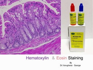
- 1. Hematoxylin & Eosin Staining By Dr.Varughese George
- 2. The Hematoxylin and Eosin stain (H&E) is the most widely used histological stain because : comparative simplicity Ability to demonstrate clearly an enormous number of different tissue structures. Hematoxylin stains cell nuclei blue black shows good intranuclear detail. Eosin stains cell cytoplasm and most connective tissue fibers in varying shades and intensities of pink, orange, and red.
- 3. Stains chemical substances used to achieve visible color contrast in the microscopic picture of a prepared tissue Staining treating tissues or cells with a series of reagents so that it acquires a color; no particles of dye are seen and the stained element is transparent.
- 4. Outlines tissues and cellular components. Identification of tissues. Establishes the presence or absence of disease processes.
- 5. Most commonly used staining methods are – Hematoxylin and Eosin staining in Histopathology Gram’s Stain and Ziehl-Neelson staining in Microbiology. Romanowsky staining in Hematology. Papanicoloau staining in Cytology
- 6. Essentially Aromatic benzene ring compounds or derivatives that possess the twin properties of color and ability to bind to tissue.
- 7. Categories based on Examples Origins Natural (Hematoxylin & Carmine) Synthetic Aniline Physiochemical Properties Fluorescent (acridine orange) Leuco (Leuco methylene blue) Metachromatic (toluidiene blue) Neutral (azure-eosinate) Structure Azo (orange) Metal complex (Al or Fe complexes of hematein) Xanthene (pyronine Y) Use in biological staining Fat (oil red O) Flourescent probe(YOYO-1) Mucin (alcian blue) Use in textile dyeing Acid (eosin) Basic (safrarine) Direct (Congo Red) Supposed mode of action of dye Mordant (Gallacyarine chrome alum Reactive(mercury orange)
- 8. Acidic Dyes Stains basic components such as cytoplasm, acidiophil granules, etc Eosin, Acid Fuchsin Basic Dyes Stains acidic components such as nucleus, basophil granules,etc. Hematoxylin, Basic Fuchsin, Methylene Blue. Neutral Dyes Consists of mixtures of basic and acidic dyes. Both cations and anions contain chromophoric groups and both have colored radicals. Romanowsky dyes formed by interaction of polychrome methylene blue and eosin
- 9. A polyvalent metal ion which forms coordination complexes with certain dyes. A substance which acts as an intermediary between dye and tissue, thus increasing the affinity between them. Strictly applicable to salts and hydroxides of divalent and trivalent metals. Should not be used to indicate any substance that improves in staining in some other manner (accentulators and accelerators)
- 10. The mordant dye complex ‘lake’ combines with tissue to form tissue-mordant-dye complex, which is insoluble.
- 11. Mordant is applied first, followed by the dye. e.g Heidenhain’s iron hematoxylin Mordant and dye are mixed together and then applied. Commonly done in histotechnology e.g Alum hematoxylin solutions Dye applied first, followed by the mordant. Hardly done in histotechnology.
- 12. Accentuators: Substances which increase the staining power of dye. They increase the intensity & selectivity of stain. e.g KOH in Lofflers methylene blue phenol in carbol fuschin & carbol thionin. Accelerators Accentuators used in metallic impregnation technique for the nervous system. e.g chloral hydrate Trapping agents Agents which holds dye combination with tissue or bacteria . e.g tannic acid/iodine
- 13. Absorption or direct staining – tissue penetrated by dye solution. Indirect staining – using intermediate treatment with mordant Physical staining – simple solubility of dye in element of tissue. Chemical staining – formation of new substance e.g. PAS Adsorption phenomenon – accumulation on the surface of the compound.
- 15. Vital Staining Applied to living tissue Accomplished by injecting staining solution into some part of the body Mixing of stain with living cells Primarily used for research. Routine Staining Stains tissues with minimal differentiation except between nucleus and cytoplasm. Demonstrates general relationship among cells, tissues and organs. e.g Hematoxylin and Eosin stains Special Staining Demonstrates selective features of tissues : Particular cell products. Microscopic intracellular and intercellular structure. e.g PAS stain for mucopolysaccharide.
- 16. Regressive Staining •Tissue is initially overstained and then partially decolorized (differentiated) until the proper endpoint is reached. • Sharper degree of differentiation is obtained •The differentiation is controlled visually by microscopic examination. •Faster and more convenient . Progressive Staining •Tissue is stained for a predetermined time for adequate staining of the nuclei and leaves the background tissue relatively unstained. •Once the dye is taken up by the tissues, it is not removed. •The tissue is left in the dye solution until it retains the desired amount of coloration. •The differentiation solely relies on the selective affinity of dyes for different tissue elements.
- 17. Removal or washing out of excess stain until the color is retained only by the tissue component that is to be studied. Done with acid alcohol or ethyl alcohol Exposure to air may oxidize and improve the process.
- 18. All glassware should be thoroughly cleaned. Correct solvent should be used. Silver and osmic acid solutions should be kept in dark bottles. Solutions like dilute ammonia should be freshly prepared. Constituents of stain dissolved should follow the formula. Alcoholic solutions of the stain should be kept in dark stoppered bottles. All dyes should be filtered before use.
- 19. Specially designated bench Staining bench should be facing the window. Slide washing tray made of stainless steel. Bunsen burner to heat up the stain. Thermostatically controlled hot place to melt the wax. Microscope to control staining reaction.
- 20. Slides are stained using – Using staining dishes Small grooved coplin jars with lids. Large staining troughs Using staining racks 2 pieces of stout rods 2-4 cm apart. Using staining machine Same as processing machine but carry slide racks.
- 21. The word Hematoxylin is derived from old Greek word Haimato meaning blood & Xylon meaning wood. A natural dye extracted from the core or heartwood of tree Haematoxylon campechianum. The hematoxylin is extracted from logwood with hot water, and then precipitated out from the aqueous solution using urea. The major oxidization product of hematoxylin is hematein, a natural dye that is responsible for the color properties.
- 22. Waldeyer firmly established the use of hematoxylin in histology in 1862. Bohmer combined hematoxylin with alum as a mordant and obtained more specific staining in 1864. Heidenhan introduced his classical Iron- Alum- Hematoxylin method used as a standard technique in cytology. Ehrlich overcame the instability of hematoxylin and alum by adding glacial acteic acid and simultaneously produced his formula for hematoxylin used today.
- 23. The process of oxidation of hematoxylin. The major oxidization product is hematein, a natural dye that is responsible for the color properties. It is a poor dye but metallic mordant and forms the most powerful stain. Carried out in 2 ways – Natural oxidation. Chemical oxidation.
- 24. Natural Oxidation Carried out by exposure to light and air Slow process – takes about 3-4 months Resultant solutions seem to retain its staining ability for a long time. Advantages Disadvantage Once oxidation has reached an acceptable level, the staining solution may be used and it lasts for longer. •Requires a considerable period of time. •Two batches of naturally ripened product may not produce the same staining qualities. e.g Ehrlich’s & Delafield’s hematoxylin solutions
- 25. Achieved by addition of oxidizing agents such as mercuric oxide, sodium iodate and potassium permanganate. This process converts the hematoxylin haematin almost instantaneously, so these hematoxylin solutions are ready for use after preparation.
- 26. Properties of chemically oxidized hematoxylin Shorter useful life than the naturally oxidized haematoxylins. The process of over-oxidation of hematoxylin has established that the production of oxyhaematein inhibits successful staining. Glycerol has been incorporated in many formulas as it’s a stabiliser to prevent over-oxidation and prevent evaporation.
- 27. Properties of chemically oxidized hematoxylin Haematein is anionic, having a poor affinity for tissue, and is inadequate as a nuclear stain without the presence of a mordant. The mordant/metal cation confers a net positive charge to the dye-mordant complex and enables it to bind to anionic tissue sites, such as nuclear chromatin. The type of mordant used influences strongly the type of tissue components stained and their final color. The most useful mordants for hematoxylin are salts of aluminum, iron, and tungsten, although hematoxylin solutions using lead as a mordant are occasionally used in the demonstration of argyrophil cells).
- 28. Hematoxylin solutions can be arbitrarily classified according to which mordant is used: Alum hematoxylins Iron hematoxylins Tungsten hematoxylins Molybdenum hematoxylins Lead hematoxylins Hematoxylin without mordant.
- 29. Used routinely in the hematoxylin and eosin stain and produce good nuclear staining. The mordant is aluminum in the form of ‘potash alum - aluminum potassium sulfate or ‘ammonium alum -aluminum ammonium sulfate Alum Hematoxylin can be used progressively or regressively.
- 30. Ehrilch’s haematoxylin Mayer’s haematoxylin Harris’ haematoxylin Gill’s haematoxylin Cole’s haematoxylin Delafield’s haematoxylin Carazzi’s haematoxylin
- 31. Naturally ripened strong alum hematoxylin. Stains nuclei intensely and crisply - stained sections fade much more slowly . Stains mucin in salivary glands, cartilage and cement lines of bones Suitable for tissues subjected to acid decalcification. Suitable for tissues that have been stored for a long period in formalin fixatives which have gradually become acidic over the storage period. Suitable for Bouin’s fixed tissue. not ideal for frozen sections.
- 32. Preparation : Hematoxylin 2 g Absolute alcohol 100 ml Glycerol 100 ml Distilled water 100 ml Glacial acetic acid 10 ml Potassium alum 15 g approx.
- 33. Dissolve the hematoxylin in alcohol Incorporate glycerol to slow down the oxidation process and prolong shelf time. Add potassium alum till saturation. The stain may be ripened naturally by allowing to stand in a large flask, loosely stoppered with cotton wool – takes about 2 months. In case of emergency, the stain could be chemically ripened by the addition of sodium iodate using 50mg for every gram of hematoxylin. Filter before use.
- 34. A widely used hematoxylin stain. Chemically ripened with sodium iodate. More vigorous in action than Ehrlich’s hematoxylin. Used as both progressive and regressive stain. Used as a nuclear counterstain in the demonstration of glycogen (PAS,mucicarmine) in various enzyme histological techniques. Stain applied for a short period - 5-10 mins until nuclei are stained and then blued without any differentiation which might destroy/decolor the stained cytoplasmic components.
- 35. Preparation Hematoxylin 1 g Distilled water 1000 ml Potassium or ammonium alum 50 g Sodium iodate 0.2 g Citric acid 1 g Chloral hydrate SLR 50 g or Chloral hydrate AR 30 g
- 36. The hematoxylin, potassium alum, and sodium iodate are dissolved in distilled water by warming and stirring, or by allowing to stand at room temperature overnight. Chloral hydrate and citric acid are added & the mixture is boiled for 5 minutes, then cooled and filtered. Chloral hydrate acts as a preservative and citric acid sharpens nuclear staining
- 37. This alum hematoxylin was traditionally chemically ripened with mercuric oxide (sodium or potassium iodate is frequently used as a substitute for oxidation). It gives particular clear nuclear staining. It is used as a regressive stain in routine histology practice. It is used as a progressive stain in diagnostic exfoliative cytology. When using it as a progressive stain, an acetic acid-alcohol rinse provides a more controllable method in removing excess stain from tissue components and the glass slide.
- 38. Preparation Hematoxylin 2.5 g Absolute alcohol 25 ml Potassium alum 50 g Distilled water 500 ml Mercuric oxide 1.25 g or Sodium iodate 0.5 g Glacial acetic acid 20 ml
- 39. Dissolve hematoxylin in alcohol Add it to alum previously dissolved in warm distilled water The mixture is rapidly brought to boil Mercuric oxide is then slowly and carefully added, when the solution turns dark purple. The stain is rapidly cooled under tap water. Optional addition of glacial acetic acid. Filter before use.
- 40. Available in 3 concentrations – ◦ Gill’s I (normal) ◦ Gill’s II (double) ◦ Gill’s III (triple) most concentrated. More frequently used than Mayer’s hematoxylin for routine H&E staining. More stable than Harris’s hematoxylin, as auto-oxidation is inhibited to the extent.
- 41. Preparation of solution Hematoxylin 2 g Sodium iodate 0.2 g Aluminum sulfate 17.6 g Distilled water 750 ml Ethylene glycol (ethandiol) 250 ml Glacial acetic acid 20 ml
- 42. Advantages Fast in action. Stable for at least months. Produce little or no surface precipitate. Their preparation doesn’t involve boiling the solution. Disadvantages Staining of gelatin is adhesive and even the glass itself. Some mucus may also stain darkly, as compared to Harris’s hematoxylin.
- 43. Alum hematoxylin, artificially ripened with an alcoholic iodine solution. Has good keeping qualities and is suitable for use especially in sequence with celestine blue unlike Ehrlich’s hematoxylin
- 44. Preparation Hematoxylin 1.5 g Saturated aqueous potassium alum 700 ml 1% iodine in 95% alcohol 50 ml Distilled water 250 ml
- 45. The hematoxylin is dissolved in warm distilled water and mixed with iodine solution. The alum solution is added and the mixture brought to boil then cooled quickly and filtered. The solution is ready for immediate use, but may need on occasion filtering after storage.
- 46. Alum hematoxylin which is chemically ripened using potassium iodate Used as a progressive nuclear counterstain. Largely confined to use with frozen sections because it gives excellent and clear nuclear staining with a very short staining time.
- 47. Preparation – Hematoxylin 5 g Glycerol 100 ml Potassium alum 25 g Distilled water 400 ml Potassium iodate 0.1 g
- 48. Hematoxylin is dissolved in the glycerol Alum is dissolved in most of the water overnight. The alum solution is added slowly to the hematoxylin solution. Potassium iodate is dissolved in the rest of the water with gentle warming & is then added to the haematoxylin-alum-glycerol mixture. The final staining solution is mixed well and is then ready for immediate use; It remains usable for about 6 months.
- 49. A naturally ripened alum hematoxylin with similar longevity to Ehrlich’s hematoxylin. Preparation Hematoxylin 4 g 95% alcohol 125 ml Saturated aqueous ammonium alum 400 ml (15 g/100 ml) Glycerin 100 ml
- 50. The hematoxylin is dissolved in 25 ml of alcohol, and then added to the alum solution. This mixture is allowed to stand in light and air for 5 days and then filtered. Glycerin and a further 100 ml of 95% alcohol are added to this mixture. Allow the stain to stand exposed to light and air for about 3–4 months or until sufficiently dark in color. Filter before use.
- 51. Staining time varies according to various factors: Type of hematoxylin used e.g. Ehrlich’s hematoxylin - 20–45 minutes Mayer’s hematoxylin - 10–20 minutes. Age of stain. As the stain ages, the staining time will need to be increased. Intensity of use of stain. A heavily used hematoxylin will lose its staining powers more rapidly and longer staining times will be necessary. Whether the stain is used progressively or regressively e.g. Mayer’s hematoxylin used progressively 5–10 minutes and used regressively 10–20 minutes.
- 52. Pre-treatment of tissues or sections. e.g. length of time in fixative or acid decalcifying solution or whether paraffin or frozen sections. Post-treatment of sections e.g. subsequent acid stains such as van Gieson. Personal preference.
- 53. Oxazine Dye Has little useful coloring property of its own It forms an additional strong mordant with certain hematoxylins. Used as a preliminary to alum hematoxylin staining. Resistant to the effects of acid. Ferric salt in the prepared celestine blue solution strengthens the bond between the nucleus and the alum hematoxylin to provide a strong nuclear stain which is reasonably resistant to acid.
- 54. Preparation Celestine blue B 2.5 g Ferric ammonium sulfate 25 g Glycerin 70 ml Distilled water 500 ml
- 55. Ferric ammonium sulphate is dissolved in cold distilled water with stirring. The celestine blue B is added to this solution and the mixture is boiled for few minutes. Filtered Glycerine is added Filter before use
- 56. Weigert’s Hematoxylin. Heidenhain’s Hematoxylin. Verhoeff’s Hematoxylin. Loyez Hematoxylin.
- 57. In these hematoxylins, iron salts such as ferric chloride and ferric ammonium sulfate are used both as the oxidizing agent and as mordant. Over-oxidation of the hematoxylin is a problem with these stains. Mordant/oxidant and hematoxylin solution are prepared separately and mixed before use. Capable of demonstrating wider range of tissue structures compared to alum haematoxylin. Techniques are more time consuming and needs microscopic control for accuracy.
- 58. An iron hematoxylin used as a nuclear stain in techniques where acidic staining solutions are applied to the sections subsequently e.g Van Gieson stain – picric acid is a constituent which have marked decolorizing action on nuclei stained with alum hematoxylin. It is a useful stain, with eosin, for CNS tissues.
- 59. Preparation a) Hematoxylin solution Hematoxylin 1 g Absolute alcohol 100 ml This is allowed to ripen naturally for 4 weeks before use. b) Iron solution 30% aqueous ferric chloride (anhydrous) 4 ml Hydrochloric acid (concentrated) 1 ml Distilled water 95 ml
- 60. This iron hematoxylin uses ferric ammonium sulfate as oxidant/mordant It is used as the differentiating fluid. It is a cytological stain. It is used regressively. After staining, all components are black or dark gray-black. The hematoxylin staining is removed progressively from different tissue structures at different rates using the iron alum solution. It may be used to demonstrate Chromatin Chromosomes Nuclei Centrosomes Mitochondria Muscle striations Myelin
- 61. Preparation a. Hematoxylin solution Hematoxylin 0.5 g Absolute alcohol 10 ml Distilled water 90 ml The hematoxylin is dissolved in the alcohol, and the water is then added. The solution is allowed to ripen naturally for 4 weeks before use. b. Iron solution (5% iron alum) Ferric ammonium sulfate 5 g Distilled water 100 ml It is important that only the clear violet crystals of ferric ammonium sulfate be used.
- 62. This iron hematoxylin uses ferric ammonium sulfate as the mordant. Differentiation is by Weigert’s differentiator (borax and potassium ferricyanide). Used to demonstrate myelin. Can be applied to paraffin, frozen, or nitrocellulose sections.
- 63. This iron hematoxylin is used to demonstrate elastic fibers after all routine fixative. Ferric chloride is included in the hematoxylin staining solution, together with Lugol’s iodine, and 2% aqueous ferric chloride is used as the differentiator. Coarse elastic fibres stain black, but the staining of fine fibers may be less than satisfactory. The differentiation step is critical to the success of this method.
- 64. Widely used tungsten hematoxylin is PTAH (Phosphotungstic acid hematoxylin Technique). Used to demonstrate fibrin, muscle striations, cilia and glial fibres. Myelin can also be demonstrated Widely used as a CNS stain.
- 65. Preparation PTAH solution using haematin Haematin 0.59 g Phosphotungstic acid 5g Distilled water 500ml Stain is ready to use immediately, but short-lived. PTAH Solution (KMnO4) Haematoxylin 0.59 g Phosphotungstic acid 5g Distilled water 500ml 0.25% Aqueous KMnO4 25 ml Peak staining activity after 7 days
- 66. Results Muscle striations / neuroglia fibres / fibrin / amoeba – Dark Blue. Nuclei/cilia/RBC – Blue Myelin – Lighter blue Collagen /Osteoid / Cartilage / Elastic fibres – Deep brownish red. Cytoplasm – Pale pinkish brown.
- 67. Hematoxylin solution using molybdic acid as mordant. Rare stain Used in demonstration of collagen, coarse reticulin. Also stains Argentaffin cell granules.
- 68. Preparation a. Hematoxylin solution Hematoxylin 2.5 g Dioxane 49 ml Hydrogen peroxide 1 ml b. Phosphomolybdic acid solution Phosphomolybdic acid 16.5 g Distilled water 44 ml Diethylene glycol 11 ml The resultant dark violet solution is allowed to stand for 24 hours before use.
- 69. Results – Collagen and coarse reticulin - violet to black Argentaffin cells - black Nuclei pale - blue Paneth cells - orange Tissue fixed in dichromate do not give good results.
- 70. Used in demonstration of granules in endocrine cells of ailmentary tract and other regions. Most practical diagnostic application is in identification of endocrine cells in tumors of doubtful origin. Also used in localisation of gastrin secreting cells in stomach.
- 71. Freshly prepared hematoxylin is used to demonstrate various minerals in tissue sections. These methods are now replaced with more specific techniques.
- 72. Xanthine dyes which stains connective tissue and cytoplasm in varying intensity and shades (red to pink). Available in the following types : Eosin Y ( Eosin Yellowish, Eosin water soluble) – most widely available. Ethyl Eosin (Eosin S, eosin alcohol soluble). Eosin B ( Eosin Bluish, Erythrosine B). Ethyl eosin and eosin B are now rarely used, although occasional old methods specify their use – e.g the Harris stain for Negri bodies.
- 73. Eosin Y Most commonly used eosin. Readily soluble in water. Satisfactorily soluble in alcohol. Preparation Eosin Y, water soluble 5 gm Distilled water 1000 ml Crystals of Thymol added to inhibit fungal growth. Addition of little acetic acid (0.5 -1000 ml stain) sharpens the staining.
- 74. Principle Hematoxylin and Eosin are principle stains used for demonstration of nucleus and cytoplasm. Alum acts as a mordant and the hematoxylin containing alum stains the nucleus light blue which turns red in the presence of acid. The cell differentiation is achieved by treating the tissue with acid solution. The counterstaining is performed using eosin which imparts pink color to cytoplasm.
- 75. 1. REMOVAL OF WAX. 2. HYDRATION WITH GRADED ALCOHOLS. 3. STAINING. 4. DIFFERENTIATION 5. BLUEING 6. COUNTERSTAIN WITH EOSIN 7. DEHYDRATION THROUGH GRADED ALCOHOL. 8. CLEARING IN XYLENE 9. MOUNTING UNDER A COVER SLIP.
- 76. REMOVAL OF WAX Removal of wax with xylene (impermeable to stains). 2-3 minutes of xylene immersion sufficient for sections of 10µ thickness. Facilitated by warming the slides at 60 degrees oven to melt the wax.
- 77. HYDRATION WITH GRADED ALCOHOLS Sections are transferred to absolute alcohol for 1-2 minutes – til it becomes opaque. Sections rinsed in 2nd bath of alcohol, drained and taken to water. Any pigments or deposits should be removed at this stage.
- 78. STAINING Slides immersed in hematoxylin (Mayers / Harris/ Gills) If regressive stain is used, longer time is required to overstain the structures
- 79. DIFFERENTIATION Sections are dipped in acid alcohol, agitated and washed in tap water. Observed under microscope If underdifferentiated – returned to acid alcohol. If overdifferentiated – returned to hematoxylin and differentiation repeated.
- 80. BLUEING Slides after draining off hematoxylin is transferred to ammonia water for 2 minutes. Section when removed from hematoxylin or acid alcohol are pink in color. Washing turns them blue Blueing. Blueing solutions are usually preferred alkaline . a)Ammonium hydroxide in 70% alcohol b) lithium carbonate stock solution c) Scott’s tap water Magnesium sulfate (MgSO4) 30.0 gm Sodium bicarbonate 2.0 gm Tap water 3000.0 ml d) tap water pH - 7 COUNTERSTAIN WITH EOSIN Transfer the slides to 1% aqueous eosin for 2 minutes. Wash in running water.
- 81. DEHYDRATION THROUGH GRADED ALCOHOL. After staining the sections are transferred to 90% alcohol and agitated for 10 seconds followed by then to absolute alcohol 1for (10-15 sec) followed by absolute alcohol 2 for 30 sec.
- 82. Slides are transferred to xylene 1 and left until completely clear . It should betestedfor clarity. Then its transferred to xylene 2which they be mounted.
- 83. MOUNTING Its required to maintain high refractive index necessary for microscopy and to protect the sections during storage. METHOD : A drop the mountant is placed on the section , place the convenient sized coverslip into position. (Air bubbles may be removed by gentle pressure on the coverslip) MOUNTANTS : Aqueous mountants Resinous mountants
- 84. USES : • To be used with metachromatic dyes. • Standard mountant for fat tissues. Low refracting index of 1.4 – 1.42. Glycerin is added to prevent cracking and splitting of dyes. Bacteriostatic agent should be added (thymol). Types : • Gelatin media • Gum arabic media Apathy’s medium – fluorescent microscopy Highman’s modified apathy’s medium Farrant’s medium Fructose syrup
- 85. Its composed of natural or synthetic resins. Stained preparations are most transparent when the refractive index is 1.54. Natural resins : ◦ Canada balsam dissolved in xylol to 55-70%. ◦ Dammar balsam. ◦ Colophonium resin – used in alcoholic solutions. ◦ Terpene resin. Synthetic resins : ◦ Plasticizers such as tricreysl phosphate or dibutyl phatalate is added. ◦ Commonly used is kirkpatrick and lendrum’s DPX
- 86. DIESTRENE PLASTIZISER XYLENE Distrene 80 10 gms Dibutylphthalate 5 ml Xylene 35 ml The slides can be cleaned of excess mountant
- 87. To coat the edges of coverslip , so that no air bubbles develop. Commonly used are : ◦ Paraffin wax ◦ Cement ◦ Varnish
- 88. Results Nuclei appear blue/black in color Cytoplasm appears in varying shades of pink Muscle fibers appear deep pink/red in color Red blood cells appear orange/red in color Fibrin appears deep pink in color.
- 89. 1. HYDRATION WITH GRADED ALCOHOLS. 2. STAINING. 3. DIFFERENTIATION 4. BLUEING 5. COUNTERSTAIN WITH EOSIN 6. DEHYDRATION THROUGH GRADED ALCOHOL. 7. CLEARING IN XYLENE 8. MOUNTING UNDER A COVER SLIP.
- 90. 1. Remove polyethylene glycol fixative in 50% alcohol, 2 minutes. 2. Hydrate in 95% alcohol, 2 minutes, and 70% alcohol, minutes. 3. Rinse in water, 1 minute. 4. Stain in Harris’s hematoxylin, 5 minutes. 5. Rinse in water, 2 minutes. 6. Differentiate in 0.5% aqueous hydrochloric acid,10 seconds ,approx. 7. Rinse in water, 2 minutes. 8. ‘Blue’ in Scott’s tap water substitute, 2 minutes.
- 91. 9. Rinse in water, 2 minutes. 10. Dehydrate, 70% alcohol for 2 minutes. 11. Dehydrate, 95% alcohol, 2 minutes. 12. Dehydrate, 95% alcohol, 2 minutes. 13. Stain in OG 6, 2 minutes. 14. Rinse in 95% alcohol, 2 minutes. 15. Rinse in 95% alcohol, 2 minutes. 16. Stain in EA 50, 3 minutes. 17. Rinse in 95% alcohol, 1 minute.
- 92. Results Nuclei appear blue/black in color. Cytoplasm (non-keratinizing squamous cells) appear blue/green in color. Keratinizing cells appear pink/orange in color.
- 93. 1. Freeze suitable tissue block onto a chuck. 2. Cut cryostat sections at 3–6 μm thickness. 3. Fix section in 10% neutral buffered formalin at room temperature for 20 seconds. 4. Rinse in tap water. 5. Stain in double strength Carazzi’s hematoxylin for 1 minute. 6. Wash well in tap water for 10–20 seconds. 7. Stain in 1% aqueous eosin for 10 seconds. 8. Rinse in tap water. 9. Dehydrate, clear, and mount.
- 94. 1. 2. 3. 4. 5. 6. 7. 8. 9. Dewax and hydrate paraffin sections, removing mercury precipitate if indicated. Oxidize for 5 minutes in 0.5% aqueous periodic acid. Rinse in tap and then in distilled water. Place in Schiff’s reagent for 15 minutes ( 10 minutes for frozen sections). Rinse for 2 minutes in each of three changes of freshly made sulfite rinse. Wash 5 to 10 minutes in running tap water. Optional counterstain with Harris’ hematoxylin for 1 -3 minutes or in light green (0.1% in 0.1% acetic acid) for 5-20 seconds. Light green is especially useful when searching for fungi. If hematoxylin is used, differentiate by means of 3-5 quick dips in 1% acid alcohol, wash in tap water and blue in Scott’s tap water substitute; then wash 5 minutes in running water. Dehydrate, clear and mount.
- 95. Results Nuclei appear blue in color. PAS +ve materials appear magneta (purple-red) in color.