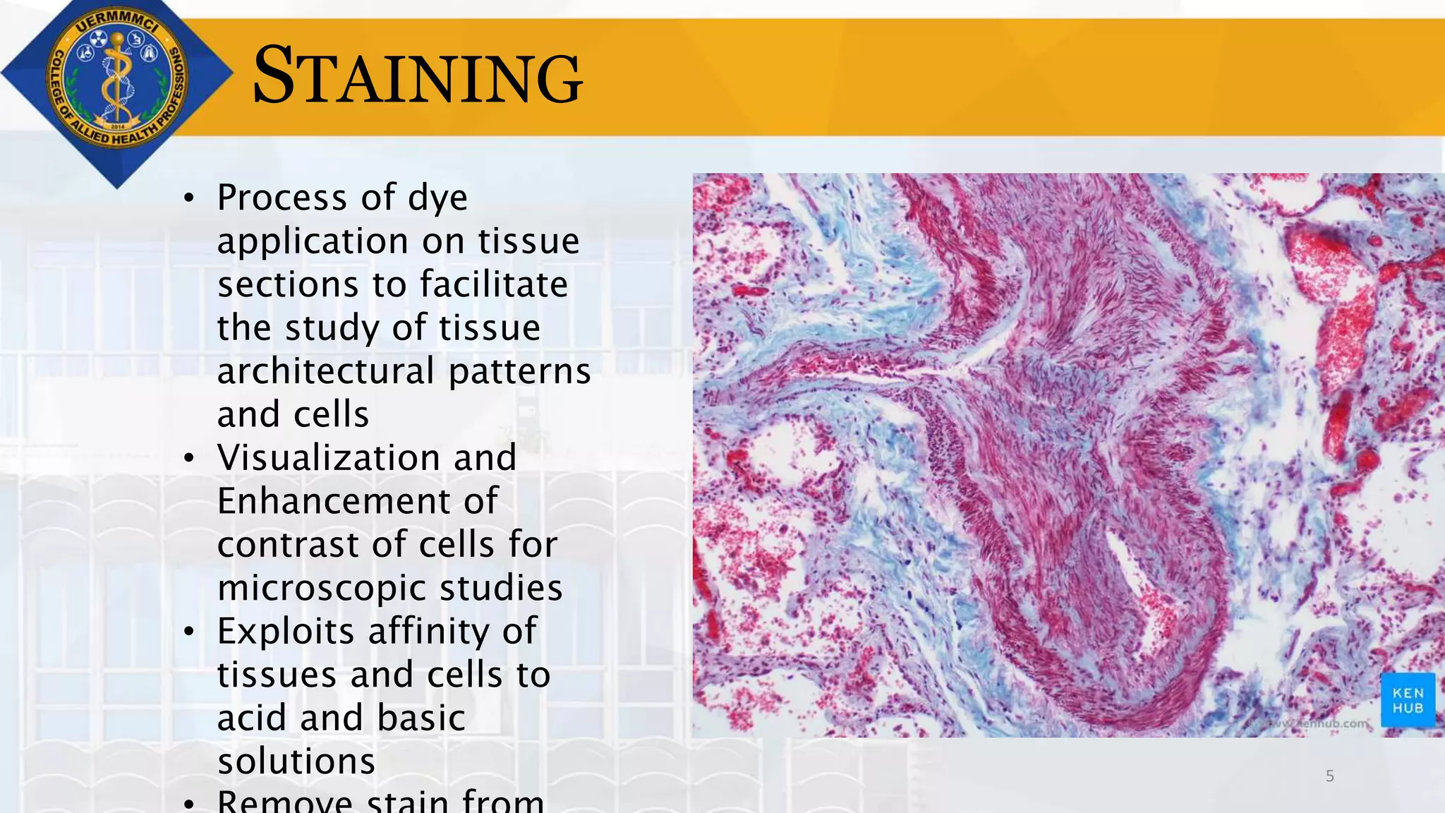
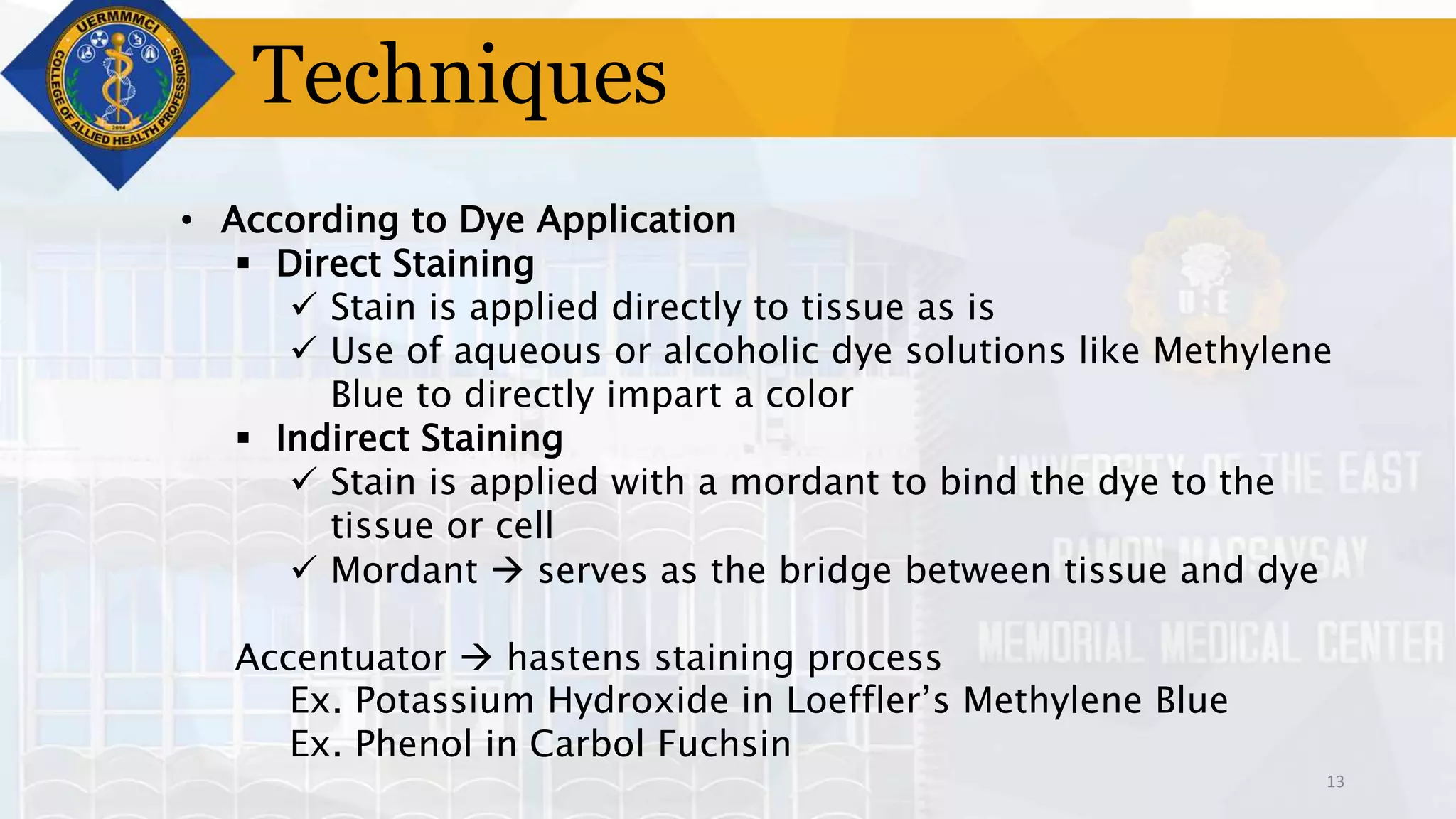
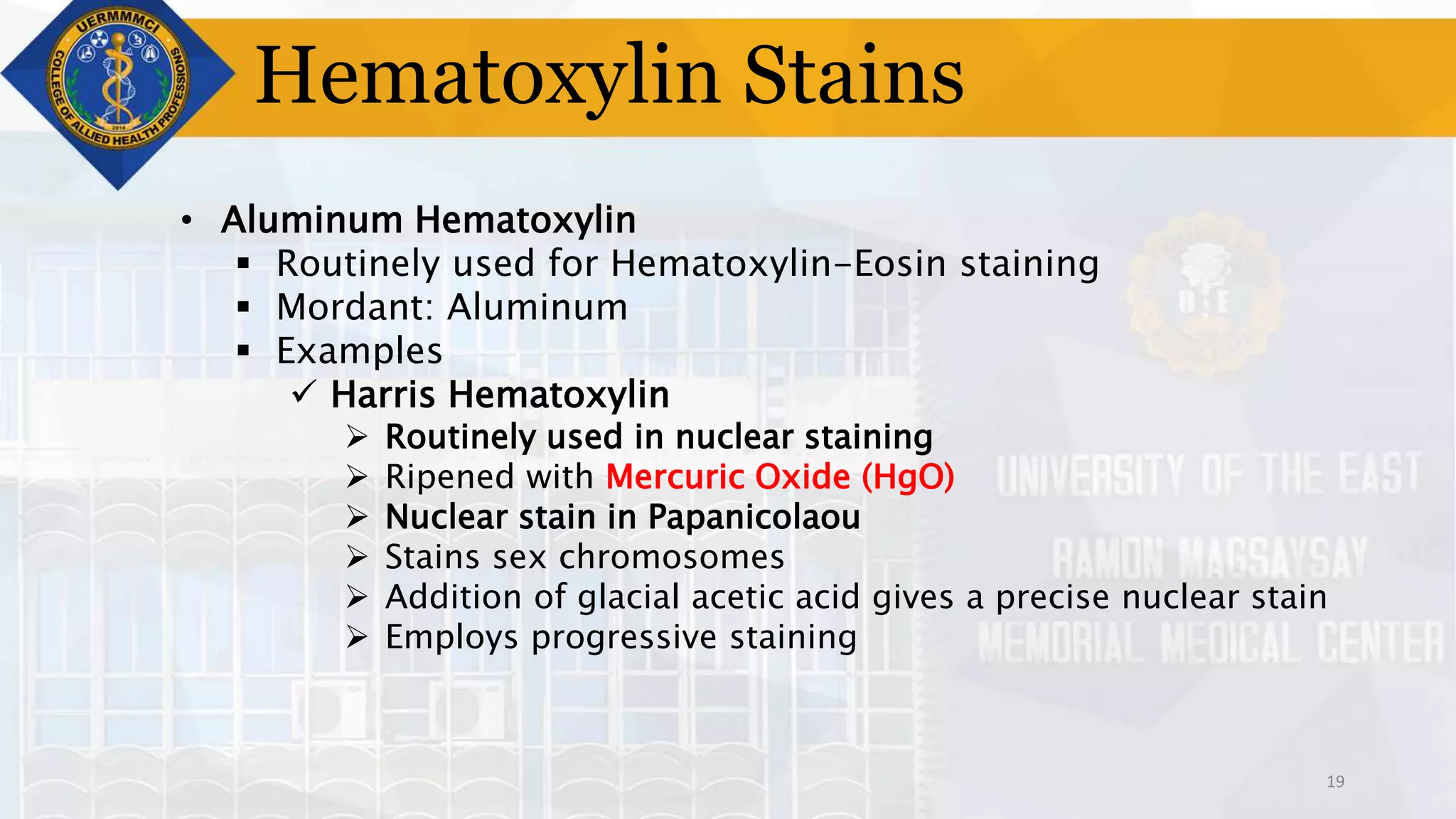
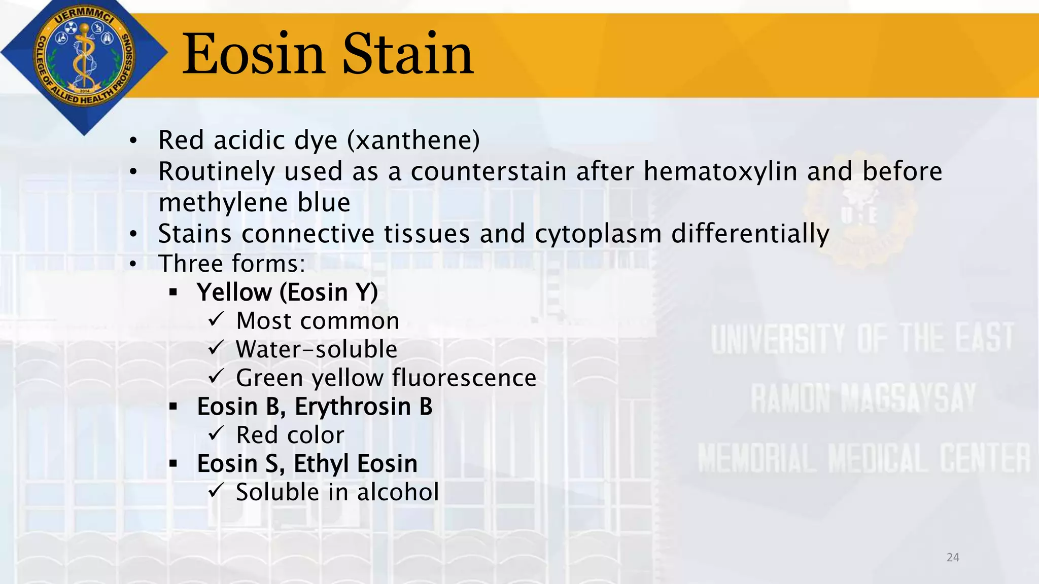
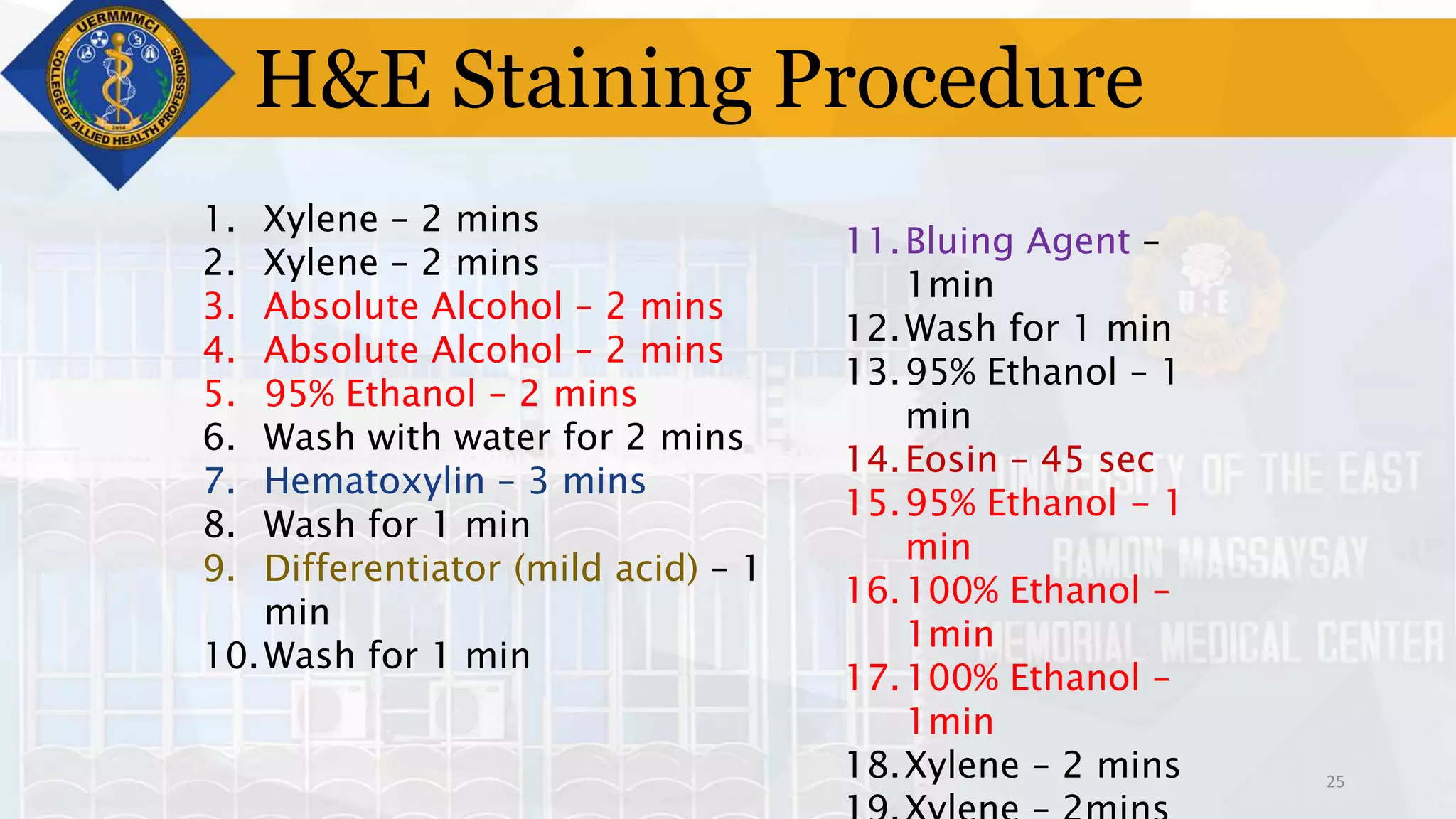
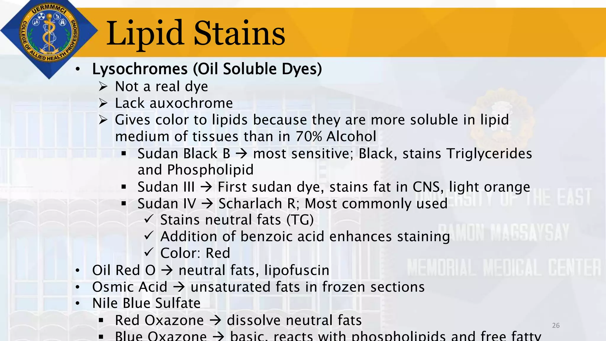
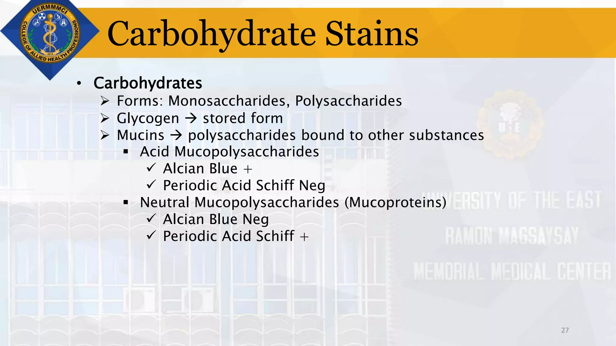
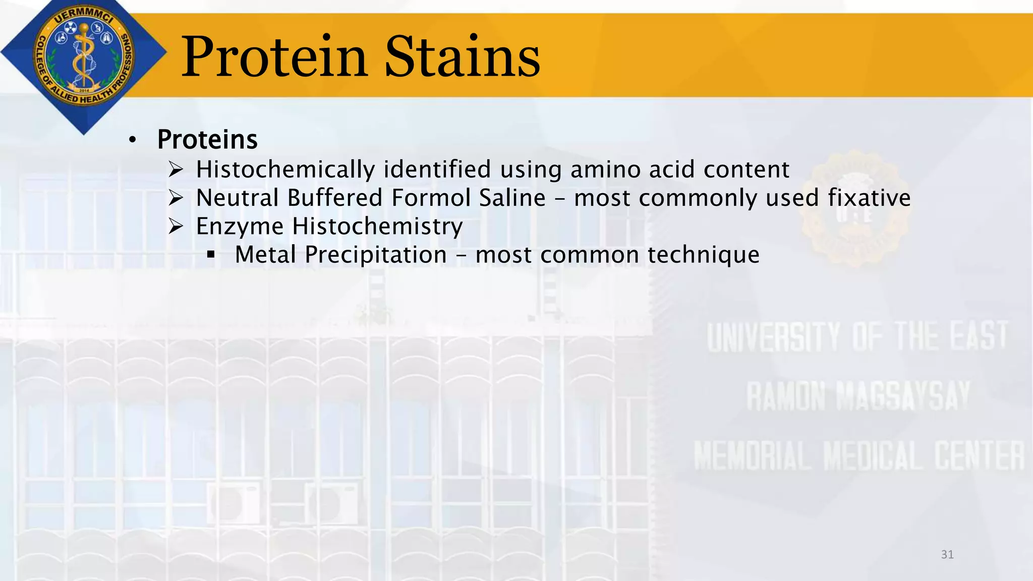
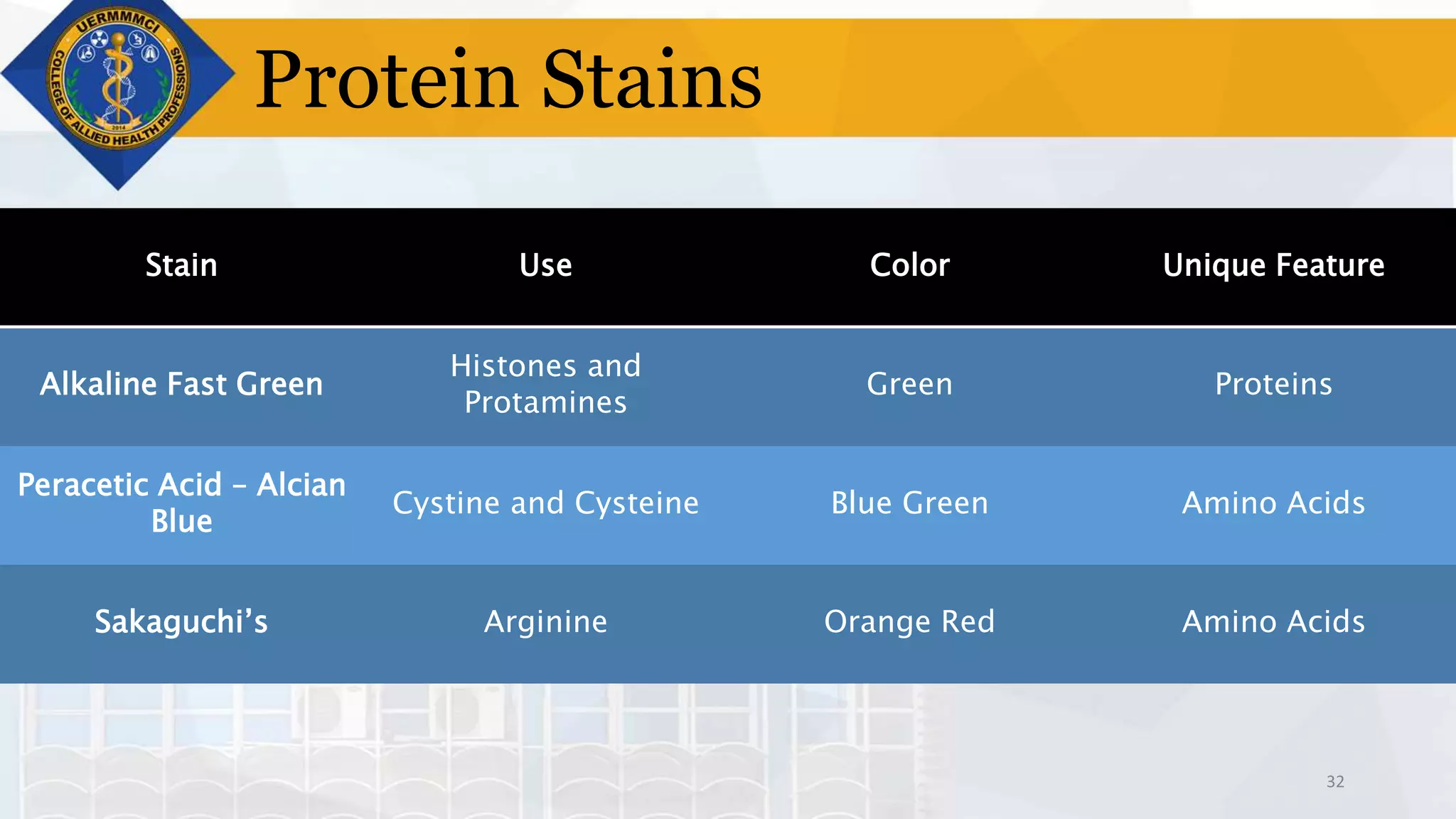
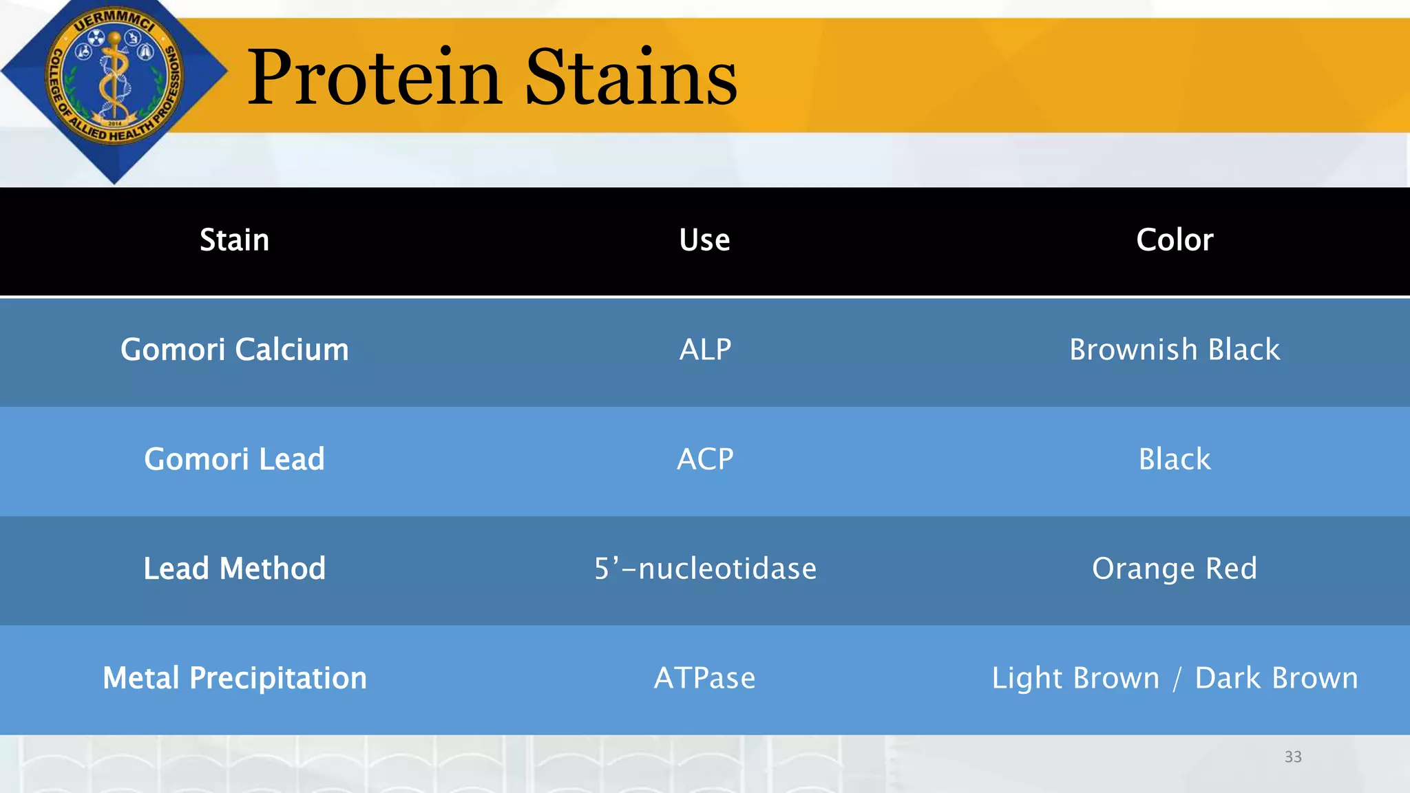
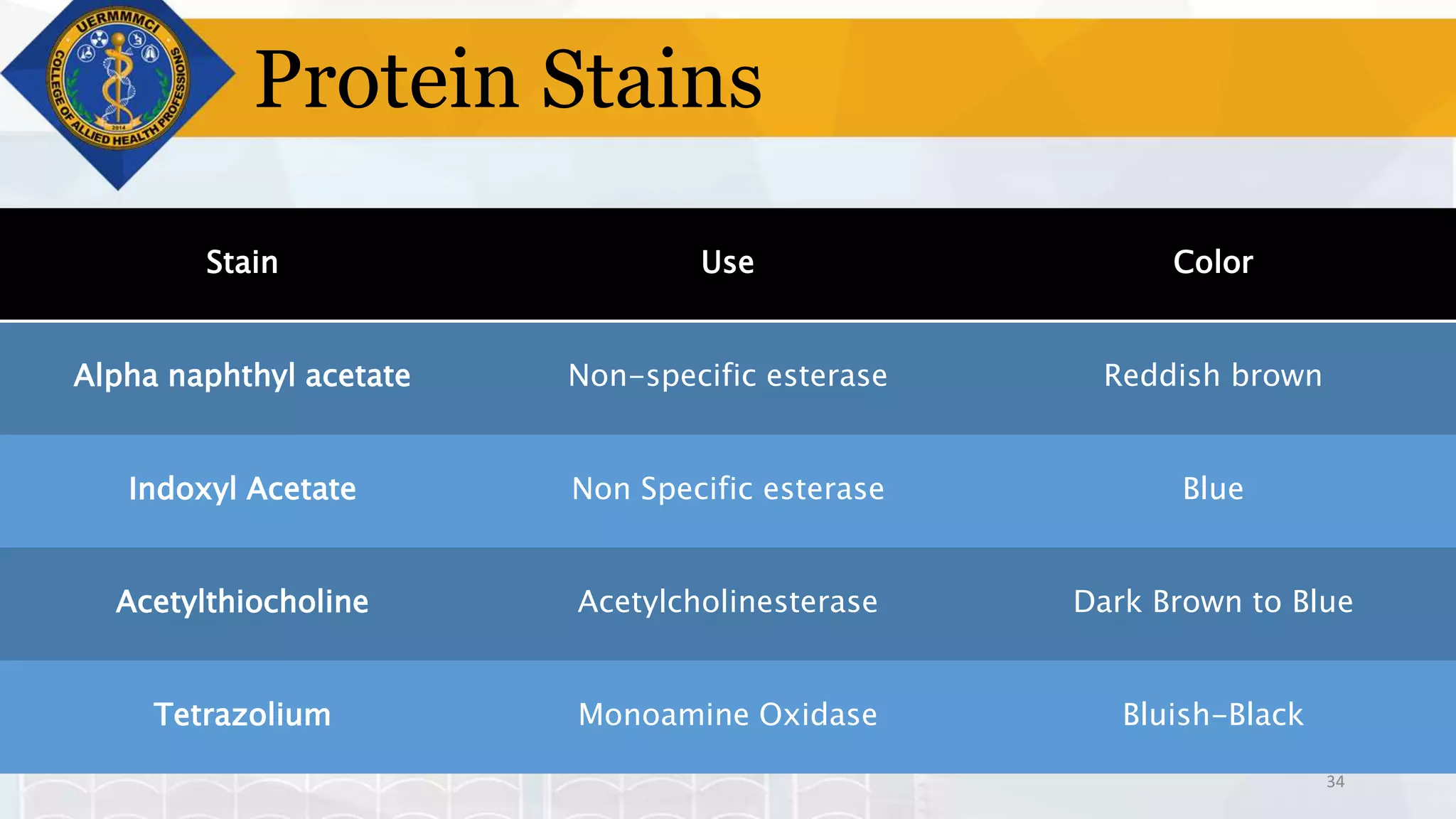
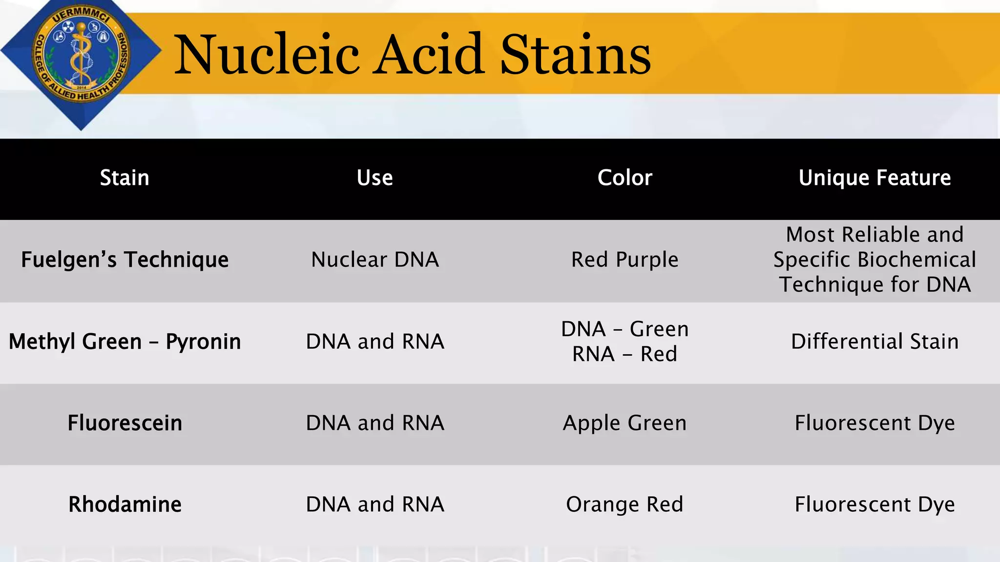
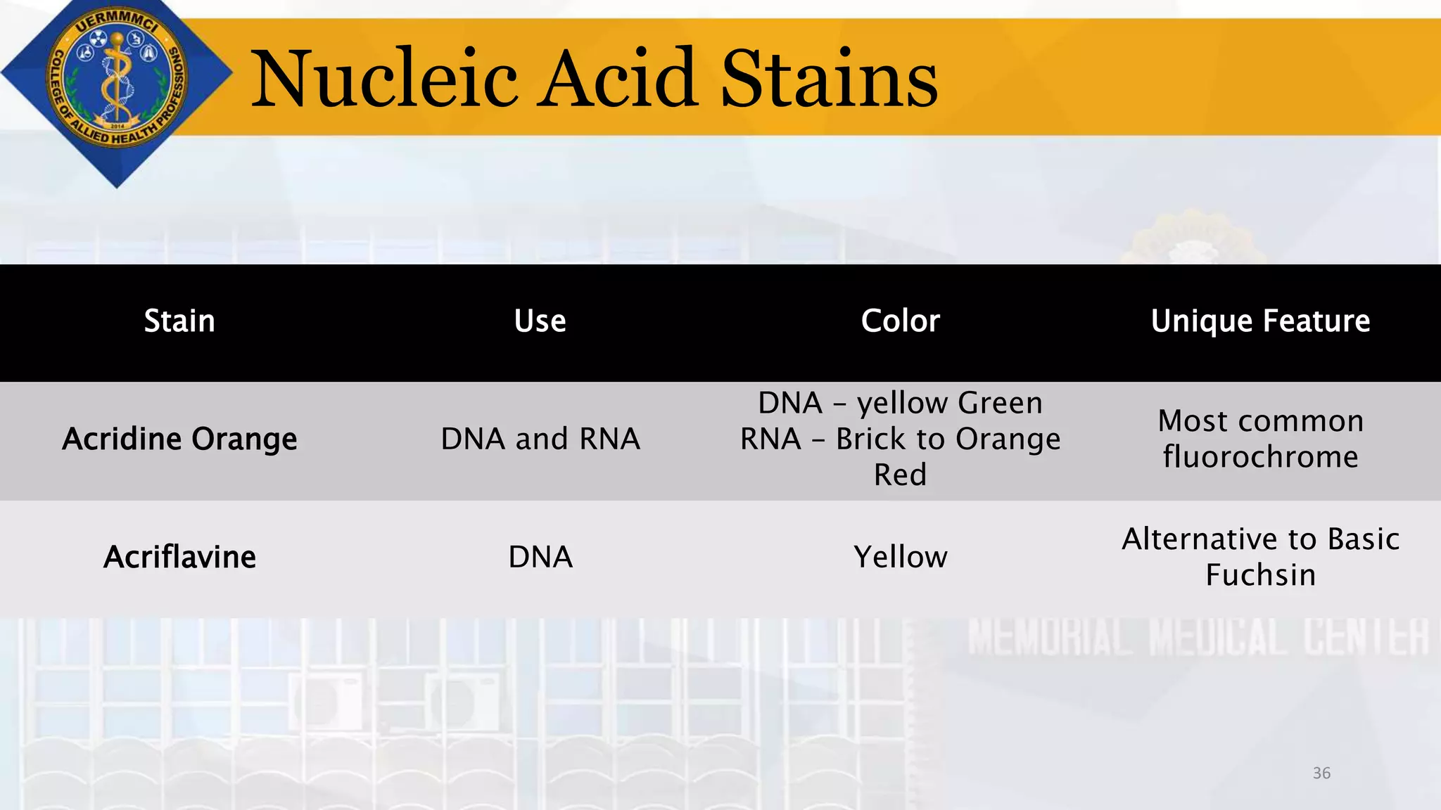
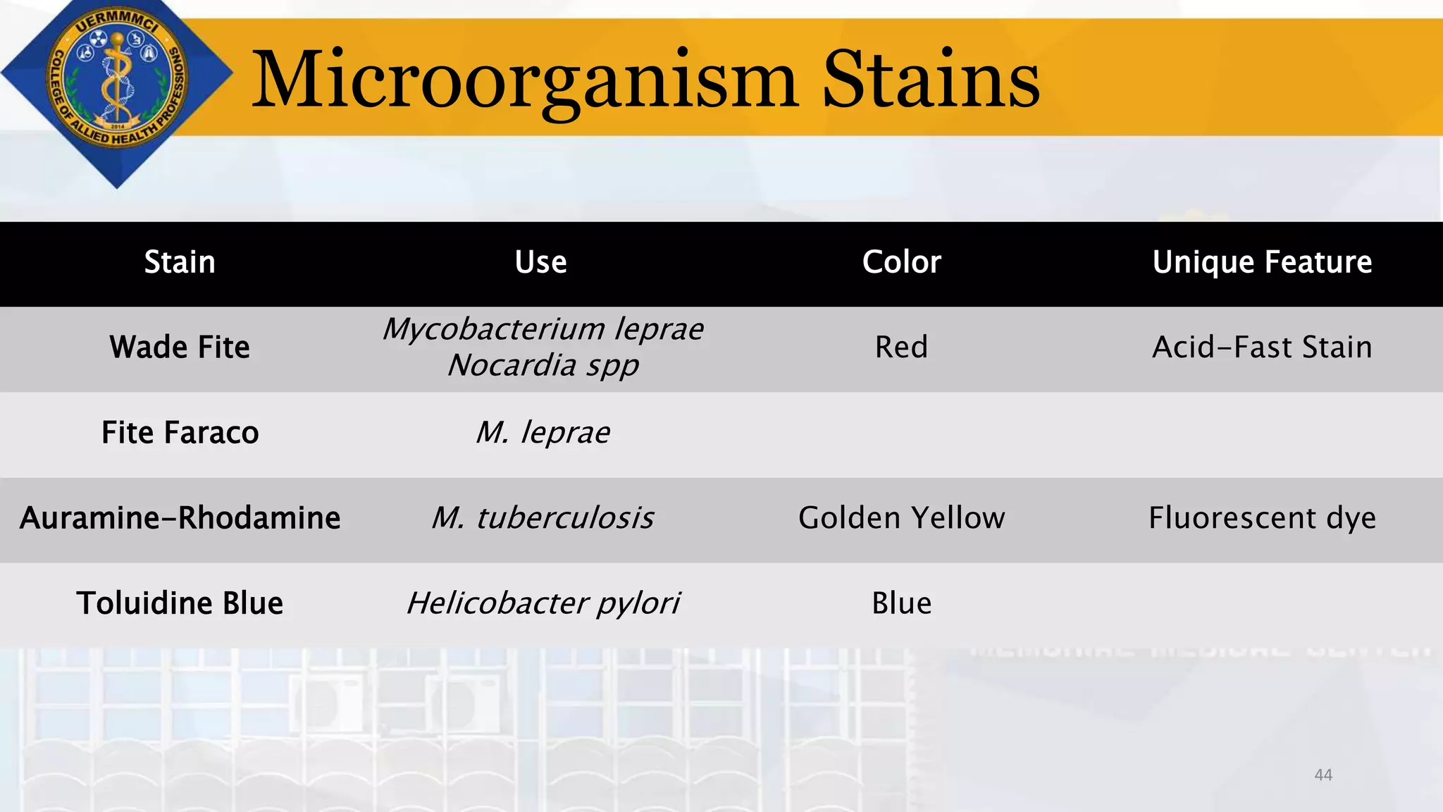
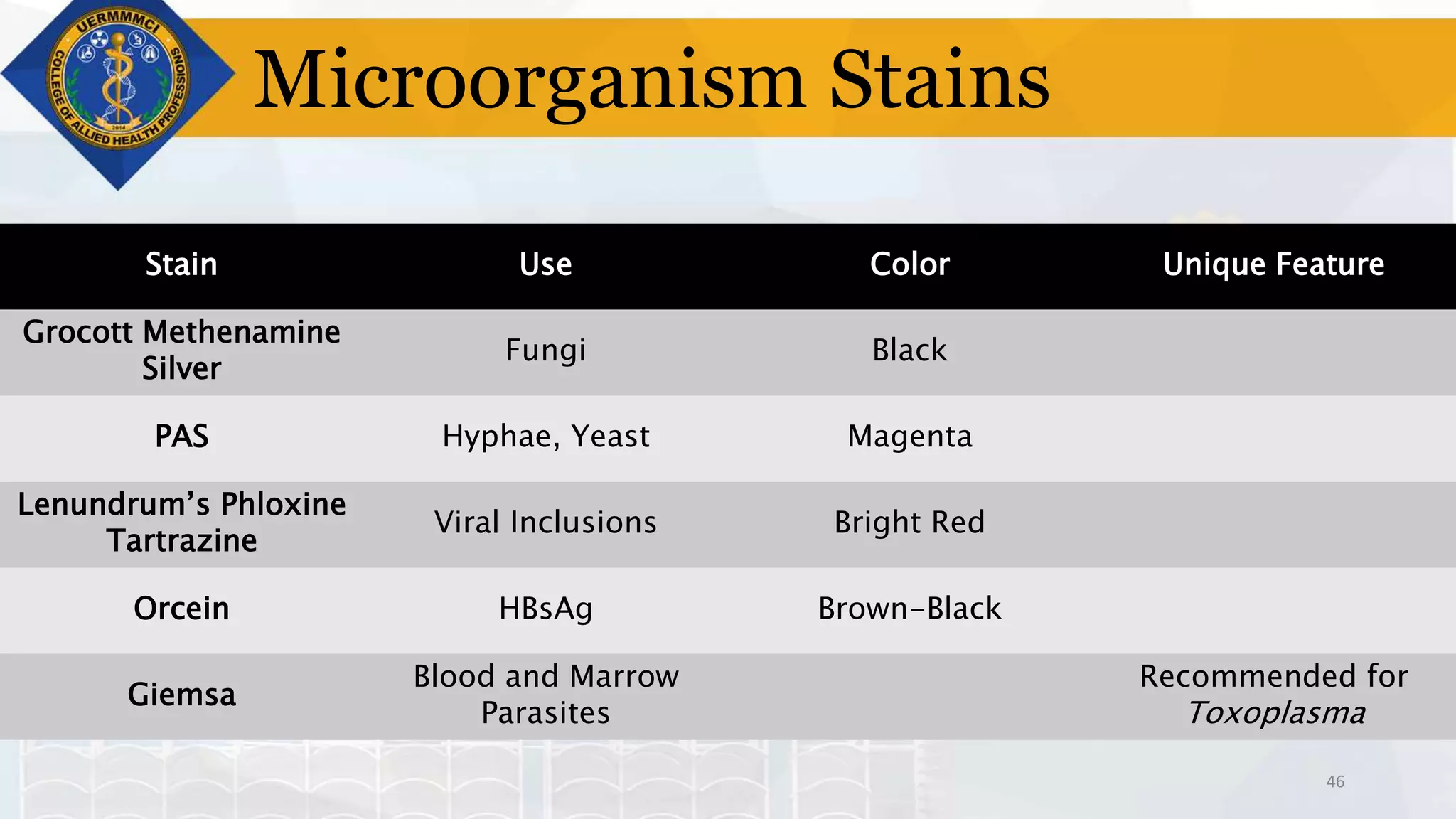
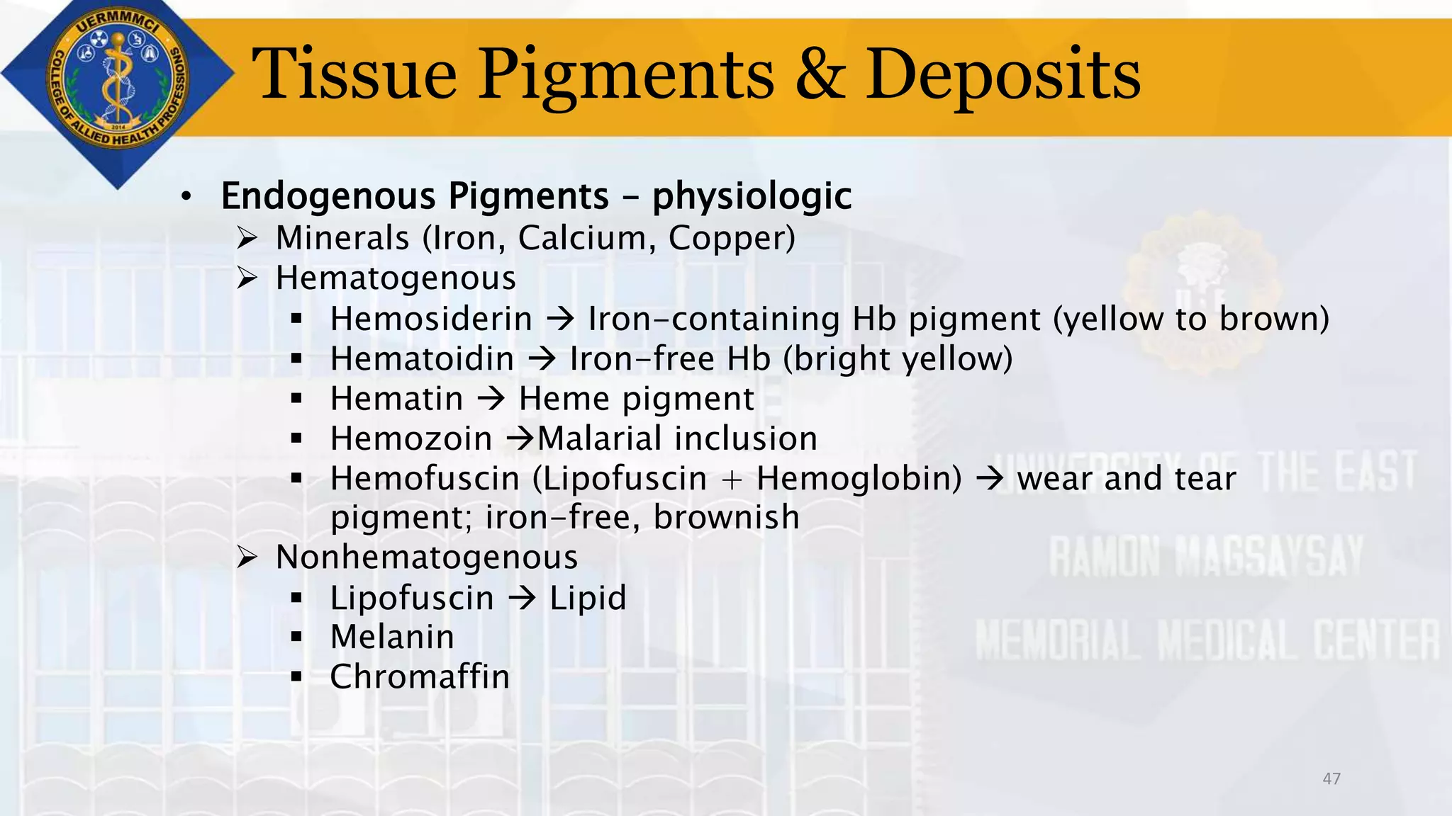
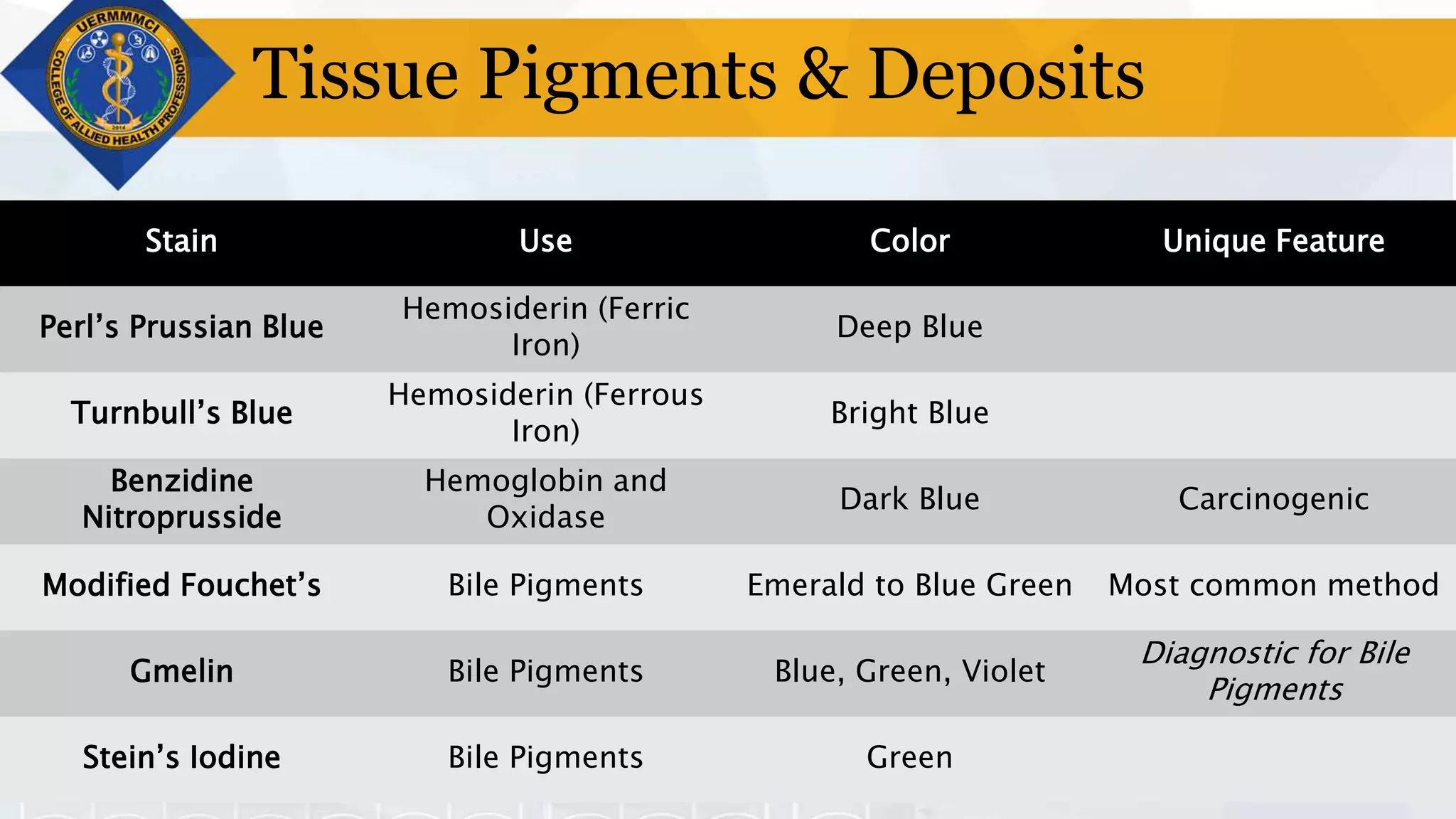
1
TISSUE PROCESSING
1. NUMBERING
2. FIXATION
3. DECALCIFICATION
4. DEHYDRATION
5. CLEARING
6. IMPREGNATION
7. EMBEDDING
8. TRIMMING
9. SECTIONING
10.STAINING
11.MOUNTING
12.LABELING
HISTOPATHOLOGIC TECHNIQUES
STAINING
Presented by : Ronel Wynlor D. Abarca, RMT
Creator of all things, true source of Light and Wisdom, lofty
source of all being, graciously let a ray of light of Your brilliance
penetrate into the darkness of our understanding and take from
us the double darkness in which we have been born, the
darkness of sin and ignorance.
Give us a sharp sense of understanding, a retentive memory,
and the ability to grasp things correctly, and fundamentally.
Grant us the talent of being exact in our explanations, and the
ability to express ourselves with thoroughness and charm.
Point out the beginning, direct the progress, help in the
completion.
We ask these through Christ, your Son, our Lord, Amen.
4
LEARNING OUTCOMES
1. Define and Differentiate an Accentuator from a Mordant.
2. Enumerate the different types of stains.
3. List the stains used for carbohydrates, proteins, nucleic acid.
4. List the stains used for connective, muscle, skeletal, and
nervous tissues.
5. List the stains used to visualize microorganisms in tissues.
6. Know which stain is best used for a certain tissue specimen.
7. Explain the principles and process of H&E staining.
5
STAINING
• Process of dye
application on tissue
sections to facilitate
the study of tissue
architectural patterns
and cells
• Visualization and
Enhancement of
contrast of cells for
microscopic studies
• Exploits affinity of
tissues and cells to
acid and basic
solutions
6
Classifications
• According to pH  simplest yet most important classification
 Acidic Dyes
 Affinity towards the Cytoplasm which is a basic structure
(Acidophilic)
 Ex. Eosin, Picric Acid
 Basic Dyes
 Affinity towards the Nucleus which is an acidic structure
(Basophilic)
 Ex. Methylene Blue
 Neutral Dyes
 Combined Acidic and Basic Dyes
 Stains Cytoplasm and Nucleus simultaneously but with
different colors
 Ex. Romanowsky Stain (Eosin and Azure B or Methylene
7
Classifications
• According to Function
 Histologic Stains
 Stains that directly interact with the tissue or cell
 Histochemical Stains
 Stains that chemically react with tissue constituents
 Ex. Perl’s Prussian Blue reacts with Iron constituents
 Ex. Periodic Acid Schiff reacts with Glycogen components
 Immunohistochemical Stains
 Stains that are labelled with antibodies to target specific
tissue antigens
8
Classifications
• According to Source
 Natural Dyes
 Stains that are derived from plant and animal
products
 Synthetic Dyes
 Stains that are chemically generated, usually
from hydrocarbons like benzene
9
Natural Dyes
• Hematoxylin
 Derived from Hematoxylum campechianum (logwood)
 Most commonly used for histologic studies
 Not in itself a DYE!
 Hematin  active coloring agent obtained from ripening
 Natural Ripening
 Expose the substance to sunlight and air
 Slow, 3-4 months
 Ex. Ehrlich’s Hematoxylin, Delafield’s Hematoxylin
 Artificial Ripening
 Induced by oxidation via Hydrogen peroxide mercuric
oxide, or potassium permanganate, sodium perborate
and sodium iodate
 Ex. Mayer’s Hematoxylin, Harris Hematoxylin
10
Natural Dyes
• Hematoxylin
 BLUING  process of converting soluble red component of
hematoxylin into insoluble blue complexes
 To ensure that hematoxylin will stain the nucleus blue.
 Bluing Agents
 Ammonia Water
 Scott’s Tap Water
 Lithium Carbonate
11
Natural Dyes
• Cochineal Dyes
 Extracted from Coccus cacti bug
 With alum
 Carmine dye
 Chromatin and nuclear stain for fresh tissue and smear
preparation
 With picric acid
 Picrocarmine
 Neuropathological stain
 With aluminum chloride
 Best’s carmine
 Demonstration of glycogen
12
Natural Dyes
• Orcein Dyes
 Extracted from lichens
 Used to stain elastic
fibers especially on the
skin (brown color)
 Colorless, treated with
ammonia, exposed to
air to produce
blue/violet color
 Ex. Taenzer-Unna’s
 Tenzer’s orcein +
Unna’s Polychrome
methylene blue
13
Techniques
• According to Dye Application
 Direct Staining
 Stain is applied directly to tissue as is
 Use of aqueous or alcoholic dye solutions like Methylene
Blue to directly impart a color
 Indirect Staining
 Stain is applied with a mordant to bind the dye to the
tissue or cell
 Mordant  serves as the bridge between tissue and dye
Accentuator  hastens staining process
Ex. Potassium Hydroxide in Loeffler’s Methylene Blue
Ex. Phenol in Carbol Fuchsin
14
Techniques
• According to Sequence
 Progressive Staining
 Tissues are stained in a definite and specific sequence
 Staining proceeds with specific time intervals until
desired color is achieved.
 Differentiator is not REQUIRED (may be present)
 Differentiator is used to remove background staining
only
 Retrogressive Staining
 Tissue is overstained and a differentiator is applied until
the desired color is achieved
 Alters the nuclear stain
 Differentiation process observed under microscope
15
Techniques
• According to Color Contrast
 Simple Staining
 Tissues are stained with a single dye and therefore will
all appear similar in color
 Differential Staining
 Use of two dyes, the primary stain which imparts color to
the tissue element of concern, and the counterstain
which imparts a contrasting color to the other
structures.
16
Counterstains
Cytoplasmic Nuclear
Red Yellow Green Red Blue
Eosin Y Picric Acid Light Green SF Neutral Red Methylene
Blue
Eosin B Orange G Lissamine
Green
Safranin O Toluidine Blue
Phloxine B Rose Bengal Carmine Celestine Blue
Hematoxylin
17
Techniques
• Metachromatic Staining
 Impartment of a color to the tissue that is different from the
original color of the dye
• Vital Staining  staining of cells or tissues without killing them
 Intravital Staining
 Dye is injected to a body part
 Ex. India Ink, Lithium, and Carmine
 Supravital Staining
 Dye is applied to the removed tissue specimen
 Ex. Neutral Red, Janus Green, Trypan Blue, Thionine, and
Toluidine Blue
18
Solvents
• Distilled Water
• Alcohol
• Aniline Water
• Phenol
19
Hematoxylin Stains
• Aluminum Hematoxylin
 Routinely used for Hematoxylin-Eosin staining
 Mordant: Aluminum
 Examples
 Harris Hematoxylin
 Routinely used in nuclear staining
 Ripened with Mercuric Oxide (HgO)
 Nuclear stain in Papanicolaou
 Stains sex chromosomes
 Addition of glacial acetic acid gives a precise nuclear stain
 Employs progressive staining
20
Hematoxylin Stains
Aluminum Hematoxylin
 Ehrlich’s Hematoxylin
 Recommended for bone and cartilage
 Recommended nuclear stain for immunohistochemistry and
cytochemical staining
 Not applicable for frozen sections
 Ripened with Sodium Iodate
 Glycerin  slows oxidation; prolongs shelf-life
 Mayer’s Hematoxylin
 Ripened with Sodium Iodate
 Primarily a regressive stain
21
Hematoxylin Stains
Aluminum Hematoxylin
 Delafield’s Hematoxylin
 Recommended for blood films
 Recommended stain for Microfilariae following Knott’s
Concentration
 Naturally Ripened (Sunlight)
 Corazzi’s Hematoxylin
 Used for Frozen Sections
 Artificially Ripened with Potassium Iodide
 Cole’s Hematoxylin
 Ripened with Alcoholic Iodine
22
Hematoxylin Stains
• Iron Hematoxylin
 Iron  Mordant and Ripening Agent
 All are employed in regressive staining
 Examples
 Weigert’s Hematoxylin
 Mordant/Oxidizer: Ferric Ammonium Chloride
 Standard Iron Hematoxylin solution
 Used in demonstrating muscle fibers and connective
tissues
 Heidenhain’s Hematoxylin
 Mordant/Oxidizer: Iron Alum
 For Nuclear and cytoplasmic inclusions
 Cytological stain
 Result: Gray-Black
 Loyez Hematoxylin  for frozen sections
23
Hematoxylin Stains
• Tungsten Hematoxylin
 Mordant: 1% aqueous phosphotungstic acid
 Oxidizer: Potassium Permanganate
 Examples
 Phosphotungstic Acid Hematoxylin
 Naturally Ripened
 Progressive stain
 For CNS and general staining of tissues
• Lead Hematoxylin
 For demonstration of granules of endocrine cells of alimentary
tract
• Copper Hematoxylin
 For spermatogenesis studies
24
Eosin Stain
• Red acidic dye (xanthene)
• Routinely used as a counterstain after hematoxylin and before
methylene blue
• Stains connective tissues and cytoplasm differentially
• Three forms:
 Yellow (Eosin Y)
 Most common
 Water-soluble
 Green yellow fluorescence
 Eosin B, Erythrosin B
 Red color
 Eosin S, Ethyl Eosin
 Soluble in alcohol
25
H&E Staining Procedure
1. Xylene – 2 mins
2. Xylene – 2 mins
3. Absolute Alcohol – 2 mins
4. Absolute Alcohol – 2 mins
5. 95% Ethanol – 2 mins
6. Wash with water for 2 mins
7. Hematoxylin – 3 mins
8. Wash for 1 min
9. Differentiator (mild acid) – 1
min
10.Wash for 1 min
11.Bluing Agent –
1min
12.Wash for 1 min
13.95% Ethanol – 1
min
14.Eosin – 45 sec
15.95% Ethanol - 1
min
16.100% Ethanol –
1min
17.100% Ethanol –
1min
18.Xylene – 2 mins
26
Lipid Stains
• Lysochromes (Oil Soluble Dyes)
 Not a real dye
 Lack auxochrome
 Gives color to lipids because they are more soluble in lipid
medium of tissues than in 70% Alcohol
 Sudan Black B  most sensitive; Black, stains Triglycerides
and Phospholipid
 Sudan III  First sudan dye, stains fat in CNS, light orange
 Sudan IV  Scharlach R; Most commonly used
 Stains neutral fats (TG)
 Addition of benzoic acid enhances staining
 Color: Red
• Oil Red O  neutral fats, lipofuscin
• Osmic Acid  unsaturated fats in frozen sections
• Nile Blue Sulfate
 Red Oxazone  dissolve neutral fats
27
Carbohydrate Stains
• Carbohydrates
 Forms: Monosaccharides, Polysaccharides
 Glycogen  stored form
 Mucins  polysaccharides bound to other substances
 Acid Mucopolysaccharides
 Alcian Blue +
 Periodic Acid Schiff Neg
 Neutral Mucopolysaccharides (Mucoproteins)
 Alcian Blue Neg
 Periodic Acid Schiff +
28
Carbohydrate Stains
Stain Use Color Unique Feature
Periodic Acid Schiff
Stains glycogen,
mucoproteins, glomerular
basement membrane
Magenta
Intensity of PAS is
proportional to content
PAS with Diastase Control
Method of Choice for
Glycogen
Red Control – only nuclei is
stained
Addition of Diastase serves
as control
Best Carmine Glycogen Bright Red
Selective and Highly
Specific
Langhans Iodine Glycogen Mahogany Brown Oldest Glycogen Stain
29
Carbohydrate Stains
Stain Use Color Unique Feature
Alcian Blue Acid MPS Blue
Most popular method for
Acid MPS
Azure A Acid MPS Crimson or Red Violet
Most useful metachromatic
dye; Fixative: Mercuric
Chloride
Uranyl Acetate Azure Acid MPS Crimson or Red Violet Metachromatic Stain
Toluidine Blue Acid MPS Red Purple Metachromatic Stain
30
Carbohydrate Stains
Stain Use Color Unique Feature
Combine Alcian Blue PAS
Technique
Acid and Neutral MPS
Acid Mucin  Blue
Neutral Mucin  Magenta
Separate Acid and Neutral
Mucin
Combined Gomori’s
Aldehyde Fuchsin – Alcian
Blue
Mucins
Sulfated Mucins  Purple
Carboxylated Mucin 
Blue
Greater Affinity for Sulfated
Mucins
Mucicarmine Mucins Red
Addition of Aluminum
Hydroxide - Southgate’s
Mucicarmine Technique
stains encapsulated fungi
such as Cryptococcus
neoformans
Hale’s Colloidal (Dialyzed)
Iron Technique
Mucin Acid Mucin – Dark blue
Greater Sensitivity and
Intensity compared to
Alcian Blue
Fluorescent Acridine
Orange
Acid Mucins
Brilliant Orange
Fluorescence
Uses Fluorochrome Stains
Temporarily
31
Protein Stains
• Proteins
 Histochemically identified using amino acid content
 Neutral Buffered Formol Saline – most commonly used fixative
 Enzyme Histochemistry
 Metal Precipitation – most common technique
32
Protein Stains
Stain Use Color Unique Feature
Alkaline Fast Green
Histones and
Protamines
Green Proteins
Peracetic Acid – Alcian
Blue
Cystine and Cysteine Blue Green Amino Acids
Sakaguchi’s Arginine Orange Red Amino Acids
33
Protein Stains
Stain Use Color
Gomori Calcium ALP Brownish Black
Gomori Lead ACP Black
Lead Method 5’-nucleotidase Orange Red
Metal Precipitation ATPase Light Brown / Dark Brown
34
Protein Stains
Stain Use Color
Alpha naphthyl acetate Non-specific esterase Reddish brown
Indoxyl Acetate Non Specific esterase Blue
Acetylthiocholine Acetylcholinesterase Dark Brown to Blue
Tetrazolium Monoamine Oxidase Bluish-Black
35
Nucleic Acid Stains
Stain Use Color Unique Feature
Fuelgen’s Technique Nuclear DNA Red Purple
Most Reliable and
Specific Biochemical
Technique for DNA
Methyl Green – Pyronin DNA and RNA
DNA – Green
RNA - Red
Differential Stain
Fluorescein DNA and RNA Apple Green Fluorescent Dye
Rhodamine DNA and RNA Orange Red Fluorescent Dye
36
Nucleic Acid Stains
Stain Use Color Unique Feature
Acridine Orange DNA and RNA
DNA – yellow Green
RNA – Brick to Orange
Red
Most common
fluorochrome
Acriflavine DNA Yellow
Alternative to Basic
Fuchsin
37
Connective Tissue Stains
• Connective Tissue
 Produced by Fibroblasts
 3 Types of CT Fibers
 Reticular
 Collagen
 Elastic
 Deposits
 Firbrin
 Fibrinoid – eosinophilic, seen in hypersentivity
 Hyaline
 Amyloid
 Semi-translucent, ground-glass, or hyaline eosinophilic
substance
 Composed of chondroitin sulfate protein complex
38
Connective Tissue Stains
Stain Use Color Unique Feature
Gomori’s Silver Reticulin Black
Van Gieson’s Collagen Pink or Deep Red Simplest Method
Masson’s Trichrome Collagen Blue
Mallory’s Aniline Blue
Collagen, Elastic, Reticular,
Hyaline, Amyloid
Collagen – Red
Elastic – Pale Pink or Yellow
Not specific for collagen;
excellent and colorful
method
39
Connective Tissue Stains
Stain Use Color Unique Feature
Azocarmine Amyloid and Mucus Colloid Deep Blue
Renal Glomerular
Membrane
Weigert’s Elastic Dark Blue
Verhoeff’s Elastic Black
Orcein (Tanzer-Unna) Elastic Dark Brown Elastic Fibers of skin
Krajian;s Elastic, Fibrin, Amyloid
Elastic – Bright Red
Fibrin and CT – Dark Blue
Rapid Method for Elastic
Fibers
40
Connective Tissue Stains
Stain Use Color Unique Feature
Martius Scarlet Blue Fibrin Red
Mallory’s Phosphotungstic
Acid Hematoxylin
Firbin Dark Blue
Gram’s Iodine Amyloid Delicate Purple or Blue
Congo Red Amyloid Red
Alkaline Congo Red –
method of choice
Methyl Violet – Crystal
Violet
Amyloid Purplish Metachromatic Staining
Thioflavine Amyloid
Silver Blue / Yellow
Fluorescence
41
Muscle and Bone Stains
Stain Use Color
Modified Gomori’s Trichrome Muscle Fibers Red
Mallory’s PTAH Muscle Fibers Blue
Heidenhain’s Iron Hematoxylin Muscle Fibers Grey Black
Lissamine Fast Red Tartrazine Muscle Fibers Red
42
CNS Stains
Stain Use Color Unique Feature
Bielschowsky’s
Neurons, Axons,
Neurofibrils
Black on Grayish
Background
Bodian’s
Nerve Fibers and Nerve
Endings
Black
Demonstrates neuritic
plaques and neurofibrillary
tangles for Alzheimer’s
Sevier Munger
Neuritic Plaques and
Neurofibriliary Tangles
Black
Cresyl Fast Violet Nissl Granules Purple – Dark Blue
Weigert Pal Myelin Sheath Blue Black
Brilliant stain but time
consuming
43
CNS Stains
Stain Use Color Unique Feature
Kluver and Barrera Luxol
Fast Blue
Myelin Blue or Green
Has counterstain for Nissl
Granules
Luxol Fast Blue Myelin Blue Green
Weil’s Myelin Black
Cajal’s Gold Sublimate Astrocytes Black on a light background
Schleifsteins Negri Bodies Deep Magenta
Victoria Blue Neuroglia For frozen section
44
Microorganism Stains
Stain Use Color Unique Feature
Wade Fite
Mycobacterium leprae
Nocardia spp
Red Acid-Fast Stain
Fite Faraco M. leprae
Auramine-Rhodamine M. tuberculosis Golden Yellow Fluorescent dye
Toluidine Blue Helicobacter pylori Blue
45
Microorganism Stains
Stain Use Color Unique Feature
Dieterle
Spirochetes
Legionella pnemophila
Dark Brown to Black
Levaditi’s Spirochetes Black
Modified Steiner-
Steiner
Spirochetes Black
Also stains Donovan
Bodies and Fungi
Warthin Starry Spirochetes Black
46
Microorganism Stains
Stain Use Color Unique Feature
Grocott Methenamine
Silver
Fungi Black
PAS Hyphae, Yeast Magenta
Lenundrum’s Phloxine
Tartrazine
Viral Inclusions Bright Red
Orcein HBsAg Brown-Black
Giemsa
Blood and Marrow
Parasites
Recommended for
Toxoplasma
47
Tissue Pigments & Deposits
• Endogenous Pigments – physiologic
 Minerals (Iron, Calcium, Copper)
 Hematogenous
 Hemosiderin  Iron-containing Hb pigment (yellow to brown)
 Hematoidin  Iron-free Hb (bright yellow)
 Hematin  Heme pigment
 Hemozoin Malarial inclusion
 Hemofuscin (Lipofuscin + Hemoglobin)  wear and tear
pigment; iron-free, brownish
 Nonhematogenous
 Lipofuscin  Lipid
 Melanin
 Chromaffin
48
Tissue Pigments & Deposits
• Exogenous – foreign
 Carbon  most common
 Het black on lungs and adjacent lymph nodes (Anthracotic)
 Anthracosis  carbon deposition in lungs
 Can be confused with melanin
 Melanin dissolves in bleaching agents
 Silica
 Coal Worker’s Pneumoconiosis
49
Tissue Pigments & Deposits
Stain Use Color Unique Feature
Perl’s Prussian Blue
Hemosiderin (Ferric
Iron)
Deep Blue
Turnbull’s Blue
Hemosiderin (Ferrous
Iron)
Bright Blue
Benzidine
Nitroprusside
Hemoglobin and
Oxidase
Dark Blue Carcinogenic
Modified Fouchet’s Bile Pigments Emerald to Blue Green Most common method
Gmelin Bile Pigments Blue, Green, Violet
Diagnostic for Bile
Pigments
Stein’s Iodine Bile Pigments Green
50
Tissue Pigments & Deposits
Stain Use Color Unique Feature
Schmorl’s Ferric
Ferricyanide
Reducing Substances
(bile, lipofuscin,
melanin)
Dark Blue
Gomori’s Aldehyde
Fuchsin
Lipofuscin Purple
Mallory’s Fuchsin Hemofuscin Red
Masson Fontana
Melanin and
Argentaffin
Black
Calcium Dye Lake Calcium
With H&E  Deep
Purplish Blue
With Sodium Alizarin
Red S  Orange Red
Skeletal system of
Embryos
51
Tissue Pigments & Deposits
Stain Use Color Unique Feature
Gypsum and Oxalate Soluble Calcium
Von Kossa’s Silver
Nitrate
Calcium Black Indirect
Lindquist’s Modified
Rhodamine
Copper Red to Orange-Red
Apathy’s Mountant –
recommended
mounting medium to
prevent copper fading
Birefringence
Monosodium Urate
Calcium
Pyrophosphate
MSU (-)
Pyrophosphate (+)
Not a stain but a
phenomenon observed
under Polarizing
Microscope
52
Electron Microscopy Stains
• Phosphotungstic Acid
 General
• Uranyl Acetate
 Best stain
• Lead
 Primary or Secondary Stain
HISTOPATHOLOGIC TECHNIQUES
Mounting and Labelling
Presented by : Ronel Wynlor D. Abarca, RMT
54
LEARNING OUTCOMES
1. Understand the process of mounting.
2. Know the refractive index of the glass slide and the different
mounting media discussed.
3. Differentiate an aqueous mounting media from a resinous
mounting media.
4. Explain the process and importance of ringing.
5. Explain the process and importance of labelling.
6. Perform mounting properly.
55
Mounting & Labelling
• Mounting  use of a medium and a coverslip to facilitate
handling, storage, protection of the tissue section
 Media must have a RI almost near to RI of Glass (1.510)
• Ringing
 Sealing of margins of coverslips to prevent escape of fluid
and sticking of slidrd
 Kronig Cement (2 parts paraffim mixed with 4-9 parts
powdered colophonium resin
 Durofix or Dunoyer’s (Cellulose Adhesives)
• Labelling
 Year
 Specimen Number
56
Mounting Media
• Aqueous
 Composed of Gelatin, Glycerin Jelly, or gum Arabic
(Solidifiers)
 Glycerol prevents drying and cracking
 Sugar  increases refractive index
 Merthiolate  Preservative
• Resinous
 More Viscous
 Mostcommon type
57
Aqueous Mounting Media
• Water
• Glycerol (semi-permanent)
 Sections are kept preserved for years if sealed with paraffin
 Standard mounting Medium
 1.46 RI
 Requires ringing
• Farrant’s Medium (Gum Arabic Medium)
 1.44 RI
• Apathy’s Medium
 For methylene blue stained nerve preparations
• Brun’s Fluid  frozen sections
• Levulose (Fructose)
58
Resinous Mounting Media
• Canada Balsam - natural
 Transparent, colorless, for whole mounts and thick sections
 1.524 RI
 Diluted in Xylene/Toluene
• DPX
 Synthetic, For small tissue sections; dries rapidly
 1.52 RI
• XAM
 Synthetic resin mixture dissolved in xylene (75:25)
• Clarite
 Synthetic, Preferred over DPX
• Permount, HSR, Clearmount
59
Resinous Mounting Media
• Eukitt®
• Poly(butyl methacrylate-co-
methyl methacrylate)
• 45% resin, 55% xylene
• 1.50 RI
• Routinely used in both manual
and automated coverslipping
60
Coverslip
• Histopath Coverslip is rectangular and longer.
 1.0 mm thick
 24 mm x 50 mm to 24 mm x 60 mm
61
Mounting Process
• Soak the stained slide in Xylene.
• Pick a slide to be mounted, slightly remove excess xylene, just
enough in which the slide is not dripping with xylene.
• Add a drop of mounting medium at the a bit lower from the center
of the section.
• Place cover slip.
 Either drop the coverlip and let the medium spread, or
 Start at the edge of the slide (away from the frosted end) and
gently guide the cover slip by slightly bending it and gently
adding pressure as you press the cover slip against the glass
slide.
• Wipe the edges and the bottom.
• Remove excess mounting media and bubbles using applicator
stick.
62
Mounting Process
63
Remounting
• Soak the mounted slide in Xylene.
• Check from time to time if the cover slip can be easily detached
from the slide.
• Remove the cover slip.
• Soak again the slide in Xylene.
• Begin the remounting process.
HISTOPATH Staining.pptx

HISTOPATH Staining.pptx

  • 1.
    1 TISSUE PROCESSING 1. NUMBERING 2.FIXATION 3. DECALCIFICATION 4. DEHYDRATION 5. CLEARING 6. IMPREGNATION 7. EMBEDDING 8. TRIMMING 9. SECTIONING 10.STAINING 11.MOUNTING 12.LABELING
  • 2.
  • 3.
    Creator of allthings, true source of Light and Wisdom, lofty source of all being, graciously let a ray of light of Your brilliance penetrate into the darkness of our understanding and take from us the double darkness in which we have been born, the darkness of sin and ignorance. Give us a sharp sense of understanding, a retentive memory, and the ability to grasp things correctly, and fundamentally. Grant us the talent of being exact in our explanations, and the ability to express ourselves with thoroughness and charm. Point out the beginning, direct the progress, help in the completion. We ask these through Christ, your Son, our Lord, Amen.
  • 4.
    4 LEARNING OUTCOMES 1. Defineand Differentiate an Accentuator from a Mordant. 2. Enumerate the different types of stains. 3. List the stains used for carbohydrates, proteins, nucleic acid. 4. List the stains used for connective, muscle, skeletal, and nervous tissues. 5. List the stains used to visualize microorganisms in tissues. 6. Know which stain is best used for a certain tissue specimen. 7. Explain the principles and process of H&E staining.
  • 5.
    5 STAINING • Process ofdye application on tissue sections to facilitate the study of tissue architectural patterns and cells • Visualization and Enhancement of contrast of cells for microscopic studies • Exploits affinity of tissues and cells to acid and basic solutions
  • 6.
    6 Classifications • According topH  simplest yet most important classification  Acidic Dyes  Affinity towards the Cytoplasm which is a basic structure (Acidophilic)  Ex. Eosin, Picric Acid  Basic Dyes  Affinity towards the Nucleus which is an acidic structure (Basophilic)  Ex. Methylene Blue  Neutral Dyes  Combined Acidic and Basic Dyes  Stains Cytoplasm and Nucleus simultaneously but with different colors  Ex. Romanowsky Stain (Eosin and Azure B or Methylene
  • 7.
    7 Classifications • According toFunction  Histologic Stains  Stains that directly interact with the tissue or cell  Histochemical Stains  Stains that chemically react with tissue constituents  Ex. Perl’s Prussian Blue reacts with Iron constituents  Ex. Periodic Acid Schiff reacts with Glycogen components  Immunohistochemical Stains  Stains that are labelled with antibodies to target specific tissue antigens
  • 8.
    8 Classifications • According toSource  Natural Dyes  Stains that are derived from plant and animal products  Synthetic Dyes  Stains that are chemically generated, usually from hydrocarbons like benzene
  • 9.
    9 Natural Dyes • Hematoxylin Derived from Hematoxylum campechianum (logwood)  Most commonly used for histologic studies  Not in itself a DYE!  Hematin  active coloring agent obtained from ripening  Natural Ripening  Expose the substance to sunlight and air  Slow, 3-4 months  Ex. Ehrlich’s Hematoxylin, Delafield’s Hematoxylin  Artificial Ripening  Induced by oxidation via Hydrogen peroxide mercuric oxide, or potassium permanganate, sodium perborate and sodium iodate  Ex. Mayer’s Hematoxylin, Harris Hematoxylin
  • 10.
    10 Natural Dyes • Hematoxylin BLUING  process of converting soluble red component of hematoxylin into insoluble blue complexes  To ensure that hematoxylin will stain the nucleus blue.  Bluing Agents  Ammonia Water  Scott’s Tap Water  Lithium Carbonate
  • 11.
    11 Natural Dyes • CochinealDyes  Extracted from Coccus cacti bug  With alum  Carmine dye  Chromatin and nuclear stain for fresh tissue and smear preparation  With picric acid  Picrocarmine  Neuropathological stain  With aluminum chloride  Best’s carmine  Demonstration of glycogen
  • 12.
    12 Natural Dyes • OrceinDyes  Extracted from lichens  Used to stain elastic fibers especially on the skin (brown color)  Colorless, treated with ammonia, exposed to air to produce blue/violet color  Ex. Taenzer-Unna’s  Tenzer’s orcein + Unna’s Polychrome methylene blue
  • 13.
    13 Techniques • According toDye Application  Direct Staining  Stain is applied directly to tissue as is  Use of aqueous or alcoholic dye solutions like Methylene Blue to directly impart a color  Indirect Staining  Stain is applied with a mordant to bind the dye to the tissue or cell  Mordant  serves as the bridge between tissue and dye Accentuator  hastens staining process Ex. Potassium Hydroxide in Loeffler’s Methylene Blue Ex. Phenol in Carbol Fuchsin
  • 14.
    14 Techniques • According toSequence  Progressive Staining  Tissues are stained in a definite and specific sequence  Staining proceeds with specific time intervals until desired color is achieved.  Differentiator is not REQUIRED (may be present)  Differentiator is used to remove background staining only  Retrogressive Staining  Tissue is overstained and a differentiator is applied until the desired color is achieved  Alters the nuclear stain  Differentiation process observed under microscope
  • 15.
    15 Techniques • According toColor Contrast  Simple Staining  Tissues are stained with a single dye and therefore will all appear similar in color  Differential Staining  Use of two dyes, the primary stain which imparts color to the tissue element of concern, and the counterstain which imparts a contrasting color to the other structures.
  • 16.
    16 Counterstains Cytoplasmic Nuclear Red YellowGreen Red Blue Eosin Y Picric Acid Light Green SF Neutral Red Methylene Blue Eosin B Orange G Lissamine Green Safranin O Toluidine Blue Phloxine B Rose Bengal Carmine Celestine Blue Hematoxylin
  • 17.
    17 Techniques • Metachromatic Staining Impartment of a color to the tissue that is different from the original color of the dye • Vital Staining  staining of cells or tissues without killing them  Intravital Staining  Dye is injected to a body part  Ex. India Ink, Lithium, and Carmine  Supravital Staining  Dye is applied to the removed tissue specimen  Ex. Neutral Red, Janus Green, Trypan Blue, Thionine, and Toluidine Blue
  • 18.
    18 Solvents • Distilled Water •Alcohol • Aniline Water • Phenol
  • 19.
    19 Hematoxylin Stains • AluminumHematoxylin  Routinely used for Hematoxylin-Eosin staining  Mordant: Aluminum  Examples  Harris Hematoxylin  Routinely used in nuclear staining  Ripened with Mercuric Oxide (HgO)  Nuclear stain in Papanicolaou  Stains sex chromosomes  Addition of glacial acetic acid gives a precise nuclear stain  Employs progressive staining
  • 20.
    20 Hematoxylin Stains Aluminum Hematoxylin Ehrlich’s Hematoxylin  Recommended for bone and cartilage  Recommended nuclear stain for immunohistochemistry and cytochemical staining  Not applicable for frozen sections  Ripened with Sodium Iodate  Glycerin  slows oxidation; prolongs shelf-life  Mayer’s Hematoxylin  Ripened with Sodium Iodate  Primarily a regressive stain
  • 21.
    21 Hematoxylin Stains Aluminum Hematoxylin Delafield’s Hematoxylin  Recommended for blood films  Recommended stain for Microfilariae following Knott’s Concentration  Naturally Ripened (Sunlight)  Corazzi’s Hematoxylin  Used for Frozen Sections  Artificially Ripened with Potassium Iodide  Cole’s Hematoxylin  Ripened with Alcoholic Iodine
  • 22.
    22 Hematoxylin Stains • IronHematoxylin  Iron  Mordant and Ripening Agent  All are employed in regressive staining  Examples  Weigert’s Hematoxylin  Mordant/Oxidizer: Ferric Ammonium Chloride  Standard Iron Hematoxylin solution  Used in demonstrating muscle fibers and connective tissues  Heidenhain’s Hematoxylin  Mordant/Oxidizer: Iron Alum  For Nuclear and cytoplasmic inclusions  Cytological stain  Result: Gray-Black  Loyez Hematoxylin  for frozen sections
  • 23.
    23 Hematoxylin Stains • TungstenHematoxylin  Mordant: 1% aqueous phosphotungstic acid  Oxidizer: Potassium Permanganate  Examples  Phosphotungstic Acid Hematoxylin  Naturally Ripened  Progressive stain  For CNS and general staining of tissues • Lead Hematoxylin  For demonstration of granules of endocrine cells of alimentary tract • Copper Hematoxylin  For spermatogenesis studies
  • 24.
    24 Eosin Stain • Redacidic dye (xanthene) • Routinely used as a counterstain after hematoxylin and before methylene blue • Stains connective tissues and cytoplasm differentially • Three forms:  Yellow (Eosin Y)  Most common  Water-soluble  Green yellow fluorescence  Eosin B, Erythrosin B  Red color  Eosin S, Ethyl Eosin  Soluble in alcohol
  • 25.
    25 H&E Staining Procedure 1.Xylene – 2 mins 2. Xylene – 2 mins 3. Absolute Alcohol – 2 mins 4. Absolute Alcohol – 2 mins 5. 95% Ethanol – 2 mins 6. Wash with water for 2 mins 7. Hematoxylin – 3 mins 8. Wash for 1 min 9. Differentiator (mild acid) – 1 min 10.Wash for 1 min 11.Bluing Agent – 1min 12.Wash for 1 min 13.95% Ethanol – 1 min 14.Eosin – 45 sec 15.95% Ethanol - 1 min 16.100% Ethanol – 1min 17.100% Ethanol – 1min 18.Xylene – 2 mins
  • 26.
    26 Lipid Stains • Lysochromes(Oil Soluble Dyes)  Not a real dye  Lack auxochrome  Gives color to lipids because they are more soluble in lipid medium of tissues than in 70% Alcohol  Sudan Black B  most sensitive; Black, stains Triglycerides and Phospholipid  Sudan III  First sudan dye, stains fat in CNS, light orange  Sudan IV  Scharlach R; Most commonly used  Stains neutral fats (TG)  Addition of benzoic acid enhances staining  Color: Red • Oil Red O  neutral fats, lipofuscin • Osmic Acid  unsaturated fats in frozen sections • Nile Blue Sulfate  Red Oxazone  dissolve neutral fats
  • 27.
    27 Carbohydrate Stains • Carbohydrates Forms: Monosaccharides, Polysaccharides  Glycogen  stored form  Mucins  polysaccharides bound to other substances  Acid Mucopolysaccharides  Alcian Blue +  Periodic Acid Schiff Neg  Neutral Mucopolysaccharides (Mucoproteins)  Alcian Blue Neg  Periodic Acid Schiff +
  • 28.
    28 Carbohydrate Stains Stain UseColor Unique Feature Periodic Acid Schiff Stains glycogen, mucoproteins, glomerular basement membrane Magenta Intensity of PAS is proportional to content PAS with Diastase Control Method of Choice for Glycogen Red Control – only nuclei is stained Addition of Diastase serves as control Best Carmine Glycogen Bright Red Selective and Highly Specific Langhans Iodine Glycogen Mahogany Brown Oldest Glycogen Stain
  • 29.
    29 Carbohydrate Stains Stain UseColor Unique Feature Alcian Blue Acid MPS Blue Most popular method for Acid MPS Azure A Acid MPS Crimson or Red Violet Most useful metachromatic dye; Fixative: Mercuric Chloride Uranyl Acetate Azure Acid MPS Crimson or Red Violet Metachromatic Stain Toluidine Blue Acid MPS Red Purple Metachromatic Stain
  • 30.
    30 Carbohydrate Stains Stain UseColor Unique Feature Combine Alcian Blue PAS Technique Acid and Neutral MPS Acid Mucin  Blue Neutral Mucin  Magenta Separate Acid and Neutral Mucin Combined Gomori’s Aldehyde Fuchsin – Alcian Blue Mucins Sulfated Mucins  Purple Carboxylated Mucin  Blue Greater Affinity for Sulfated Mucins Mucicarmine Mucins Red Addition of Aluminum Hydroxide - Southgate’s Mucicarmine Technique stains encapsulated fungi such as Cryptococcus neoformans Hale’s Colloidal (Dialyzed) Iron Technique Mucin Acid Mucin – Dark blue Greater Sensitivity and Intensity compared to Alcian Blue Fluorescent Acridine Orange Acid Mucins Brilliant Orange Fluorescence Uses Fluorochrome Stains Temporarily
  • 31.
    31 Protein Stains • Proteins Histochemically identified using amino acid content  Neutral Buffered Formol Saline – most commonly used fixative  Enzyme Histochemistry  Metal Precipitation – most common technique
  • 32.
    32 Protein Stains Stain UseColor Unique Feature Alkaline Fast Green Histones and Protamines Green Proteins Peracetic Acid – Alcian Blue Cystine and Cysteine Blue Green Amino Acids Sakaguchi’s Arginine Orange Red Amino Acids
  • 33.
    33 Protein Stains Stain UseColor Gomori Calcium ALP Brownish Black Gomori Lead ACP Black Lead Method 5’-nucleotidase Orange Red Metal Precipitation ATPase Light Brown / Dark Brown
  • 34.
    34 Protein Stains Stain UseColor Alpha naphthyl acetate Non-specific esterase Reddish brown Indoxyl Acetate Non Specific esterase Blue Acetylthiocholine Acetylcholinesterase Dark Brown to Blue Tetrazolium Monoamine Oxidase Bluish-Black
  • 35.
    35 Nucleic Acid Stains StainUse Color Unique Feature Fuelgen’s Technique Nuclear DNA Red Purple Most Reliable and Specific Biochemical Technique for DNA Methyl Green – Pyronin DNA and RNA DNA – Green RNA - Red Differential Stain Fluorescein DNA and RNA Apple Green Fluorescent Dye Rhodamine DNA and RNA Orange Red Fluorescent Dye
  • 36.
    36 Nucleic Acid Stains StainUse Color Unique Feature Acridine Orange DNA and RNA DNA – yellow Green RNA – Brick to Orange Red Most common fluorochrome Acriflavine DNA Yellow Alternative to Basic Fuchsin
  • 37.
    37 Connective Tissue Stains •Connective Tissue  Produced by Fibroblasts  3 Types of CT Fibers  Reticular  Collagen  Elastic  Deposits  Firbrin  Fibrinoid – eosinophilic, seen in hypersentivity  Hyaline  Amyloid  Semi-translucent, ground-glass, or hyaline eosinophilic substance  Composed of chondroitin sulfate protein complex
  • 38.
    38 Connective Tissue Stains StainUse Color Unique Feature Gomori’s Silver Reticulin Black Van Gieson’s Collagen Pink or Deep Red Simplest Method Masson’s Trichrome Collagen Blue Mallory’s Aniline Blue Collagen, Elastic, Reticular, Hyaline, Amyloid Collagen – Red Elastic – Pale Pink or Yellow Not specific for collagen; excellent and colorful method
  • 39.
    39 Connective Tissue Stains StainUse Color Unique Feature Azocarmine Amyloid and Mucus Colloid Deep Blue Renal Glomerular Membrane Weigert’s Elastic Dark Blue Verhoeff’s Elastic Black Orcein (Tanzer-Unna) Elastic Dark Brown Elastic Fibers of skin Krajian;s Elastic, Fibrin, Amyloid Elastic – Bright Red Fibrin and CT – Dark Blue Rapid Method for Elastic Fibers
  • 40.
    40 Connective Tissue Stains StainUse Color Unique Feature Martius Scarlet Blue Fibrin Red Mallory’s Phosphotungstic Acid Hematoxylin Firbin Dark Blue Gram’s Iodine Amyloid Delicate Purple or Blue Congo Red Amyloid Red Alkaline Congo Red – method of choice Methyl Violet – Crystal Violet Amyloid Purplish Metachromatic Staining Thioflavine Amyloid Silver Blue / Yellow Fluorescence
  • 41.
    41 Muscle and BoneStains Stain Use Color Modified Gomori’s Trichrome Muscle Fibers Red Mallory’s PTAH Muscle Fibers Blue Heidenhain’s Iron Hematoxylin Muscle Fibers Grey Black Lissamine Fast Red Tartrazine Muscle Fibers Red
  • 42.
    42 CNS Stains Stain UseColor Unique Feature Bielschowsky’s Neurons, Axons, Neurofibrils Black on Grayish Background Bodian’s Nerve Fibers and Nerve Endings Black Demonstrates neuritic plaques and neurofibrillary tangles for Alzheimer’s Sevier Munger Neuritic Plaques and Neurofibriliary Tangles Black Cresyl Fast Violet Nissl Granules Purple – Dark Blue Weigert Pal Myelin Sheath Blue Black Brilliant stain but time consuming
  • 43.
    43 CNS Stains Stain UseColor Unique Feature Kluver and Barrera Luxol Fast Blue Myelin Blue or Green Has counterstain for Nissl Granules Luxol Fast Blue Myelin Blue Green Weil’s Myelin Black Cajal’s Gold Sublimate Astrocytes Black on a light background Schleifsteins Negri Bodies Deep Magenta Victoria Blue Neuroglia For frozen section
  • 44.
    44 Microorganism Stains Stain UseColor Unique Feature Wade Fite Mycobacterium leprae Nocardia spp Red Acid-Fast Stain Fite Faraco M. leprae Auramine-Rhodamine M. tuberculosis Golden Yellow Fluorescent dye Toluidine Blue Helicobacter pylori Blue
  • 45.
    45 Microorganism Stains Stain UseColor Unique Feature Dieterle Spirochetes Legionella pnemophila Dark Brown to Black Levaditi’s Spirochetes Black Modified Steiner- Steiner Spirochetes Black Also stains Donovan Bodies and Fungi Warthin Starry Spirochetes Black
  • 46.
    46 Microorganism Stains Stain UseColor Unique Feature Grocott Methenamine Silver Fungi Black PAS Hyphae, Yeast Magenta Lenundrum’s Phloxine Tartrazine Viral Inclusions Bright Red Orcein HBsAg Brown-Black Giemsa Blood and Marrow Parasites Recommended for Toxoplasma
  • 47.
    47 Tissue Pigments &Deposits • Endogenous Pigments – physiologic  Minerals (Iron, Calcium, Copper)  Hematogenous  Hemosiderin  Iron-containing Hb pigment (yellow to brown)  Hematoidin  Iron-free Hb (bright yellow)  Hematin  Heme pigment  Hemozoin Malarial inclusion  Hemofuscin (Lipofuscin + Hemoglobin)  wear and tear pigment; iron-free, brownish  Nonhematogenous  Lipofuscin  Lipid  Melanin  Chromaffin
  • 48.
    48 Tissue Pigments &Deposits • Exogenous – foreign  Carbon  most common  Het black on lungs and adjacent lymph nodes (Anthracotic)  Anthracosis  carbon deposition in lungs  Can be confused with melanin  Melanin dissolves in bleaching agents  Silica  Coal Worker’s Pneumoconiosis
  • 49.
    49 Tissue Pigments &Deposits Stain Use Color Unique Feature Perl’s Prussian Blue Hemosiderin (Ferric Iron) Deep Blue Turnbull’s Blue Hemosiderin (Ferrous Iron) Bright Blue Benzidine Nitroprusside Hemoglobin and Oxidase Dark Blue Carcinogenic Modified Fouchet’s Bile Pigments Emerald to Blue Green Most common method Gmelin Bile Pigments Blue, Green, Violet Diagnostic for Bile Pigments Stein’s Iodine Bile Pigments Green
  • 50.
    50 Tissue Pigments &Deposits Stain Use Color Unique Feature Schmorl’s Ferric Ferricyanide Reducing Substances (bile, lipofuscin, melanin) Dark Blue Gomori’s Aldehyde Fuchsin Lipofuscin Purple Mallory’s Fuchsin Hemofuscin Red Masson Fontana Melanin and Argentaffin Black Calcium Dye Lake Calcium With H&E  Deep Purplish Blue With Sodium Alizarin Red S  Orange Red Skeletal system of Embryos
  • 51.
    51 Tissue Pigments &Deposits Stain Use Color Unique Feature Gypsum and Oxalate Soluble Calcium Von Kossa’s Silver Nitrate Calcium Black Indirect Lindquist’s Modified Rhodamine Copper Red to Orange-Red Apathy’s Mountant – recommended mounting medium to prevent copper fading Birefringence Monosodium Urate Calcium Pyrophosphate MSU (-) Pyrophosphate (+) Not a stain but a phenomenon observed under Polarizing Microscope
  • 52.
    52 Electron Microscopy Stains •Phosphotungstic Acid  General • Uranyl Acetate  Best stain • Lead  Primary or Secondary Stain
  • 53.
    HISTOPATHOLOGIC TECHNIQUES Mounting andLabelling Presented by : Ronel Wynlor D. Abarca, RMT
  • 54.
    54 LEARNING OUTCOMES 1. Understandthe process of mounting. 2. Know the refractive index of the glass slide and the different mounting media discussed. 3. Differentiate an aqueous mounting media from a resinous mounting media. 4. Explain the process and importance of ringing. 5. Explain the process and importance of labelling. 6. Perform mounting properly.
  • 55.
    55 Mounting & Labelling •Mounting  use of a medium and a coverslip to facilitate handling, storage, protection of the tissue section  Media must have a RI almost near to RI of Glass (1.510) • Ringing  Sealing of margins of coverslips to prevent escape of fluid and sticking of slidrd  Kronig Cement (2 parts paraffim mixed with 4-9 parts powdered colophonium resin  Durofix or Dunoyer’s (Cellulose Adhesives) • Labelling  Year  Specimen Number
  • 56.
    56 Mounting Media • Aqueous Composed of Gelatin, Glycerin Jelly, or gum Arabic (Solidifiers)  Glycerol prevents drying and cracking  Sugar  increases refractive index  Merthiolate  Preservative • Resinous  More Viscous  Mostcommon type
  • 57.
    57 Aqueous Mounting Media •Water • Glycerol (semi-permanent)  Sections are kept preserved for years if sealed with paraffin  Standard mounting Medium  1.46 RI  Requires ringing • Farrant’s Medium (Gum Arabic Medium)  1.44 RI • Apathy’s Medium  For methylene blue stained nerve preparations • Brun’s Fluid  frozen sections • Levulose (Fructose)
  • 58.
    58 Resinous Mounting Media •Canada Balsam - natural  Transparent, colorless, for whole mounts and thick sections  1.524 RI  Diluted in Xylene/Toluene • DPX  Synthetic, For small tissue sections; dries rapidly  1.52 RI • XAM  Synthetic resin mixture dissolved in xylene (75:25) • Clarite  Synthetic, Preferred over DPX • Permount, HSR, Clearmount
  • 59.
    59 Resinous Mounting Media •Eukitt® • Poly(butyl methacrylate-co- methyl methacrylate) • 45% resin, 55% xylene • 1.50 RI • Routinely used in both manual and automated coverslipping
  • 60.
    60 Coverslip • Histopath Coverslipis rectangular and longer.  1.0 mm thick  24 mm x 50 mm to 24 mm x 60 mm
  • 61.
    61 Mounting Process • Soakthe stained slide in Xylene. • Pick a slide to be mounted, slightly remove excess xylene, just enough in which the slide is not dripping with xylene. • Add a drop of mounting medium at the a bit lower from the center of the section. • Place cover slip.  Either drop the coverlip and let the medium spread, or  Start at the edge of the slide (away from the frosted end) and gently guide the cover slip by slightly bending it and gently adding pressure as you press the cover slip against the glass slide. • Wipe the edges and the bottom. • Remove excess mounting media and bubbles using applicator stick.
  • 62.
  • 63.
    63 Remounting • Soak themounted slide in Xylene. • Check from time to time if the cover slip can be easily detached from the slide. • Remove the cover slip. • Soak again the slide in Xylene. • Begin the remounting process.

Editor's Notes

  • #2 AFTER SECTIONING  SECTION IS CLEARED WITH XYLENE  DEHYDRATED IN DESCENDING GRADE OF ALCOHOL - STAINING  dehydration, clearing  mounting
  • #13 Also the source of litmus paper (pH paper indicators)
  • #66 https://youtu.be/uazEYTbMPZ0