Downloaded 249 times


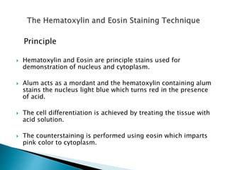
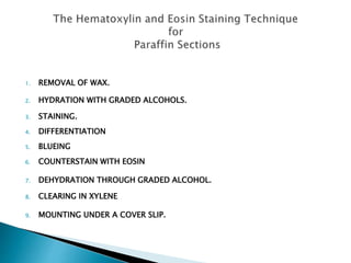
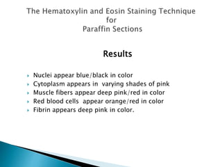
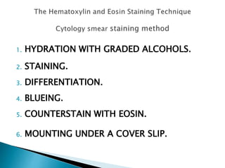
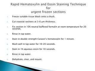


The Hematoxylin and Eosin stain is the most widely used histological stain. It clearly demonstrates many tissue structures using a simple method. Hematoxylin stains cell nuclei blue-black, while Eosin stains cytoplasm and connective tissues in shades of pink, orange, and red. This allows for the identification of tissues and the detection of disease processes under the microscope. The stain involves coloring the sample with hematoxylin, differentiating with an acid, and counterstaining with eosin.


