Download as PDF, PPTX


This document discusses staining techniques used in histopathology. It describes the purpose of staining as outlining tissues and cellular components to identify tissues and establish the presence or absence of disease. The most common staining technique is hematoxylin and eosin (H&E) staining. Hematoxylin stains nuclei blue and eosin stains cytoplasm pink, allowing clear visualization of tissue structures. The document provides details on the preparation and use of hematoxylin and eosin stains as well as other specialized staining techniques.
Presentation by Dr. N. Manjula on various staining techniques in histopathology.
Staining applies dyes to tissue sections to outline structures, identify tissues, and detect diseases.
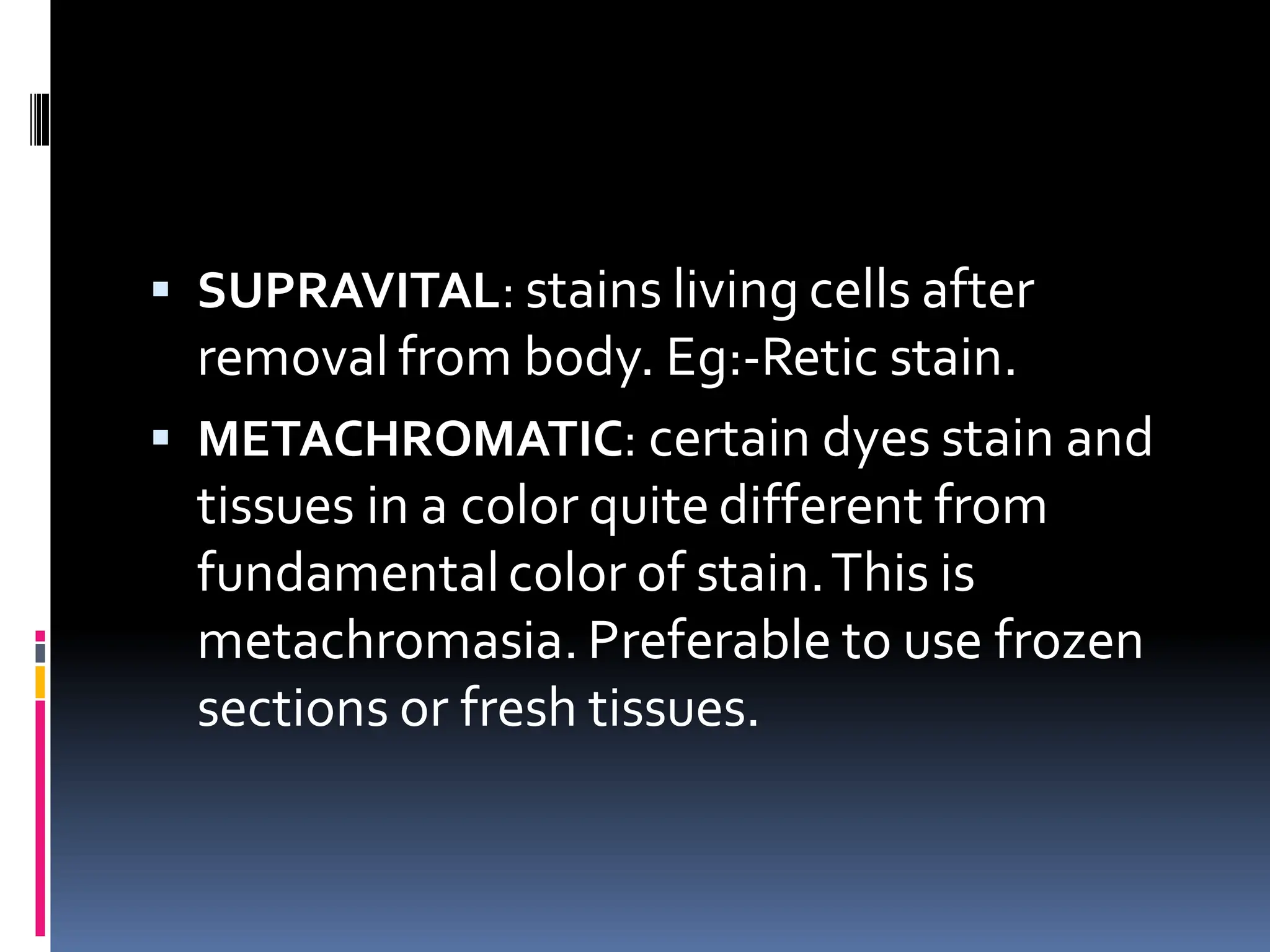
Stains categorized into routine, special, vital, supravital, metachromatic, basic, acid, and neutral.
Natural and synthetic dyes; direct and indirect staining methods using mordants.
Notable staining methods: Hematoxylin & Eosin, Gram's stain, Romanowsky stain, and their significance.
Hematoxylin's origins, extraction process, oxidation (ripening), and types including Alum and Iron hematoxylin.
Detailed discussion on Alum hematoxylin, stability issues, and progressive vs. regressive staining methods.
Types of Iron and other hematoxylins, along with usage details for specific staining applications.
Eosin as the primary counterstain, its types, solutions, and preparation necessary for staining.
Detailed preparation and staining procedure including steps from dewaxing to mounting the samples.
Visual results of various stains on different tissue components post staining process.
Brief mention of the methodology utilized for automatic staining processes.
Daily quality control measures for staining efficacy and guidelines for acceptable results.
Overview of the strengths and limitations of staining procedures in histopathology.
Key references and literature supporting the staining techniques discussed in the presentation.


