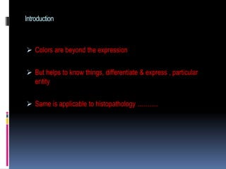
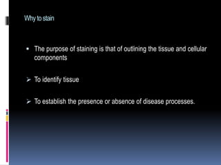
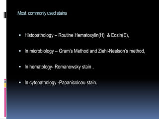
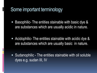
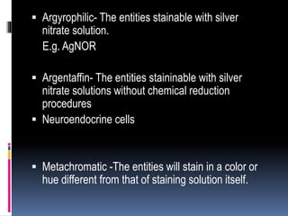
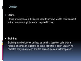
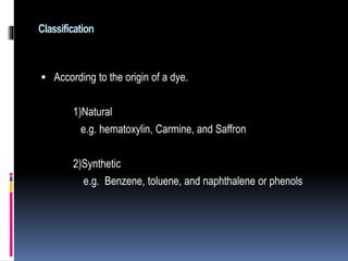
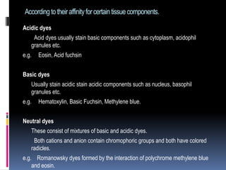
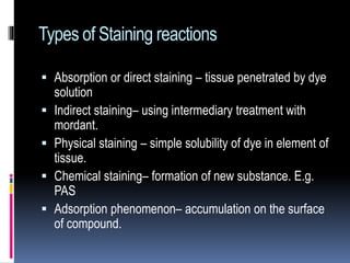
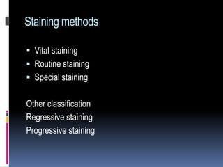
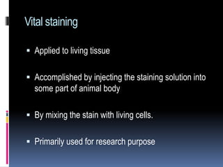
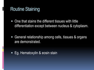
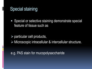
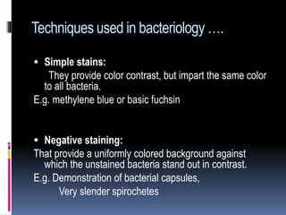
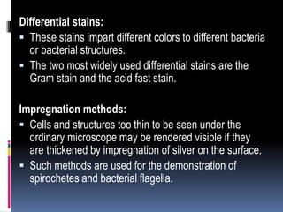
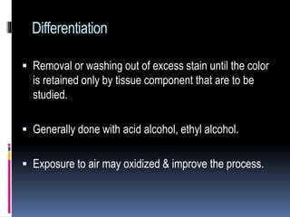
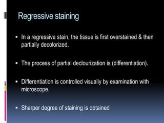
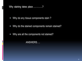
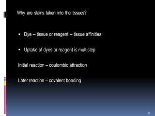
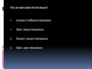
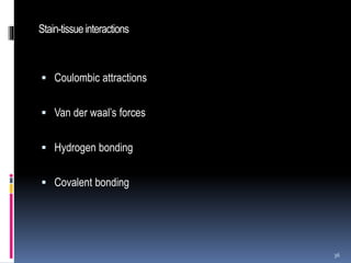
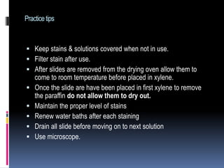
H and E staining is most important part of the histopathological diagnosis, this presentation is to highlight some important basic concept of the Staining.
The document provides information about Hematoxylin and Eosin staining. It states that H&E staining is widely used because of its simplicity, ability to clearly demonstrate tissue structures, and how it specifically stains cell nuclei blue-black and cytoplasm pink to red. Hematoxylin stains nuclei while Eosin stains cytoplasm. The document then discusses the chemistry and preparation of hematoxylin solutions, including how it is extracted from logwood and oxidized to its active form hematein. It also covers the use of mordants like aluminum, iron, and tungsten to improve hematoxylin's staining ability.
The Hematoxylin and Eosin stain is the most widely used histological stain. It clearly demonstrates many tissue structures using a simple method. Hematoxylin stains cell nuclei blue-black, while Eosin stains cytoplasm and connective tissues in shades of pink, orange, and red. This allows for the identification of tissues and the detection of disease processes under the microscope. The stain involves coloring the sample with hematoxylin, differentiating with an acid, and counterstaining with eosin.
This document discusses hematoxylin and eosin stains. It provides details on:
1) Hematoxylin is extracted from logwood and oxidized to hematin, which is responsible for staining properties. It requires a mordant like aluminum or iron salts to bind to tissues.
2) Alum hematoxylin is commonly used, with potassium or ammonium alum as the mordant. Sections can be overstained and differentiated, or stained for a predetermined time.
3) After differentiation, sections are "blued" in a weak alkaline solution to convert the hematin stain from red to blue-black in the cell nuclei.
This document provides information on techniques for collecting, preparing, preserving, and displaying specimens in a pathology museum. It discusses receiving specimens from hospitals and laboratories, preparing them by washing and fixing in formalin-based solutions, restoring color with alcohol, and long-term preservation by mounting in glycerin-based solutions. Special techniques are described for various organs and structures like lungs, brains, and calculi. The goal is to maintain specimens in a lifelike state for teaching and display over many years.
This document discusses various staining techniques used in cytology, including both routine and special stains. It provides details on the principles, procedures, and applications of stains such as May-Grunwald Giemsa, Diff-Quik, Papanicolaou, hematoxylin and eosin, periodic acid Schiff, mucicarmine, Alcian blue, Oil red O, Congo red, Feulgen, and Ziehl-Neelsen. The stains are used to demonstrate cellular and extracellular components, identify infectious organisms, and examine DNA content. Proper staining allows visualization of structures like glycogen, mucin, lipids, amyloid, and acid-fast bacteria under the microscope.
The tissue section is colourless because the fixed protein has the same refractive index as that of glass. We use dyes that have specific affinity with the different tissue proteins and colour them differently.
Colour is seen by the eye as a result of the effect of certain electromagnetic waves on the rods and cones of the retina. These waves, which have a varying length, will determine the colour that is seen.
White light being composed of all the colours of the visible spectrum varies in wavelength from 4,000 Â to 8,000 Â.
If light of a specific wavelength is absorbed from white light the resultant light will then be coloured, the colour being dependent upon the particular wavelength that has been removed.
The document discusses fixatives used in histopathology. It describes the process of fixation and how fixatives preserve tissue by denaturing or precipitating proteins. The ideal properties of a fixative are also outlined, including preventing autolysis and allowing for staining. Common fixatives are described such as formalin, Bouin's fluid, Zenker's solution and their mechanisms and appropriate uses. Factors that affect fixation like temperature, size, volume ratio, time and choice of fixative are also summarized.
The document discusses various techniques used in histopathology sample processing including decalcification, fixation, dehydration, clearing, embedding and sectioning. It covers different chemical agents used for each step along with their properties and advantages. Various methods are described such as paraffin, celloidin and vacuum embedding for optimal tissue preservation and section quality. Automatic tissue processors and freeze drying are also mentioned as techniques to reduce processing time.
Histopathology specimen processing involves several key steps: specimen identification and labeling, grossing and fixation, processing including dehydration and clearing, embedding, microtomy, and staining. Specimens are examined grossly, relevant sections are selected for histology based on findings, and blocks are prepared for microscopic examination. Proper grossing involves accurate description and oriented sampling to allow for histologic diagnosis.
The document discusses the hematoxylin and eosin stain, which is the most widely used histological stain. It stains cell nuclei blue or black using hematoxylin, and stains cell cytoplasm and connective tissue fibers pink using eosin. The purpose of staining is to identify tissue structures and the presence or absence of disease. Common stains discussed include hematoxylin and eosin, Gram's method, Ziehl-Neelson's method, and Papanicolaou stain. The document also provides details on the chemistry and procedures for hematoxylin and eosin staining.
The document provides an overview of the department of histopathology and its various benches. It describes histopathology as the microscopic examination of tissue to study disease manifestations. The key benches mentioned are processing, gross sectioning, tissue processing, embedding, cutting, staining including H&E, immunohistochemistry, special stains, cytology, cytogenetics, and semen analysis. The roles of each bench are briefly outlined.
THEORIES OF STAINING Biological Staining
Structural Components (Nature) Of Stains
Staining Mechanism
Metachromasia
Types Of Staining
Staining of Paraffin Section
A stain is any colouring organic compound that combined with another substance imparts a colour to that substance.
The term ‘dye’ is used to refer to a colouring agent that is used for general purposes, whereas the term ‘stain’ is used to refer to that dye which is used for biological purposes.
The stains used for bacteria are aniline dyes they are derived from aniline (C6H5NH2).
The most commonly used aniline dyes are crystal violet, methylene blue, basic fuchsin, safranin, eosin, etc.FACTORS INFLUENCING METACHROMASIA
The document discusses H&E (hematoxylin and eosin) staining, which is the most widely used staining technique in histopathology. H&E staining differentially colors tissue components, with hematoxylin staining nuclei blue and eosin staining cytoplasm and other tissues shades of pink. The process involves deparaffinizing tissue sections, staining with hematoxylin, differentiating with acid, counterstaining with eosin, and mounting for examination under a microscope. H&E staining allows clear visualization and analysis of cells and structures to enable histopathological diagnosis.
The Indian Dental Academy is the Leader in continuing dental education , training dentists in all aspects of dentistry and
offering a wide range of dental certified courses in different formats.for more details please visit
www.indiandentalacademy.com
The PAS stain identifies polysaccharides, mucus substances, basement membranes, and some fungi by causing them to appear magenta under the microscope. It works by first using periodic acid to oxidize carbohydrate groups, then exposing the tissue to Schiff's reagent, which causes aldehyde groups produced in the first step to appear magenta. The PAS stain is used to identify conditions involving abnormal glycogen storage or mucus production, such as certain tumors, infections, and genetic diseases. It helps diagnose issues in tissues from the liver, kidney, lung, muscle, and other organs.
1. Cytology of body fluids involves examining fluids from various body cavities including cerebrospinal fluid, pleural fluid, peritoneal fluid, pericardial fluid, and synovial fluid. Specimen collection and laboratory analysis includes gross examination, cell counts, biochemical analysis, and microscopic examination.
2. Transudates and exudates are distinguished based on characteristics like protein content and cell differentials. Infection, inflammation, and malignancy can be identified by analyzing changes in fluid characteristics.
3. Cytology of body fluids provides diagnostic information for conditions affecting various organ systems. Proper collection and analysis of physical and chemical properties aids in differential diagnosis.
This document outlines the histotechnique process which tissues undergo before microscopic examination. Key steps include: fixation to preserve tissue structure; processing involving dehydration, clearing, and impregnation to allow sectioning; embedding tissues in paraffin blocks for microtomy; sectioning samples and staining, typically with hematoxylin and eosin, for visualization under the microscope. Finally, samples are mounted on slides and labeled for storage and pathological examination.
This document discusses decalcification, which is the process of removing calcium from bone and other calcified tissues prior to sectioning and microscopic examination. It defines decalcification and lists the criteria for an ideal decalcifying agent. Various factors that affect the rate of decalcification are described, including concentration, temperature, agitation, and suspension of the tissue. The main methods of decalcification are outlined as well as the principles, types, compositions, and procedures for different decalcifying agents such as acids, ion exchange resins, and chelating agents.
This document discusses the process of decalcification, which is the removal of calcium salts from bones and calcified tissues. There are four main methods: 1) using simple dilute mineral acids like nitric acid or formic acid; 2) ion exchange resins with acid fluids; 3) electrolytic decalcification using electrodes; and 4) chelating agents like EDTA that bind calcium ions. The document provides details on procedures and advantages/disadvantages of each method.
Introduction to Histopathology and Lab organization.pptxsandeep singh
This document provides an introduction to histopathology and laboratory organization. It defines key terms like histology, histopathology, and histotechniques. It explains that histopathology involves examining tissues under a microscope to diagnose diseases. The organization of a histopathology laboratory has three parts: construction and equipment, staff, and protocols. Laboratories need specific rooms and equipment for receiving samples, processing, staining, and storage. Staff roles include pathologists, technicians, and attendants. Laboratories must establish standard operating procedures and safety measures to properly handle tissues and chemicals.
The document discusses cryostats, which are devices used to cut thin frozen sections of tissues for examination under a microscope. Cryostats contain a microtome inside a freezer unit that can rapidly freeze tissue samples and cut sections as thin as 1 micrometer at temperatures below freezing. The cryostat process allows for quick diagnosis by freezing and sectioning tissues within minutes rather than having to dehydrate, embed in paraffin, and section as with traditional microtomes.
Hematoxylin and eosin staining is a common histological technique that uses hematoxylin, which stains cell nuclei blue, and eosin, which stains cytoplasm and connective tissue pink. The document describes the full H&E staining procedure, including dewaxing, hydration, staining, differentiation, dehydration, clearing and mounting of tissue sections. It also discusses the principles and properties of hematoxylin, including how it is extracted from logwood and requires oxidation or "ripening" to become an effective nuclear stain. Commonly used hematoxylin formulations including Harris's, Mayer's, and Ehrlich's are compared.
This document discusses techniques for collecting, preparing, fixing, and preserving pathological specimens for display in a museum. Some key points covered include:
- Specimens should be collected from hospitals and received with patient details, then washed in saline and fixed within 2 hours to prevent autolysis.
- Fixation using Kaiserling's formalin-based solutions arrests decay and stabilizes tissues. It is important to ensure even penetration and the correct volume, temperature, pH and time of fixation.
- After fixation, specimens may be treated with alcohol to restore color before long-term preservation in mounting fluid, such as a glycerin-based solution.
- Hollow organs should be inflated or packed during fixation to
This document discusses frozen sections and cryostats. Frozen sections are prepared without dehydration or embedding to enable rapid diagnosis within 10 minutes. They have applications in intraoperative diagnosis, enzyme histochemistry, immunohistochemistry, and other techniques. Tissue is frozen using liquid nitrogen or other cryogenic methods, turning water within the tissue to ice which acts as an embedding medium for sectioning. Cryostats maintain low temperatures, typically -20 to -30°C, for sectioning frozen tissue blocks. Optimal cutting temperatures vary by tissue type and whether the tissue is fixed.
This document provides information on staining blood films and smears. It discusses the different types of stains used including Romanowsky stains like Leishman stain, Giemsa stain, Wright stain, and Field stain. Specimens should be collected in EDTA and smears prepared within an hour then fixed in methanol or ethanol to preserve cell morphology before staining. Romanowsky stains use methylene blue and eosin dyes to reveal subtle differences in cell structures and components.
This document discusses the process of decalcification, which is the removal of calcium from tissues to make them suitable for section cutting. It outlines the key steps: selection of tissue, fixation, decalcification using mineral acids, chelating agents, or electrophoresis, detection of endpoint, neutralization, and washing. Common decalcifying agents discussed include Gooding and Stewart's fluid, von Ebner's fluid, citrate-citric acid buffer, and chelating agents like EDTA. The factors that influence decalcification speed and the importance of determining the endpoint are also summarized.
Embedding is the process of enclosing tissue specimens in an embedding medium such as paraffin wax to support the specimen for sectioning. The choice of embedding medium depends on the type of tissue, microscope, and microtome being used. Common embedding mediums include paraffin wax, celloidin, resin, and gelatin. Paraffin wax is most widely used due to its hardness and ability to produce high quality sections. Proper orientation of the specimen in the embedding block is important for pathological examination and diagnosis.
This document outlines the steps for histology lab procedures which include fixation, processing, sectioning, staining, and mounting of tissues. It lists the required chemicals, glassware, and instruments. The key steps are:
1. Tissues are fixed in formalin or Bouin's fixative for 24 hours to preserve cells and structures.
2. Processing involves dehydration using graded alcohols, clearing with benzene, and infiltration with paraffin wax for embedding.
3. Sections are cut from the wax blocks using a microtome, mounted on slides, and stained with H&E stain.
4. Stained sections are viewed under a microscope after dewaxing and mounting.
This document provides an overview of microtomy, which is the process of cutting thin sections of tissue for microscopic examination. It discusses the history and types of microtomes, including rocking, rotary, base-sledge, sliding, vibrating, freezing, saw, cryostat, ultra, and laser microtomes. For each type, the key features and mechanisms are described. It also covers the different parts of microtomes like the knife, block and knife holders. Finally, it discusses the various knife profiles, materials, angles and their applications in microtomy.
This document describes the hematoxylin and eosin stain (H&E stain) procedure performed at a 125-bed community hospital. Over a 30-day period, 291 specimens were analyzed, with 15 found to contain cancerous cells and 276 containing benign cells. The H&E stain involves hematoxylin staining cell nuclei blue-black and eosin staining cell cytoplasm and connective tissues shades of pink and red. This allows differentiation of tumor/cancer cells from normal cells under a microscope.
The document outlines the steps in a histology procedure: 1) Specimens are accessioned and given an identifying number; 2) The specimen is described and parts are placed in cassettes during gross examination; 3) Tissue fixation preserves the specimen's structure and prevents loss of constituents; 4) Tissue processing dehydrates, clears, and impregnates the tissue to allow for embedding in wax; 5) Embedded tissue is sectioned into thin slices and stained with dyes to enhance contrast for microscopic examination.
Hematoxylin and eosin (H&E) staining is the most common histology stain. Hematoxylin stains cell nuclei blue by binding to DNA and RNA, while eosin stains cytoplasm and extracellular components pink. The staining process involves deparaffinizing tissue sections, staining with hematoxylin, differentiating with acid to remove excess stain, staining with eosin, and mounting for examination. Hematoxylin is extracted from logwood and oxidized to hematin, which binds tissue as a cationic dye with a mordant like alum. Eosin Y is the typical counterstain used to visualize cytoplasm. Together, H&E staining provides excellent contrast to study cell and
This document describes various smear preparation techniques used in cytology, including direct smears, blood smear technique, squash technique, large volume centrifugation, small volume centrifugation, membrane filtration, cell blocks, density gradient centrifugation, and gravity sedimentation. Direct smears involve spreading the specimen directly onto a slide. Blood smear technique produces a thin, uniform smear for staining. Squash technique results in a thin, uniform preparation. Large volume centrifugation concentrates fluid specimens by separating the buffy coat layer. Small volume centrifugation uses a cyto-centrifuge to deposit cells directly onto a slide. Membrane filtration uses a filter to collect cells on a slide. Cell blocks allow processing samples as histopath
This document provides information about linen and fabric handling for housekeeping. It discusses the importance of linen and fabric for hotel operations. Proper selection and handling of linen is the housekeeping department's responsibility to protect the hotel's investment. The document defines what linen and fabric are made of and discusses factors to consider when choosing linens such as fiber content, thread count, weave, and size. It also covers natural fibers like wool, silk, cotton and linen as well as man-made fibers like acetate, acrylic, polyester, rayon, nylon and spandex. The document discusses label identification and provides washing symbols to indicate care instructions. It also gives examples of common stain types and cleaning procedures.
The H&E stain is the most common stain used in histology. It involves staining tissue samples with hematoxylin, which stains nuclei blue, followed by a counterstain with eosin, which stains cytoplasm and other tissue structures pink. The staining process involves deparaffinization, hydration, hematoxylin staining, differentiation, bluing, eosin counterstaining, dehydration, clearing, and coverslipping. Both automated and manual methods can be used to perform the H&E stain, and quality control measures help ensure consistent, high-quality results.
The document discusses the key steps in histological techniques which are used to prepare tissue samples for microscopic examination. The techniques include fixation, dehydration, clearing, embedding, sectioning, and staining of tissues. Fixation preserves the tissue structure using chemical fixatives. The tissue then undergoes dehydration, clearing and infiltration with paraffin wax before being embedded in wax blocks and sectioned for microscopic analysis. Staining helps to visualize different structures within the tissue sample under the microscope.
The document discusses various histological staining techniques. It begins by explaining hematoxylin and eosin staining, which provides basic diagnostic information. It then covers special stains that highlight specific tissue components, categorized by the structures they identify such as carbohydrates, amyloid, lipids, nucleic acids, and microorganisms. Carbohydrate stains discussed include periodic acid Schiff, alcian blue, mucicarmine, and others. Amyloid identification using Congo red and methyl violet is explained. Lipid stains using Sudan dyes are also summarized. The document provides details on techniques for staining nucleic acids and identifying bacteria by Gram staining.
The document provides an introduction to ELISA (enzyme-linked immunosorbent assay), which is a biochemical technique used mainly in immunology to detect the presence of an antibody or antigen in a sample. It describes the basic principles and steps of the ELISA process, which involves detecting antibodies or antigens using an enzyme-labeled secondary antibody and color changing reaction. Key aspects covered include antigen-antibody binding, use of enzyme labels, substrate conversion, and quantitative/qualitative applications of ELISA for detecting various molecules.
Histological staining methods aim to differentiate tissues and cells by altering contrast or color. There are several staining techniques including vital staining of living cells, staining by selective solubility of dyes in lipids, chemical reactions between dyes and tissues, metallic impregnation, and staining with synthetic or natural dyes. Dyes contain chromophores for color and auxochromes that determine staining action. Staining can be progressive, with sequential coloring of elements, or regressive with initial overstaining followed by differentiation. Factors like dye concentration, temperature, pH, fixation influence dye binding to tissues.
GENERAL PRINCIPLES OF STAINING AND H & E STAIN.pptxDr.Shubham Patel
This document provides an overview of general principles of staining and the hematoxylin and eosin (H&E) stain. It discusses why staining is necessary, common terminology used, theories of staining, types of stains and dyes, the roles of mordants and accentuators, and metachromasia. It then focuses on H&E stain, including the history and development of hematoxylin, the staining process, and different types of hematoxylin classifications based on the oxidation procedure and mordant used.
The Hematoxylin and Eosin stain (H&E) is the most widely used histological stain due to its comparative simplicity and ability to clearly demonstrate many different tissue structures. Hematoxylin stains cell nuclei blue-black and shows good intranuclear detail, while Eosin stains cell cytoplasm and connective tissue in varying shades of pink, orange, and red. The H&E stain outlines tissues and cellular components and is essential for identifying tissues and establishing the presence or absence of disease processes. Several hematoxylin solutions are described in the document, varying in their mordants, methods of oxidation, and intended uses in histology.
This document discusses hematoxylin and eosin (H&E) staining, which is the most commonly used staining method in histopathology. It introduces the purpose of staining tissues, which is to outline cellular and tissue components to identify tissues and detect disease processes. The key terminology related to staining is defined, such as basophilic, acidophilic, sudanophilic. Dyes are classified based on their origin (natural vs synthetic) and affinity for tissue components (acidic, basic, neutral). H&E staining specifically uses the basic dye hematoxylin to stain nuclei purple and the acidic dye eosin to stain cytoplasm and other tissue components pink.
This document discusses staining techniques used to visualize bacteria under a microscope. It begins by explaining why staining is necessary given that bacteria are colorless and microscopic. It then describes different types of stains including basic stains that directly stain bacteria and acidic stains used for negative staining of the background. Key differential staining techniques are also summarized, including Gram staining which separates bacteria into Gram-positive and Gram-negative groups based on cell wall structure, and acid-fast staining used to identify Mycobacterium species. The document provides details on staining methods, the mechanisms of different staining techniques, and their importance in classifying and identifying bacterial specimens.
Notes for STAINING AND ANALYSIS of HISTOLOGICAL PREPARATIONSimprovemed
This document provides an overview of histological staining techniques. It discusses how histological preparations are stained using interactions between dyes, solvents, and tissue components. Different staining methods result in different colors that highlight various structures. A classic example is hematoxylin and eosin staining, where hematoxylin stains acidic components blue and eosin stains basic components pink. Specialized staining techniques also exist, such as immunohistochemistry. Proper staining selection depends on the tissue and research goals. Histological preparations are then analyzed under a microscope to study cell and tissue morphology.
This document discusses histopathology staining techniques. It defines histopathology as staining tissue samples to examine cellular and intracellular structures microscopically. The two main categories of stains are natural dyes derived from natural resources, and artificial dyes produced through chemical reactions. Hematoxylin is a commonly used natural dye that stains nuclei blue, while eosin is an artificial dye that counterstains cytoplasm pink. The document outlines hematoxylin and eosin staining as well as other histological stains and their characteristics.
The document discusses staining techniques used in histopathology. It defines staining as using substances to color tissue components to aid differentiation under a microscope. Staining involves chemical reactions between dyes and tissue constituents, resulting in colored end products. Proper staining reveals tissue morphology and enables diagnosis. It discusses various staining methods like direct, indirect, progressive, regressive staining and the use of mordants, facilitators and vital staining. The document also covers staining theories of physical adsorption and chemical reactions between dyes and tissue acids or bases.
Dental Anatomy and Dental Histology Project on Hematoxylin & Eosin Stain - 1S...deepupadhyaya
The document discusses the Hematoxylin and Eosin staining technique. It begins with the presenter's information and acknowledgments. Hematoxylin and Eosin staining is then introduced as the most widely used histological staining technique. It allows for clear demonstration of numerous tissue structures as hematoxylin stains nuclei blue and eosin stains cytoplasm and connective tissues pink. The document proceeds to describe the individual stains hematoxylin and eosin in depth, including their properties and types. It concludes with an overview of the hematoxylin and eosin staining procedure for paraffin sections and cytology smears.
Glossary of staining methods, reagents, immunostaining, terminology and eponymsPravin Amabade
This document provides a glossary of terms related to biological staining methods, reagents, immunostaining, and related terminology. It contains over 150 entries defined in black bold, with cross-references in underlined blue italic. The glossary aims to define terms for researchers across sub-disciplines of microtechnique to increase understanding of staining rationales, reagents, and eponyms. It is updated periodically to expand coverage of dyes, fixatives, and other procedures.
The document discusses various staining methods used in histology. It describes how stains are classified as basic, acid or neutral dyes based on their chemical properties. Staining methods can be simple, compound, indirect, direct, progressive, regressive or selective depending on the technique. Commonly used stains include hematoxylin, eosin, safranin and Mallory's trichrome. The hematoxylin and eosin stain is also described in detail, outlining the staining process and factors that can influence results. Different types of mounting media are discussed for permanent or temporary coverslipping of tissue samples.
The Indian Dental Academy is the Leader in continuing dental education , training dentists in all aspects of dentistry and
offering a wide range of dental certified courses in different formats.for more details please visit
www.indiandentalacademy.com
Staining techniques are used to enhance contrast and visibility of structures under the microscope. There are several types of staining including simple staining using one dye, differential staining using multiple dyes to distinguish structures, and special staining for specific structures like capsules, endospores, and flagella. The choice of stain depends on factors like the chemical composition and charge of the dye. Common stains used are basic dyes that bind to negatively charged cell components and acid dyes that stain the background. Differential stains include Gram stain and acid-fast stain that classify bacteria based on cell wall differences in staining.
1. Hematoxylin is a natural dye extracted from logwood that is used as a nuclear stain. It requires oxidation to hematein and use of a mordant like aluminum or iron to effectively stain nuclei.
2. Alum hematoxylin is commonly used, with Ehrlich's, Harris', and Mayer's being examples. It stains nuclei blue-black but is sensitive to acid. Iron hematoxylin is more resistant.
3. Oxidation can be done naturally over months or chemically instantly using sodium iodate or mercuric oxide. This affects the staining properties and lifespan.
This document provides information on tissue processing and histopathologic techniques, focusing on staining. It defines staining as applying dyes to tissue sections to facilitate microscopic study. It then classifies stains based on pH, function, source, dye application technique, sequence, and color contrast. Specific staining techniques and commonly used stains are described for carbohydrates, lipids, proteins, and nucleic acids. Hematoxylin and eosin staining is explained as the most widely used staining procedure.
procedure for Staining on routine histopath.pptIvanaUngajan2
This document discusses hematoxylin and eosin (H&E) staining techniques. It describes the appearance of the nucleus and cytoplasm in H&E stained slides. Key components that stain with H&E are described such as the nucleolus, chromatin, ribosomes and endoplasmic reticulum. The mechanisms of nuclear and cytoplasmic staining are explained in relation to pH and the isoelectric point of proteins. Common nuclear and plasma stains used in H&E, including hematoxylin, hematein and eosin, are outlined with details on their preparation, use and staining properties.
By definition dyes can be said to be colouredHamdi Abdalnabi
Dyes are colored, ionizing, aromatic organic compounds that are applied in an aqueous solution and may require a mordant. They are commonly used to identify tissue components under microscopy and are applied to numerous substrates like textiles. The first synthetic dye, mauve, was discovered accidentally in 1856 by William Perkin, launching the modern chemical industry. Dyes get their color from chromophores, delocalized electron structures that absorb different wavelengths of light. Color can be altered by adding chemical modifiers that change electron energy levels. Dyes require auxochromes to impart solubility and cohesiveness through their ability to ionize.
This document provides an overview of hematoxylin and eosin staining techniques. It discusses the structures of dyes, classifications of dyes, staining mechanisms, hematoxylin, eosin, and the hematoxylin and eosin staining procedure. The key steps of the H&E staining procedure are deparaffinization, hydration, hematoxylin staining, differentiation, blueing, and eosin counterstaining to color nuclei blue and cytoplasm pink, respectively. The document also covers dye origins, properties, affinities, and terms used in biological staining.
The document is an acknowledgement thanking various people who helped with completing a project, including the project guide, parents, classmates, and others who assisted directly or indirectly. It also includes sections on the definition, classification, and production process of dyes. Dyes are colored substances that are applied to materials to change their color. They work by absorbing certain wavelengths of light. Dyes can be classified in several ways, including by their chemical structure and the products they are applied to. The document provides examples of dye production processes and uses.
This document provides information about organic chemistry concepts including synthetic dyes, synthetic drugs, and synthetic polymers. It discusses the classification of dyes based on their chemical constitution and provides examples of important synthetic dyes like Congo red, Crystal violet, Phenolphthalein, and Alizarin. It also summarizes theories on the relationship between color and chemical structure, including Witt's theory and the modern electronic concept. Classification of dyes as natural or synthetic is described.
The document discusses various hematological investigations and artifacts. It describes the process of a differential count, which involves examining a peripheral blood smear under a microscope to determine percentages of different white blood cells. The blood smear can also reveal abnormal red blood cell and white blood cell morphologies. The document then discusses the steps to make a good blood smear, including using a clean slide and proper angulation and force when spreading the blood. Potential artifacts from improper smear preparation or staining are also described. The document concludes with discussing various hematological tests including complete blood count, erythrocyte sedimentation rate, coagulation tests, and factors that could affect the results.
The document discusses various investigations and artifacts in hematology. It covers topics like complete blood count, hemogram tests, tests of hemostatic function, blood collection methods, and common errors in hematological tests. The complete blood count includes tests like hemoglobin concentration, total erythrocyte count, total leukocyte count, and blood film examination. Hemostatic function tests include bleeding time, clotting time, prothrombin time, and fibrinogen determination. Proper blood collection and anticoagulant use are important to avoid hemolysis and other errors in test results.
The document provides an overview of immunohistochemical (IHC) techniques. It discusses the basic principles of IHC, including antigen-antibody reactions and the use of primary and secondary antibodies. It also describes different IHC staining methods such as direct, indirect, and peroxidase-antiperoxidase methods. Key enzymes and chromogens used in IHC are discussed, as well as factors that influence antibody binding such as dilution, incubation time and temperature.
This document discusses the history and techniques of immunohistochemistry. It covers:
- The early development of immunohistochemistry from 1941 to the present.
- Common fixatives used such as formalin, Bouin's solution, B5, Zenker's solution, and modifications like PLDP.
- The benefits and drawbacks of different fixatives for preserving tissue morphology and antigenicity.
- Methods for improving antigen retrieval after fixation, like proteolytic enzymes and heat.
- The importance of fixation for maintaining tissue structure while not blocking epitopes.
This document provides an overview of red lesions that can occur in the oral cavity. It discusses normal variations in oral mucosa color and various factors that can affect color. Red lesions are classified and several common types are described in detail, including traumatic erythematous macules, purpuric macules, inflammatory fibrous hyperplasia, nicotine stomatitis, erythroplakia, carcinoma, and candidiasis. Diagnostic features, histopathology, differential diagnoses, and management are covered for key red lesions. The document aims to guide clinicians in identifying and diagnosing different oral red lesions.
This document provides a review of midline diastemas. It discusses the prevalence and various etiological factors that can cause midline diastemas, such as an enlarged labial frenum, tooth size discrepancies, missing teeth, supernumerary teeth, oral habits, and certain medical conditions. The management involves orthodontic treatment to close the space as well as potentially frenectomy surgery. Retention is important after treatment to prevent relapse of the diastema.
This document reviews non-invasive techniques for age estimation using dental development and changes. It discusses:
1. Several non-invasive dental age estimation techniques including Schour and Masler's atlas method, Demirjian's scoring method, and Kvaal's radiographic method of measuring secondary dentin deposition.
2. Factors used for dental age estimation including tooth emergence, mineralization, root development, attrition, and changes in dental tissue composition.
3. Applications of dental age estimation in different age groups from prenatal to adults. The most accurate methods vary depending on the age group.
4. Comparison of different dental age estimation methods finding that methods combining multiple teeth and examiners tend
This document describes a case report of a solitary angiokeratoma lesion found on the tongue of a 38-year-old male patient. Solitary angiokeratomas of the oral mucosa are rare. The lesion was a well-circumscribed, dark brown growth on the dorsal surface of the tongue. Histopathological examination revealed numerous dilated blood vessels in the papillary dermis along with hyperkeratosis and acanthosis of the epithelium, consistent with angiokeratoma. Immunohistochemical staining was positive for CD34, confirming the lesion contained proliferating blood vessels. No other lesions were found on the patient's body. The lesion was completely excised with no recurrence after 6 months of follow up.
Hematoxylin and eosin staining is the most widely used histological staining technique. It uses hematoxylin, which stains cell nuclei blue, and eosin, which stains cytoplasm and connective tissue pink. The process involves dewaxing tissue sections, hydrating them, staining with hematoxylin, differentiating with acid alcohol, counterstaining with eosin, dehydrating, and mounting. Hematoxylin requires a mordant like aluminum or iron to stain tissues. Commonly used hematoxylins include Harris', Mayer's, Ehrlich's, and Delafield's.
This document provides an overview of the histology of the major salivary glands, including the parotid, submandibular, and sublingual glands. It describes the secretory end pieces composed of serous and mucous cells, as well as the ductal system including intercalated, striated, and excretory ducts. The minor salivary glands are also briefly discussed. The roles of myoepithelial cells and the different cell types involved in saliva production are summarized.
This document provides an overview of hematoxylin and eosin staining. It discusses the theory behind staining, including how dyes interact with tissues through various bonding mechanisms. It also describes factors that influence staining results, such as rates of dye uptake and loss, binding site affinities, and tissue modification during fixation. The document highlights how hematoxylin and eosin work as the most commonly used routine stain in histopathology.
This document provides information about the anatomy and development of the major and minor salivary glands. It discusses the parotid gland, submandibular gland, sublingual gland, and minor salivary glands. For each gland it describes the location, structure, duct system, blood supply, nerve innervation, and other key details. It also covers the embryonic development of the salivary glands from the initial bud formation through branching and lumen development.
This document provides an overview of tissue fixation. It begins with an introduction to fixation and its importance in histology. The main functions of fixatives are to prevent autolysis and putrefaction of tissue. Various fixation methods and types of fixatives are described, including aldehydes, metallic salts, picric acid, alcohols, chromates, and osmium tetroxide. The document discusses the reactions of fixatives with proteins, nucleic acids, lipids, and carbohydrates. Common fixatives like formaldehyde and glutaraldehyde are compared. Factors affecting fixation and the effects of fixation are also summarized.
This document provides an overview of oral epithelium, including its structure and function. It discusses the four main layers of stratified squamous epithelium found in the oral cavity - stratum basale, stratum spinosum, stratum granulosum, and stratum corneum. It describes the cells found in each layer, such as stem cells, transit amplifying cells, and keratinocytes, and the changes they undergo during differentiation and keratinization. The document also discusses non-keratinocyte cells like melanocytes, Langerhans cells, and Merkel cells. It provides details on cytoskeletal components, basement membrane, and the specialized oral mucosas.
Epithelium is a tissue that covers external surfaces and lines internal cavities and tubes of the body. It has several key characteristics including being a continuous sheet of cells attached to a basement membrane and generally being avascular.
There are several types of epithelium classified by the number of cell layers - simple (one cell layer), pseudostratified (cells appear in multiple layers but are all attached to the basement membrane), and stratified (multiple cell layers). Epithelium also includes glandular epithelium specialized for secretion. The seminar covered the classification, structure, and functions of the main types of epithelium.
TEST BANK For An Introduction to Brain and Behavior, 7th Edition by Bryan Kol...rightmanforbloodline
TEST BANK For An Introduction to Brain and Behavior, 7th Edition by Bryan Kolb, Ian Q. Whishaw, Verified Chapters 1 - 16, Complete Newest Versio
TEST BANK For An Introduction to Brain and Behavior, 7th Edition by Bryan Kolb, Ian Q. Whishaw, Verified Chapters 1 - 16, Complete Newest Version
Adhd Medication Shortage Uk - trinexpharmacy.comreignlana06
The UK is currently facing a Adhd Medication Shortage Uk, which has left many patients and their families grappling with uncertainty and frustration. ADHD, or Attention Deficit Hyperactivity Disorder, is a chronic condition that requires consistent medication to manage effectively. This shortage has highlighted the critical role these medications play in the daily lives of those affected by ADHD. Contact : +1 (747) 209 – 3649 E-mail : sales@trinexpharmacy.com
Histololgy of Female Reproductive System.pptxAyeshaZaid1
Dive into an in-depth exploration of the histological structure of female reproductive system with this comprehensive lecture. Presented by Dr. Ayesha Irfan, Assistant Professor of Anatomy, this presentation covers the Gross anatomy and functional histology of the female reproductive organs. Ideal for students, educators, and anyone interested in medical science, this lecture provides clear explanations, detailed diagrams, and valuable insights into female reproductive system. Enhance your knowledge and understanding of this essential aspect of human biology.
Cell Therapy Expansion and Challenges in Autoimmune DiseaseHealth Advances
There is increasing confidence that cell therapies will soon play a role in the treatment of autoimmune disorders, but the extent of this impact remains to be seen. Early readouts on autologous CAR-Ts in lupus are encouraging, but manufacturing and cost limitations are likely to restrict access to highly refractory patients. Allogeneic CAR-Ts have the potential to broaden access to earlier lines of treatment due to their inherent cost benefits, however they will need to demonstrate comparable or improved efficacy to established modalities.
In addition to infrastructure and capacity constraints, CAR-Ts face a very different risk-benefit dynamic in autoimmune compared to oncology, highlighting the need for tolerable therapies with low adverse event risk. CAR-NK and Treg-based therapies are also being developed in certain autoimmune disorders and may demonstrate favorable safety profiles. Several novel non-cell therapies such as bispecific antibodies, nanobodies, and RNAi drugs, may also offer future alternative competitive solutions with variable value propositions.
Widespread adoption of cell therapies will not only require strong efficacy and safety data, but also adapted pricing and access strategies. At oncology-based price points, CAR-Ts are unlikely to achieve broad market access in autoimmune disorders, with eligible patient populations that are potentially orders of magnitude greater than the number of currently addressable cancer patients. Developers have made strides towards reducing cell therapy COGS while improving manufacturing efficiency, but payors will inevitably restrict access until more sustainable pricing is achieved.
Despite these headwinds, industry leaders and investors remain confident that cell therapies are poised to address significant unmet need in patients suffering from autoimmune disorders. However, the extent of this impact on the treatment landscape remains to be seen, as the industry rapidly approaches an inflection point.
Osteoporosis - Definition , Evaluation and Management .pdfJim Jacob Roy
Osteoporosis is an increasing cause of morbidity among the elderly.
In this document , a brief outline of osteoporosis is given , including the risk factors of osteoporosis fractures , the indications for testing bone mineral density and the management of osteoporosis
Integrating Ayurveda into Parkinson’s Management: A Holistic ApproachAyurveda ForAll
Explore the benefits of combining Ayurveda with conventional Parkinson's treatments. Learn how a holistic approach can manage symptoms, enhance well-being, and balance body energies. Discover the steps to safely integrate Ayurvedic practices into your Parkinson’s care plan, including expert guidance on diet, herbal remedies, and lifestyle modifications.
Rasamanikya is a excellent preparation in the field of Rasashastra, it is used in various Kushtha Roga, Shwasa, Vicharchika, Bhagandara, Vatarakta, and Phiranga Roga. In this article Preparation& Comparative analytical profile for both Formulationon i.e Rasamanikya prepared by Kushmanda swarasa & Churnodhaka Shodita Haratala. The study aims to provide insights into the comparative efficacy and analytical aspects of these formulations for enhanced therapeutic outcomes.
TEST BANK For Community Health Nursing A Canadian Perspective, 5th Edition by...Donc Test
TEST BANK For Community Health Nursing A Canadian Perspective, 5th Edition by Stamler, Verified Chapters 1 - 33, Complete Newest Version Community Health Nursing A Canadian Perspective, 5th Edition by Stamler, Verified Chapters 1 - 33, Complete Newest Version Community Health Nursing A Canadian Perspective, 5th Edition by Stamler Community Health Nursing A Canadian Perspective, 5th Edition TEST BANK by Stamler Test Bank For Community Health Nursing A Canadian Perspective, 5th Edition Pdf Chapters Download Test Bank For Community Health Nursing A Canadian Perspective, 5th Edition Pdf Download Stuvia Test Bank For Community Health Nursing A Canadian Perspective, 5th Edition Study Guide Test Bank For Community Health Nursing A Canadian Perspective, 5th Edition Ebook Download Stuvia Test Bank For Community Health Nursing A Canadian Perspective, 5th Edition Questions and Answers Quizlet Test Bank For Community Health Nursing A Canadian Perspective, 5th Edition Studocu Test Bank For Community Health Nursing A Canadian Perspective, 5th Edition Quizlet Test Bank For Community Health Nursing A Canadian Perspective, 5th Edition Stuvia Community Health Nursing A Canadian Perspective, 5th Edition Pdf Chapters Download Community Health Nursing A Canadian Perspective, 5th Edition Pdf Download Course Hero Community Health Nursing A Canadian Perspective, 5th Edition Answers Quizlet Community Health Nursing A Canadian Perspective, 5th Edition Ebook Download Course hero Community Health Nursing A Canadian Perspective, 5th Edition Questions and Answers Community Health Nursing A Canadian Perspective, 5th Edition Studocu Community Health Nursing A Canadian Perspective, 5th Edition Quizlet Community Health Nursing A Canadian Perspective, 5th Edition Stuvia Community Health Nursing A Canadian Perspective, 5th Edition Test Bank Pdf Chapters Download Community Health Nursing A Canadian Perspective, 5th Edition Test Bank Pdf Download Stuvia Community Health Nursing A Canadian Perspective, 5th Edition Test Bank Study Guide Questions and Answers Community Health Nursing A Canadian Perspective, 5th Edition Test Bank Ebook Download Stuvia Community Health Nursing A Canadian Perspective, 5th Edition Test Bank Questions Quizlet Community Health Nursing A Canadian Perspective, 5th Edition Test Bank Studocu Community Health Nursing A Canadian Perspective, 5th Edition Test Bank Quizlet Community Health Nursing A Canadian Perspective, 5th Edition Test Bank Stuvia
1. Hematoxylin & Eosin
staining
Presented by
Dr. Shrikant Sonune
Guided by
Dr Ashok Patil,
Dr Shilpa Kandalgaonkar,
Dr Mayur Chaudhary,
Dr Suyog Tupsakhare,
Dr Mahesh Gabhane.
2. Content
Introduction
General principles of staining (theory )
Theory of staining
Method of H & E staining
Theory of H & E staining
Types of hematoxylin
3. Introduction
Colors are beyond the expression
But helps to know things, differentiate & express , particular
entity
Same is applicable to histopathology ……….
4. Whytostain
The purpose of staining is that of outlining the tissue and cellular
components
To identify tissue
To establish the presence or absence of disease processes.
5. Most commonlyusedstains
Histopathology – Routine Hematoxylin(H) & Eosin(E),
In microbiology – Gram’s Method and Ziehl-Neelson’s method,
In hematology- Romanowsky stain ,
In cytopathology -Papanicoloau stain.
6. Some important terminology
Basophilic- The entities stainable with basic dye &
are substances which are usually acidic in nature.
Acidophilic- The entities stainable with acidic dye &
are substances which are usually basic in nature.
Sudanophilic - The entities stainable with oil soluble
dyes e.g. sudan III, IV
7. Argyrophilic- The entities stainable with silver
nitrate solution.
E.g. AgNOR
Argentaffin- The entities staininable with silver
nitrate solutions without chemical reduction
procedures
Neuroendocrine cells
Metachromatic -The entities will stain in a color or
hue different from that of staining solution itself.
8. Definition
Stains:
Stains are chemical substances used to achieve visible color contrast
in the microscopic picture of a prepared tissue.
Staining:
Staining may be loosely defined as treating tissue or cells with a
reagent or series of reagents so that it acquires a color; usually, no
particles of dyes are seen and the stained element is transparent.
9. DYES
These are essentially aromatic benzene ring compounds
or derivatives that possess the twin properties of color and
ability to bind to tissue.
10. Classification
According to the origin of a dye.
1)Natural
e.g. hematoxylin, Carmine, and Saffron
2)Synthetic
e.g. Benzene, toluene, and naphthalene or phenols
11. Accordingtotheiraffinityforcertaintissuecomponents.
Acidic dyes
Acid dyes usually stain basic components such as cytoplasm, acidophil
granules etc.
e.g. Eosin, Acid fuchsin
Basic dyes
Usually stain acidic stain acidic components such as nucleus, basophil
granules etc.
e.g. Hematoxylin, Basic Fuchsin, Methylene blue.
Neutral dyes
These consist of mixtures of basic and acidic dyes.
Both cations and anion contain chromophoric groups and both have colored
radicles.
e.g. Romanowsky dyes formed by the interaction of polychrome methylene blue
and eosin.
12. Types of Staining reactions
Absorption or direct staining – tissue penetrated by dye
solution
Indirect staining– using intermediary treatment with
mordant.
Physical staining – simple solubility of dye in element of
tissue.
Chemical staining– formation of new substance. E.g.
PAS
Adsorption phenomenon– accumulation on the surface
of compound.
13. Staining methods
Vital staining
Routine staining
Special staining
Other classification
Regressive staining
Progressive staining
14. Vital staining
Applied to living tissue
Accomplished by injecting the staining solution into
some part of animal body
By mixing the stain with living cells.
Primarily used for research purpose
15. Routine Staining
One that stains the different tissues with little
differentiation except between nucleus & cytoplasm.
General relationship among cells, tissues & organs
are demonstrated.
Eg. Hematoxylin & eosin stain
16. Special staining
Special or selective staining demonstrate special
feature of tissue such as
particular cell products,
Microscopic intracellular & intercellular structure.
e.g. PAS stain for mucopolysaccharide
17. Techniques used in bacteriology ….
Simple stains:
They provide color contrast, but impart the same color
to all bacteria.
E.g. methylene blue or basic fuchsin
Negative staining:
That provide a uniformly colored background against
which the unstained bacteria stand out in contrast.
E.g. Demonstration of bacterial capsules,
Very slender spirochetes
18. Differential stains:
These stains impart different colors to different bacteria
or bacterial structures.
The two most widely used differential stains are the
Gram stain and the acid fast stain.
Impregnation methods:
Cells and structures too thin to be seen under the
ordinary microscope may be rendered visible if they
are thickened by impregnation of silver on the surface.
Such methods are used for the demonstration of
spirochetes and bacterial flagella.
19. Differentiation
Removal or washing out of excess stain until the color
is retained only by tissue component that are to be
studied.
Generally done with acid alcohol, ethyl alcohol.
Exposure to air may oxidized & improve the process.
20. Regressive staining
In a regressive stain, the tissue is first overstained & then
partially decolorized.
The process of partial declourization is (differentiation).
Differentiation is controlled visually by examination with
microscope.
Sharper degree of staining is obtained
21. Progressive staining
Once the dye taken up by the tissue it is not removed
Differentiation in progressive staining relies solely on
selective affinity of dyes for different tissue element
The tissue is left in dye solution only until it retains
the desired amount of coloration.
22. Mordant
A substance which acts as an intermediary between
dye and tissue.
The term mordant is strictly applicable to salts and
hydroxides of divalent and trivalent metals
Should not be used to indicate any substance that
improves in staining in some other manner
(accentulators and accelators).
23. Mordant can be defined as polyvalent metal ion
which forms coordination complexes with certain
dyes.
The complex of the mordant and dye is called a ‘lake’.
The mordant dye combines with tissue to form tissue-
mordant-dye complex.
This is insoluble
Allowing subsequent counter staining and dehydration.
24. Methods mordant- dye applied
Mordant and dye mixed together.
e.g. Haematoxylin with potassium alum in Ehrlich’s
Haematoxylin.
Mordant is used first, followed by the dye.
e.g. The preliminary iron alum bath before Heidenhain’s
Haematoxylin.
The dye is applied first, followed by the mordant.
e. g. application of the methylene blue dye and after that
grams iodine mordant.
25. Accentuators
They increase the staining power of the dyes with which they
are used.
They do not form lakes with dyes and they are not essential
for the chemical union of the dye with the tissue.
Accentuators are often acids or alkalies which are added to
anionic (acidic) dyes and cationic (basic) dyes respectively.
e.g. Potassium hydroxide in Loeffler’s methylene blue phenol
in carbol thionin and carbol fuchsin. This increases the
intensity and selectivity of staining.
26. Accelerators:
Accelerators are used in the metallic impregnation
techniques for the nervous system.
e.g. Chloral hydrate and Veronal in Cajal’s methods.
27. Depth of coloration is depends on….
Chemical affinity
Density of component
Permeability of component
ph of reagent
Method of fixation
Oxidation & reduction
28. Factors influencing staining reaction
The component of the fixative used
pH of fixative
pH of solutions
Mordants
Chemical or reagent which produces oxidation or
reduction
29. Historical aspect of hematoxylin
The introduction of hematoxylin is attributed to Waldeyer
in 1862 that used it as a watery extract but without very
much success.
Two years later Bohmer combined haematoxylin with
alum as a mordant and obtained more specific staining.
Ehrlich (1886) who overcame the instability of
hematoxylin and alum by the additions of glacial acetic
acid and at the same time produced his formula for
haematoxylin as it is used today.
30. Historical aspect of hematoxylin
The credit for introducing the differentiation of
stained sections goes to the Bottcher (1869) who
used alcohol when staining nuclei with solutions of
rosanilin nitrate.
In 1891 Heidenhain introduced his classical iron
alum-haematoxylin method which today is still the
standard technique of the cytologist.
31.
32. Theoryofstaining
Staining involves the visual labeling of some entity by attaching
or depositing in its vicinity, a marker of characteristic color and
shape.
Therefore, stain is the marker or the reagent used to generate
the marker .
32
33. Why staining takes place ………….?
Why do any tissue components stain ?
Why do the stained components remain stained?
Why are all the components not stained?
ANSWERS….
33
34. Why are stains taken into the tissues?
Dye -- tissue or reagent -- tissue affinities
Uptake of dyes or reagent is multistep
Initial reaction – coulombic attraction
Later reaction – covalent bonding
34
37. Coulombic attractions
Also termed as Salt links or electrostatic bonds
Arise from electrostatic attraction of unlike ions
Eg:
Basic dyes –phosphated DNA and RNA
Acid dyes – sulfated mucosubstances
37
38. Coulombic attractions
Dye ion binding depends on
Charge signs of dye and tissue
Magnitude
Amount of non dye electrolyte present in dye bath
Ability of tissue substrate to shrink or swell
38
39. Coulombic attractions
Such phenomena is important in
Bests carmine stain
When dyes go into the solution
39
They ionise and dissociate
Acid dyes provide anions - chromogen
Positively charged ions - auxochrome
40. Coulombic attractions
Reactive tissue groupings consist of
Bound moiety of one charge
Mobile moiety of opp. Charge
Tissue dissociates when immersed in dye bath
Staining occurs….
When chromogen of one charge attracts the bound tissue
moiety of other charge
40
41. Vanderwaal sforces
Occur between reagent and tissue substrates
Involves various intermolecular attractions
Dipole-dipole
Dipole induced dipole
Dispersion forces
These forces are polar attractions
Effective over a short distance
Non symmetrical molecules posses stronger dipoles than
symmetrical molecules
41
42. Vanderwaal ‘s forces
Eg: staining of elastic fibres by orceins
Orcein is a large molecular weight dye
It has stronger dipoles
Used in alcoholic solutions
Elastin is a hydrophobic protein
Has many polarisable amino acids
Hence the criteria for this force is met
42
43. Hydrogenbonding
Form of dye tissue attraction
Arises when hydrogen atom lies between two electronegative
atoms
Spontaneous thermodynamic changes towards disorganization
leads to attraction between dye molecule and tissue groups
43
44. Hydrogenbonding
Eg: Staining of amyloid and cellulose by various bi azo dyes
In the above examples, selective staining involves hydrogen
bonding substitutes in the dye molecules
44
45. Covalent bonding
Can occur between the stain and the tissue
In contrast to ionic bonding,
Covalent bonding involves sharing of electrons
Eg: in water, each of the 2 hydrogen atoms shares an electron
with oxygen, and the oxygen atom likewise shares two hydrogen
electrons
This bonding is of significance in mordant dying process
45
46. Solvent –Solvent Interaction
Hydrophobic bonding
It is the tendency of hydrophobic grouping to come together
Water molecules held in clusters - hydrogen bonding
Transient clusters stabilised –hydrophobic groups
Removing hydrophobic bonds – hydrophobic groups of
substrate and reagent becomes marked
Eg: staining of fat by sudan dyes
46
47. Stain-staininteractions
Dye molecule attracts each other – dye aggregates
These are classic sites for metachromatic staining
Eg: Toulidine blue – this effect is because dye aggregates have
spectral properties different from monomeric dyes
Eg: Microcrystals of metallic silver in silver impregnation
47
48. Whystainsremainintissueafterremovingfromstainbath?
2 possibilities…
No affinity for processing fluids or mounting media
Dissolves in these substances slowly
Sections stained with basic dyes – should be dehydrated rapidly
Sections stained with acidic or basic dyes are mounted in non
aqueous media to prevent loss of dye
48
50. Numberandaffinitiesofbinding sites
Acid dyes – affinity for basic tissues
Basic dyes- affinity for acidic tissues
This produces 2 tone staining pattern in which cytoplasm
contrast the nuclei
Affinities are influenced by
pH
Concentration of inorganic salt
50
51. Rateofreagent uptake
Dyes diffusing at different rates exhibit staining rates of varying
intensities
Eg:
Red cells- stain slowly
Collagen fibres stain rapidly
Muscle fibres are intermediate in staining rates
51
52. Rateofreaction
Selective staining depends on differential rates of reaction
At low pH, hydrolysis of an organic phosphate is rapid in tissues
containing acid phosphatases
Structures containing alkaline phosphatases, whose pH optima
are higher, the hydrolysis rates are slower.
52
53. Rateofreagent loss
Factors affecting are:
1. Variation in section thickness
2. Temperature
3. Stirring of the reagent solution
4. Presence of cavities in the tissues
Dyes are easily lost by permeable structures
Impermeable structures retain stain the longest
53
54. Effectof tissuemodificationonstaining
Effects of fixation
May enhance or reduce the ionic strength of reactive groups
Formalin and osmium tetroxide- induce basophilia
Acidic dichromate solutions – induce acidophilia
54
55. Effectsof specimengeometryonstaining?
Varied 3D features of specimen
1. Differences in few micro meter affects staining
2. Dispersed cells stain differently compared to that cut from a
block
3. Thin sections stain differently from thick
4. Sections with irregular surfaces stain differently compared to
smooth surface
55
57. Effectsof specimengeometryonstaining?
Complex effects of specimen geometry
Results from swelling of tissue components
Such swellings increase the rate of staining
This probably contributes to
High selectivity of aqueous alcian blue for mucins
High selectivity of strong acid picro trichrome stains for
collagen fibres
57
58. Effectsof resinembedding onstaining?
Resin as stain excluders
Stain penetration is slower
Biological material is occluded – crooslinking increased –
staining reduced
Resin as stain binders
Can give staining artefacts
Resin may reduce the amount of reagent reaching the target
Trap excess molecules – antiplasticising effect
58
59. Factors contributing to dye tissue affinities
Interactions Practical example
Solvent –solvent interaction
Hydrophobic bonding Staining systems using aq.solution of dyes
or other organic reagent
e.g. enzyme substrate
Stain- stain interaction Metachromatic staining with basic dyes
Reagents-tissue interaction
Coulombic attractions Acid & basic dyes
Van der waal’s forces Elastic fibre stain
Hydrogen bonding Staining of polysaccharides
Covalent bonding Method such as PAS.
61. Armamentariuminstaining
Specially designated bench
Staining bench Should be facing window
Slide washing tray made of stainless steel
Bunsen burner – to heat up the stain
Thermostatically controlled hot plate to melt the wax
Microscope to control staining reaction
62. Armamentariuminstaining
Slides are stained in following ways
1. Using staining dishes
Small grooved couplin jars with glass lids
Large staining troughs
2. Using staining racks
Two pieces of stout glass rods 2-4 cm apart
3. Using staining machine
Same as processing machine but carry slide racks
63. Armamentariuminstaining
Stock stain
Should be labelled
Pouring should be opp. side of label
Choice of containers depends on
Speed of evaporation
Frequency of use
Length of exposure of sections
Need of speedy access
Need of an elevated temperature
64. Requirementsforstaining
All glassware should be thoroughly cleaned
Correct solvent should be used
Silver and osmic acid solutions should be kept in dark bottles
Solutions like dilute ammonia should be freshly prepared
Constituents of stain dissolved should follow the formula
Alcoholic solutions of the stain should be kept in glass stoppered
bottles
All dyes should be filtered before use
65. Practicetips
Keep stains & solutions covered when not in use.
Filter stain after use.
After slides are removed from the drying oven allow them to
come to room temperature before placed in xylene.
Once the slide are have been placed in first xylene to remove
the paraffin do not allow them to dry out.
Maintain the proper level of stains
Renew water baths after each staining
Drain all slide before moving on to next solution
Use microscope.