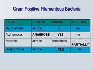
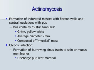
Actinomyces and Nocardia Dr Kamran Afzal Asst Prof Microbiology
Clinically important Gram positive bacilli Bacilli with branching filaments 1. Actinomyces 2. Nocardia Spore forming 1. Bacillus 2. Clostridium Non-spore forming 1. Corynebacterium 2. Listeria 3. Lactobacillus
Gram Positive Filamentous Bacteria no YES aerobe Streptomyces PARTIALLY sometimes  aerobe Nocardia no YES ANAEROBE Actinomyces yes no aerobe Mycobacteria ACID FAST GRANULE OXYGEN GENUS
Actinomycetes
Actinomycetes Genera Actinomyces and Nocardia are Gram positive non-motile and branching filamentous bacteria  Actinomyces is responsible for diseases of the oral cavity and intestines Nocardia causes pulmonary disease
Actinomyces “ Mykes” – Greek for “fungus” Thought by early microbiologists to be fungus Non-spore forming anaerobic or facultative anaerobic gram-positive bacteria Normally found in mouth, throat and GIT of humans “ Opportunistic Pathogens” Those causing disease do not exist freely in nature
Actinomyces Actinomyces A. israelii  – the commonest A. meyeri A. naeslundii A. odontolyticus A. viscosus Actinomyces species: 30; 8: human disease  Filamentous, branching, Gram-positive, pleomorphic, non-spore forming, non acid fast, anaerobic or microaerophilic bacilli
Epidemiology Geography/Season Word wide No seasonal variation Habitat Mouth, intestine and  female genital tract Who is at risk Poor oral hygiene Oral/dental surgery Trauma patients Women with IUDs Transmission From endogenous site to a sterile site
Actinomycosis Non-contagious chronic infection characterized by multiple abscesses with granuloma formation, tissue destruction and open draining sinuses Cervico-facial - commonest Thoracic Abdominal Pelvic Central nervous system Contiguous; does not respect tissue planes
Pathogenesis Not highly virulent (Opportunist) Component of Oral Flora Periodontal pockets Dental plaque Tonsillar crypts Takes advantage of injury to penetrate mucosal barriers Coincident infection Trauma Surgery
Pathogenesis Source of infection: Endogenous After local trauma, organisms invade tissues Due to low oxygen tension, organisms multiply Form hard yellow granules (sulfur granules) which are bacterial filaments solidified with tissue exudates  These granules drain outside through sinuses
Actinomycosis Formation of indurated masses with fibrous walls and central loculations with pus Pus contains "Sulfur Granules" Gritty, yellow white Average diameter 2mm Composed of “mycelial” mass Chronic infection  Formation of burrowing sinus tracts to skin or mucus membranes Discharge purulent material
Cervico-facial Actinomycosis Sinus tract originating in oral cavity has made its way to the  surface at the jawline -  “lumpy jaw” Multiple sinuses, scarring on neck and submaxillary area Contiguous spread to bone, neck, tongue etc DDX: TB, fungi, nocardia, abscess, neoplasm
Actinomycosis   Involving chest   Mycetoma foot
Lab Diagnosis Specimens  – open biopsy, aspiration material containing Sulphur granules (yellowish mycelial masses)
Lab Diagnosis Prompt  transport  to lab oratory preferably  in anaerobic   device The discharge should be mixed with sterile saline and allowed to stand, particles will separate out Place between 2 slides and crush Gram stain Fluorescence stain Histopathology of tissue
Sulfur Granule from Sinus Tract of Actinomycotic Infection The delicate filamentous bacilli (arrow) at the peripher y  of crushed granule
Laboratory Diagnosis Culture BHI, BA, Thioglycolate broth Aerobic    Anaerobic  37 0  C  37 0  C Slow growth on blood agar in 4-7 days Molar tooth colony
Histopathology
Actinomyces Identification Scheme
Treatment and Prophylaxis Surgical debridement Prolonged antibiotics (3-6 months) Penicillin Amoxicillin Tetracycline Erythromycin Clindamycin Maintenance of good oral hygiene Antibiotic prophylaxis while mouth or GI tract is penetrated
Nocardia
Nocardia 30 species, 13 of which cause human infection N. asteroides, N. brasiliensis Branching filament  < 1  µ m Fragmentation  rod, coccoid   form Gram-positive  beaded form C ell wall; mycolic acid Modified acid fast  (1%  H 2 S O 4 )  Partially acid fast
Epidemiology Nocardia is everywhere in the environment soil, organic matter, and water Human infection trauma and direct inoculation of the skin or soft tissues by inhalation Outbreaks in oncology and transplant wards from HCWs hands hospital construction with resultant contaminated dust
Risk Factors Immunocompromised patients 60% in pre-existing immune dysfunction Organ transplantation, hematologic malignancy, alcoholism, steroid use, diabetes, AIDS Patients with chronic pulmonary disorders, especially pulmonary alveolar proteinosis
Nocardiosis An acute, subacute, and chronic infection of the skin or pulmonary tract Transmitted through skin or pulmonary tract Primary, post-traumatic or post-inoculation lung disease Formation of abscesses which can metastasize
Pathology Suppurative granulomata Progressive fibrosis and necrosis Sinus formation and destruction of adjacent structures  Macroscopically visible infective granules  Mimics fungal mycetoma and actinomycetomata
Clinical Syndromes Mucocutaneous Can occur after minor trauma and animal or insect bites; may also colonize open wounds Cutaneous  Mycetoma Foot A chronic progressive, destructive disease, occurring days to months after inoculation located distally on the limbs (foot) Skin abscesses or cellulitis Lymphocutaneous infections
Pulmonary most common presentation usually indolent and progressive spread to contiguous structures, especially with soft tissue swellings or external fistulas Clinical Presentations pneumonia (often progressive in HIV) it can invade through surrounding tissues like actinomycosis lung abscess cavitary disease  empyema
CNS in 44% of cases of systemic nocardiosis classic signs and symptoms of pyogenic infections absent indolent presentations lead to a diagnosis of a brain tumour Other systems Eye:  can be hematogenous or due to eye trauma Disseminated infections  from central lines
Pulmonary Nocardiosis
Nocardia N ocardiosis ??   Specimen s : Sputum, pus, tissue biopsy   Gram’s stain   Modified acid-fast   P artially acid fast   Gram  + ve, branching filament, fragmentation   Culture and identification
Laboratory Diagnosis Culture BHI, SDA, BA, Thayer-martin agar – “Strict aerobes”  - Folded, heaped  -  Nocardia brasiliensis - White to pink, orange  -  SDA, 30 0 C, 9 days
Identification Biochemical tests Top right casein + Bottom right starch + Bottom left tyrosine + Top left xanthine  - Lysozyme test Right : resistant to lysozyme Left : does not grow in the  presence of lysozyme Nocardia ID Quad plate
Treatment I&D depending on the location Sulfas the mainstay of therapy, but susceptibilities vary For severely ill patients, CNS disease or in immunosuppressed patients use two or more drugs TMP/sulfa PLUS amikacin and a carbapenem OR third-generation cephalosporin  Oral alternatives minocycline and amoxicillin/clavulanate
Duration of Treatment Expect a clinical response in 3 - 10 days Often 3 - 6 months total Cutaneous disease usually is cured in a month or two CNS disease is treated for a year Relapses can occur up to a year after stopping therapy
Outcomes Cure rates of almost 100% are found in patients with skin or soft tissue disease 90% in pleuro-pulmonary disease 30-50% in brain abscess The longer the delay in diagnosis,  the more extensive the disease and, the worse the outcome


Actinomyces + nocardia

  • 1.
    Actinomyces and NocardiaDr Kamran Afzal Asst Prof Microbiology
  • 2.
    Clinically important Grampositive bacilli Bacilli with branching filaments 1. Actinomyces 2. Nocardia Spore forming 1. Bacillus 2. Clostridium Non-spore forming 1. Corynebacterium 2. Listeria 3. Lactobacillus
  • 3.
    Gram Positive FilamentousBacteria no YES aerobe Streptomyces PARTIALLY sometimes aerobe Nocardia no YES ANAEROBE Actinomyces yes no aerobe Mycobacteria ACID FAST GRANULE OXYGEN GENUS
  • 4.
  • 5.
    Actinomycetes Genera Actinomycesand Nocardia are Gram positive non-motile and branching filamentous bacteria Actinomyces is responsible for diseases of the oral cavity and intestines Nocardia causes pulmonary disease
  • 6.
    Actinomyces “ Mykes”– Greek for “fungus” Thought by early microbiologists to be fungus Non-spore forming anaerobic or facultative anaerobic gram-positive bacteria Normally found in mouth, throat and GIT of humans “ Opportunistic Pathogens” Those causing disease do not exist freely in nature
  • 7.
    Actinomyces Actinomyces A.israelii – the commonest A. meyeri A. naeslundii A. odontolyticus A. viscosus Actinomyces species: 30; 8: human disease Filamentous, branching, Gram-positive, pleomorphic, non-spore forming, non acid fast, anaerobic or microaerophilic bacilli
  • 8.
    Epidemiology Geography/Season Wordwide No seasonal variation Habitat Mouth, intestine and female genital tract Who is at risk Poor oral hygiene Oral/dental surgery Trauma patients Women with IUDs Transmission From endogenous site to a sterile site
  • 9.
    Actinomycosis Non-contagious chronicinfection characterized by multiple abscesses with granuloma formation, tissue destruction and open draining sinuses Cervico-facial - commonest Thoracic Abdominal Pelvic Central nervous system Contiguous; does not respect tissue planes
  • 10.
    Pathogenesis Not highlyvirulent (Opportunist) Component of Oral Flora Periodontal pockets Dental plaque Tonsillar crypts Takes advantage of injury to penetrate mucosal barriers Coincident infection Trauma Surgery
  • 11.
    Pathogenesis Source ofinfection: Endogenous After local trauma, organisms invade tissues Due to low oxygen tension, organisms multiply Form hard yellow granules (sulfur granules) which are bacterial filaments solidified with tissue exudates These granules drain outside through sinuses
  • 12.
    Actinomycosis Formation ofindurated masses with fibrous walls and central loculations with pus Pus contains &quot;Sulfur Granules&quot; Gritty, yellow white Average diameter 2mm Composed of “mycelial” mass Chronic infection Formation of burrowing sinus tracts to skin or mucus membranes Discharge purulent material
  • 13.
    Cervico-facial Actinomycosis Sinustract originating in oral cavity has made its way to the surface at the jawline - “lumpy jaw” Multiple sinuses, scarring on neck and submaxillary area Contiguous spread to bone, neck, tongue etc DDX: TB, fungi, nocardia, abscess, neoplasm
  • 14.
    Actinomycosis Involving chest Mycetoma foot
  • 15.
    Lab Diagnosis Specimens – open biopsy, aspiration material containing Sulphur granules (yellowish mycelial masses)
  • 16.
    Lab Diagnosis Prompt transport to lab oratory preferably in anaerobic device The discharge should be mixed with sterile saline and allowed to stand, particles will separate out Place between 2 slides and crush Gram stain Fluorescence stain Histopathology of tissue
  • 17.
    Sulfur Granule fromSinus Tract of Actinomycotic Infection The delicate filamentous bacilli (arrow) at the peripher y of crushed granule
  • 18.
    Laboratory Diagnosis CultureBHI, BA, Thioglycolate broth Aerobic Anaerobic 37 0 C 37 0 C Slow growth on blood agar in 4-7 days Molar tooth colony
  • 19.
  • 20.
  • 21.
    Treatment and ProphylaxisSurgical debridement Prolonged antibiotics (3-6 months) Penicillin Amoxicillin Tetracycline Erythromycin Clindamycin Maintenance of good oral hygiene Antibiotic prophylaxis while mouth or GI tract is penetrated
  • 22.
  • 23.
    Nocardia 30 species,13 of which cause human infection N. asteroides, N. brasiliensis Branching filament < 1 µ m Fragmentation rod, coccoid form Gram-positive beaded form C ell wall; mycolic acid Modified acid fast (1% H 2 S O 4 ) Partially acid fast
  • 24.
    Epidemiology Nocardia iseverywhere in the environment soil, organic matter, and water Human infection trauma and direct inoculation of the skin or soft tissues by inhalation Outbreaks in oncology and transplant wards from HCWs hands hospital construction with resultant contaminated dust
  • 25.
    Risk Factors Immunocompromisedpatients 60% in pre-existing immune dysfunction Organ transplantation, hematologic malignancy, alcoholism, steroid use, diabetes, AIDS Patients with chronic pulmonary disorders, especially pulmonary alveolar proteinosis
  • 26.
    Nocardiosis An acute,subacute, and chronic infection of the skin or pulmonary tract Transmitted through skin or pulmonary tract Primary, post-traumatic or post-inoculation lung disease Formation of abscesses which can metastasize
  • 27.
    Pathology Suppurative granulomataProgressive fibrosis and necrosis Sinus formation and destruction of adjacent structures Macroscopically visible infective granules Mimics fungal mycetoma and actinomycetomata
  • 28.
    Clinical Syndromes MucocutaneousCan occur after minor trauma and animal or insect bites; may also colonize open wounds Cutaneous Mycetoma Foot A chronic progressive, destructive disease, occurring days to months after inoculation located distally on the limbs (foot) Skin abscesses or cellulitis Lymphocutaneous infections
  • 29.
    Pulmonary most commonpresentation usually indolent and progressive spread to contiguous structures, especially with soft tissue swellings or external fistulas Clinical Presentations pneumonia (often progressive in HIV) it can invade through surrounding tissues like actinomycosis lung abscess cavitary disease empyema
  • 30.
    CNS in 44%of cases of systemic nocardiosis classic signs and symptoms of pyogenic infections absent indolent presentations lead to a diagnosis of a brain tumour Other systems Eye: can be hematogenous or due to eye trauma Disseminated infections from central lines
  • 31.
  • 32.
    Nocardia N ocardiosis?? Specimen s : Sputum, pus, tissue biopsy Gram’s stain Modified acid-fast P artially acid fast Gram + ve, branching filament, fragmentation Culture and identification
  • 33.
    Laboratory Diagnosis CultureBHI, SDA, BA, Thayer-martin agar – “Strict aerobes” - Folded, heaped - Nocardia brasiliensis - White to pink, orange - SDA, 30 0 C, 9 days
  • 34.
    Identification Biochemical testsTop right casein + Bottom right starch + Bottom left tyrosine + Top left xanthine - Lysozyme test Right : resistant to lysozyme Left : does not grow in the presence of lysozyme Nocardia ID Quad plate
  • 35.
    Treatment I&D dependingon the location Sulfas the mainstay of therapy, but susceptibilities vary For severely ill patients, CNS disease or in immunosuppressed patients use two or more drugs TMP/sulfa PLUS amikacin and a carbapenem OR third-generation cephalosporin Oral alternatives minocycline and amoxicillin/clavulanate
  • 36.
    Duration of TreatmentExpect a clinical response in 3 - 10 days Often 3 - 6 months total Cutaneous disease usually is cured in a month or two CNS disease is treated for a year Relapses can occur up to a year after stopping therapy
  • 37.
    Outcomes Cure ratesof almost 100% are found in patients with skin or soft tissue disease 90% in pleuro-pulmonary disease 30-50% in brain abscess The longer the delay in diagnosis, the more extensive the disease and, the worse the outcome
  • 38.
  • 39.
  • 40.
  • 41.
  • 42.