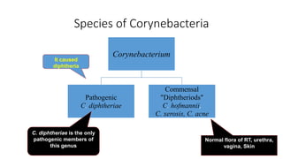
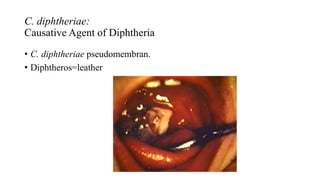
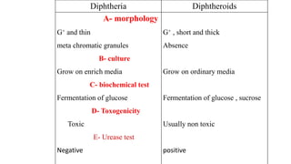
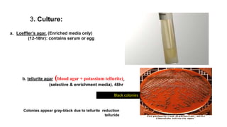
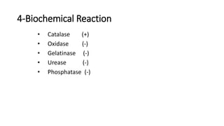
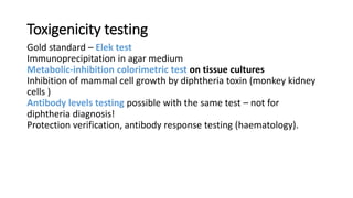
Corynebacterium
WalaHassanMustafa
M.Sc Microbiology
content
• General Characteristics
Diphtheria – epidemiology
• Species of Corynebacteria
• Diphtheria – pathogenesis
• Non pathogen Corynebacteriae Spp
• Laboratory Diagnosis
• Treatment
• vaccination
Corynebacterium is a genus of Gram-positive rod-shaped bacteria.
They are widely distributed in nature.
They are catalase positive
non-spore-forming
non-motile
rod-shaped bacteria that are straight or slightly curved.
Metachromatic granules are usually present representing stored phosphate
regions.
The bacteria group together in a characteristic way, which has been
described as the form of V or Y shape, or what is called "Chinese letters
arrangement".
General Characteristics:
Corynebacterium is a genus of Gram-positive rod-shaped bacteria.
They are widely distributed in nature.
They are catalase positive
non-spore-forming
non-motile
rod-shaped bacteria that are straight or slightly curved with irregular swelling
at one or both ends (club shaped ).
Metachromatic granules are usually present representin.
The bacteria group together in a characteristic way, which has been described
as the form of V or Y shape, or what is called "Chinese letters arrangement".
Diphtheria – epidemiology
Mainly a disease of childhood in endemic areas – uncommon below 1st year
peaks at 5.
In nature, C.diphtheriae occurs in the respiratory tract in the wounds or in
the skin of the infected person or carriers.
Nasopharyngal or cutaneous carriage of toxigenic or non toxigenic strains
can persist for life in healthy people.
Species of Corynebacteria
Corynebacterium
Pathogenic
C. diphtheriae
Commensal
"Diphtheriods"
C. hofmannii,
C. xerosis, C. acne
C. diphtheriae is the only
pathogenic members of
this genus
It caused
diphtheria
Normal flora of RT, urethra,
vagina, Skin
C. diphtheriae:
Causative Agent of Diphtheria
• C. diphtheriae pseudomembran.
• Diphtheros=leather
Diphtheria – pathogenesis :
Colonization of mucosal membrane of upper respiratory tract –
rarely invades the bloodstream
Toxin spreads through the mucosa via diffusion, cell death occurs in
superficial mucosal layers.
Pseudomembranes (coagulum of necrotic cells, fibrin and blood
elements) may spread further into teait ways and cause their obturation
Toxin may further spread into the bloodstream, affects most
susceptible cells
Myocard
Peripheral nerves (periaxonal cells)
Epithelium of renal tubules
Clinical diphtheria
Incubation period 2-4 days
Pharyngeal – catarrhal – fibrinous – pseudomembranous inflammatory reaction
Cervical lymphadenopathy, low fever Benign phase, prompt reaction to antibiotics
administration
Laryngeal (croup) – life threatening particularly in infants In the initial stage
reminds viral laryngitis Spreading of pseudomembranes to lower respiratory
tract follows Serious prognosis, tracheostomy not successful in all cases
Cutaneous diphtheria – ulcers with rolled edges, painless, grey
pseudomembranes
Skin, (conjunctiva, ear, genitals)
Toxin-mediated impairment rare
Nasal – pseudomembranes, purulent secretion
High contagiosity
Non pathogen Corynebacteriae Spp.
“Diphtheriods” :-
• Bacilli that morphology resemble diphtheria.
• It is not pathogenic.
• It is found on skin or conjunctiva and oral cavity.
• It is opportunistic pathogens.
• It is short 1-5µm with parallel sides and rounded
ends without metachromatic granules.
• Actual differentiation can be made out by biochemical
testes and virulence test .
Diphtheroids
Diphtheria
G+ , short and thick
Absence
Grow on ordinary media
Fermentation of glucose , sucrose
Usually non toxic
positive
A- morphology
G+ and thin
meta chromatic granules
B- culture
Grow on enrich media
C- biochemical test
Fermentation of glucose
D- Toxogenicity
Toxic
E- Urease test
Negative
Laboratory Diagnosis
1. Specimens : Throat swab.
Skin swab.
2. Staining:
Gram’s stain  G + ve bacilli,
Chinese letter.
Albert's stain  Metachromatic granules (dark),
bacilli (green).
3. Culture:
a. Loeffler’s agar. (Enriched media only)
(12-18hr): contains serum or egg
b. tellurite agar (blood agar + potassium tellurite)
(selective & enrichment media). 48hr
Black colonies
Colonies appear gray-black due to tellurite reduction
telluride
4-Biochemical Reaction
• Catalase (+)
• Oxidase (-)
• Gelatinase (-)
• Urease (-)
• Phosphatase (-)
Toxigenicity testing
Gold standard – Elek test
Immunoprecipitation in agar medium
Metabolic-inhibition colorimetric test on tissue cultures
Inhibition of mammal cell growth by diphtheria toxin (monkey kidney
cells )
Antibody levels testing possible with the same test – not for
diphtheria diagnosis!
Protection verification, antibody response testing (haematology).
Elek's test for toxigenicity
It is an in vitro test performed only in reference to public health
laboratories in order to know if the organism is able to produce the
diphtheria toxin or not.
Filter paper strep containing antitoxin is placed on agar plate. The
tested culture is streaked across the plate .after 48 hours the antitoxin
precipitates the toxin, resulting in the formation of bands between the
filter paper and the bacterial growth.
Gel-diffusion test (ELICK test)
Diphtheria – treatment
Antibiotics:
Penicillin – drug of choice
Macrolides for decolonization
Lincosamides, ciprofloxacin, rifampicin - alternatives
Antitoxin: (horse anti-diphtheria toxin serum)
– i.m. or i.v. for toxin
inactivation
Skin test before administration (risk of anaphylaxis)
Diphtheria – vaccination
Diphtheriae anatoxinum adsorbed to aluminium hydroxide
Vaccine protects from the effect of diphtheria toxin, not the
bacterium itself
Good immunity provided
Protective levels
Common population min. 0,01 IU/ml
Healthcere workers and other persons at risk min. 0,1 IU/ml
Booster administration every 10 years recommended
Corynebacterium.pptx

Corynebacterium.pptx

  • 1.
  • 2.
    content • General Characteristics Diphtheria– epidemiology • Species of Corynebacteria • Diphtheria – pathogenesis • Non pathogen Corynebacteriae Spp • Laboratory Diagnosis • Treatment • vaccination
  • 3.
    Corynebacterium is agenus of Gram-positive rod-shaped bacteria. They are widely distributed in nature. They are catalase positive non-spore-forming non-motile rod-shaped bacteria that are straight or slightly curved. Metachromatic granules are usually present representing stored phosphate regions. The bacteria group together in a characteristic way, which has been described as the form of V or Y shape, or what is called "Chinese letters arrangement".
  • 4.
    General Characteristics: Corynebacterium isa genus of Gram-positive rod-shaped bacteria. They are widely distributed in nature. They are catalase positive non-spore-forming non-motile rod-shaped bacteria that are straight or slightly curved with irregular swelling at one or both ends (club shaped ). Metachromatic granules are usually present representin. The bacteria group together in a characteristic way, which has been described as the form of V or Y shape, or what is called "Chinese letters arrangement".
  • 5.
    Diphtheria – epidemiology Mainlya disease of childhood in endemic areas – uncommon below 1st year peaks at 5. In nature, C.diphtheriae occurs in the respiratory tract in the wounds or in the skin of the infected person or carriers. Nasopharyngal or cutaneous carriage of toxigenic or non toxigenic strains can persist for life in healthy people.
  • 7.
    Species of Corynebacteria Corynebacterium Pathogenic C.diphtheriae Commensal "Diphtheriods" C. hofmannii, C. xerosis, C. acne C. diphtheriae is the only pathogenic members of this genus It caused diphtheria Normal flora of RT, urethra, vagina, Skin
  • 8.
    C. diphtheriae: Causative Agentof Diphtheria • C. diphtheriae pseudomembran. • Diphtheros=leather
  • 9.
    Diphtheria – pathogenesis: Colonization of mucosal membrane of upper respiratory tract – rarely invades the bloodstream Toxin spreads through the mucosa via diffusion, cell death occurs in superficial mucosal layers. Pseudomembranes (coagulum of necrotic cells, fibrin and blood elements) may spread further into teait ways and cause their obturation Toxin may further spread into the bloodstream, affects most susceptible cells Myocard Peripheral nerves (periaxonal cells) Epithelium of renal tubules
  • 10.
    Clinical diphtheria Incubation period2-4 days Pharyngeal – catarrhal – fibrinous – pseudomembranous inflammatory reaction Cervical lymphadenopathy, low fever Benign phase, prompt reaction to antibiotics administration Laryngeal (croup) – life threatening particularly in infants In the initial stage reminds viral laryngitis Spreading of pseudomembranes to lower respiratory tract follows Serious prognosis, tracheostomy not successful in all cases Cutaneous diphtheria – ulcers with rolled edges, painless, grey pseudomembranes Skin, (conjunctiva, ear, genitals) Toxin-mediated impairment rare Nasal – pseudomembranes, purulent secretion High contagiosity
  • 11.
    Non pathogen CorynebacteriaeSpp. “Diphtheriods” :- • Bacilli that morphology resemble diphtheria. • It is not pathogenic. • It is found on skin or conjunctiva and oral cavity. • It is opportunistic pathogens. • It is short 1-5µm with parallel sides and rounded ends without metachromatic granules. • Actual differentiation can be made out by biochemical testes and virulence test .
  • 12.
    Diphtheroids Diphtheria G+ , shortand thick Absence Grow on ordinary media Fermentation of glucose , sucrose Usually non toxic positive A- morphology G+ and thin meta chromatic granules B- culture Grow on enrich media C- biochemical test Fermentation of glucose D- Toxogenicity Toxic E- Urease test Negative
  • 13.
    Laboratory Diagnosis 1. Specimens: Throat swab. Skin swab. 2. Staining: Gram’s stain  G + ve bacilli, Chinese letter. Albert's stain  Metachromatic granules (dark), bacilli (green).
  • 14.
    3. Culture: a. Loeffler’sagar. (Enriched media only) (12-18hr): contains serum or egg b. tellurite agar (blood agar + potassium tellurite) (selective & enrichment media). 48hr Black colonies Colonies appear gray-black due to tellurite reduction telluride
  • 15.
    4-Biochemical Reaction • Catalase(+) • Oxidase (-) • Gelatinase (-) • Urease (-) • Phosphatase (-)
  • 16.
    Toxigenicity testing Gold standard– Elek test Immunoprecipitation in agar medium Metabolic-inhibition colorimetric test on tissue cultures Inhibition of mammal cell growth by diphtheria toxin (monkey kidney cells ) Antibody levels testing possible with the same test – not for diphtheria diagnosis! Protection verification, antibody response testing (haematology).
  • 17.
    Elek's test fortoxigenicity It is an in vitro test performed only in reference to public health laboratories in order to know if the organism is able to produce the diphtheria toxin or not. Filter paper strep containing antitoxin is placed on agar plate. The tested culture is streaked across the plate .after 48 hours the antitoxin precipitates the toxin, resulting in the formation of bands between the filter paper and the bacterial growth.
  • 18.
  • 19.
    Diphtheria – treatment Antibiotics: Penicillin– drug of choice Macrolides for decolonization Lincosamides, ciprofloxacin, rifampicin - alternatives Antitoxin: (horse anti-diphtheria toxin serum) – i.m. or i.v. for toxin inactivation Skin test before administration (risk of anaphylaxis)
  • 20.
    Diphtheria – vaccination Diphtheriaeanatoxinum adsorbed to aluminium hydroxide Vaccine protects from the effect of diphtheria toxin, not the bacterium itself Good immunity provided Protective levels Common population min. 0,01 IU/ml Healthcere workers and other persons at risk min. 0,1 IU/ml Booster administration every 10 years recommended