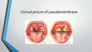
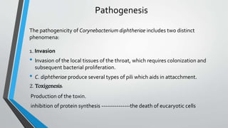
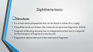
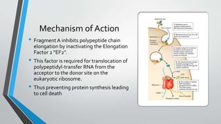
Corynebacterium
diphtheriae
H.M.Moavia Atique
University ofVeterinary and Animal Sciences Lahore
General Characters
• Corynebacterium diphtheria
(Greek: Diphtheros meaning leather)
• Gram-positive
• Rod-shaped bacterium
• Non acid fast
• Club-shaped forms = Swelled end
• Chinese characters “Cell arrangement L orV shapes
are also observed ).
• Albert Stain is used to demonstrate the metachromatic
(volutin) granules formed in the polar regions.
• Aerobic
• Non-motile
• Non-sporulating
• Non-acid fast
Biovars
Occurs in four biovars:
 gravis,
 mitis,
 intermedius
 belfanti,
based on colonial morphology and biochemical profiles
Diphtheria
• Caused by infection with toxigenic strains of gram-positive
Corynebacterium diphtheria
• Contagious and life threatening bacterial disease
• C. ulcerans and very rarely C. pseudotuberculosis produce diphtheria toxin
and also cause diphtheria.(zoonotic)
Clinical Features
• Sore throat
• Malaise
• Low-grade fever
• Grayish-white, fibrinous pseudomembrane
• Obstruction of larynx and lower air ways
• Myocarditis and peripheral neuritis
• Difficulty in swallowing.
•Sites of infection:
respiratory mucosa (respiratory diphtheria) and
the skin (cutaneous diphtheria) extra-respiratory mucosal sites, e.g., the eye, ear, or
genitals (rare)
•Reservoir of Corynaebacterium diphtheriae
Humans are the only known reservoir of C. diphtheriae
•Disease transmission
person to person by respiratory droplets
or
direct contact with respiratory secretions, discharges from skin lesions or, rarely, fomites
Incubation period is 2-5 days (range 1-10 days)
Clinical picture of pseudomembrane
Pathogenesis
The pathogenicity of Corynebacterium diphtheriae includes two distinct
phenomena:
1. Invasion
• Invasion of the local tissues of the throat, which requires colonization and
subsequent bacterial proliferation.
• C. diphtheriae produce several types of pili which aids in attacchment.
2. Toxigenesis:
Production of the toxin.
inhibition of protein synthesis ---------------the death of eucaryotic cells
Diphtheria toxin
Structure:
• It is a heat-labile polypeptide that can be lethal in a dose of 0.1 ug/kg.
• If disulfide bonds are broken, the molecule can give two fragments. (A & B).
• Fragment B (Binding domain) has no independent action but it is required
for the transport of fragment A into the cell.
• Fragment A (Active domain) is the main active fragment.
Mechanism of Action
• Fragment A inhibits polypeptide chain
elongation by inactivating the Elongation
Factor 2 "EF2".
• This factor is required for translocation of
polypeptidyl-transfer RNA from the
acceptor to the donor site on the
eukaryotic ribosome.
• Thus preventing protein synthesis leading
to cell death
Toxigenecity
Two main factors are responsible for the production of toxin:
 low extracellular concentrations of iron
The presence of a lysogenic prophage (β Phage) in the bacterial
chromosome.
Role of Iron
• Toxin is synthesized in high yield only after the exogenous supply of iron has
become exhausted.
• The tox gene is regulated by a mechanism of negative control wherein a
repressor molecule, product of the DtxR gene, is activated by iron.The
active repressor binds to the tox gene operator and prevents transcription.
• When iron is removed from the repressor,derepression occurs, the repressor
is inactivated and transcription of the tox genes can occur.
Role of β-Phage
• Only those strains of Corynebacterium diphtheriae that are
lysogenized by a specific Beta phage (β phage) produce
diphtheria toxin.
• The phage contains the structural gene for the toxin molecule.
Laboratory Diagnosis
• Isolation of C. diphtheriae by culturing
• Biochemical characterization
• Toxigenicity testing
• Polymerase chain reaction (PCR)
• Serologic testing
Specimen collection
• Throat and nasopharengeal swabs.
• When diagnosis is confirmed, patient contacts must be
subjected to the same procedures.
• Moisten swabs with Normal saline and collect in the usual
manner.
Staining method
• Gram staining
• Albert staining (metachoromatic granules)
• Appear asV and L letters, resembling
Chinese letter patterns
Growth Requirements
• Need enrichment media (blood, serum or egg)
• pH (7.4-8)
• Temp. 35- 37⁰ C
• Protein content >24g/Litre
• Iron content 0.1-0.3 ppm
• Supplementation with 0.2% yeast extract
• Aerobic/Facultative anaerobic ( good aeration…..Baffled Flask)
Culture media
Commonly used medium
•Tellurite blood agar
•Tinsdale medium
•Loffler's serum medium
Tinsdale medium
• PotassiumTellurite reduces toTellurite
• H2S is liberated through Cystine.
• H2S reacts with tellurite and produce
Black-Brown halos.
BiochemicalTests
• Urease Negative
• Nitrate Positive
• Gelatin liquefaction: Negative
• Fermentation reactions of various carbohydrates.
Toxigenecity
testing
• In-Vivo toxigenicity test:
Test performed in guinea pigs
• In-Vitro toxigenicity test:
Elek gel diffusionTest
PCR
• PCR allows for detection of
• the regulatory gene for toxin production (dtxR)
• the diphtheria toxin gene (tox) on nonviable organisms.
• tox gene (58 Kda)
Serological testing
• Skin test = Schick test
• To determine whetherThe patient is susceptible to diphtheria infection or not by
detecting the presence or absence of antibodies.
• Procedure:
1) 0.1 ml of toxin is injected intradermally in one arm.
2) 0.1 ml of heated (inactivated) toxin as a control is injected in the other arm.
3) Read after 24-48 hours.
Results
• Positive:
(Susceptible)Redness and swelling that
increases for several days and then fades,
leaving brownish pigmented area.
The control site shows no reaction.
• Negative:
(Not susceptible) Neither injection site shows any reaction
Treatment of Diphtheria
• Initially -------- small doses of the antitoxin and then gradually increasing the
dosage.
• The infection is then treated with antibiotics, such as penicillin and
erythromycin.
DiphtheriaToxoid:
Formalin inactivated diphtheria toxin.
Neutralizes only unbound toxin.
Corynebacterium diphtheriae presentation

Corynebacterium diphtheriae presentation

  • 1.
  • 2.
    General Characters • Corynebacteriumdiphtheria (Greek: Diphtheros meaning leather) • Gram-positive • Rod-shaped bacterium • Non acid fast • Club-shaped forms = Swelled end • Chinese characters “Cell arrangement L orV shapes are also observed ). • Albert Stain is used to demonstrate the metachromatic (volutin) granules formed in the polar regions.
  • 3.
    • Aerobic • Non-motile •Non-sporulating • Non-acid fast Biovars Occurs in four biovars:  gravis,  mitis,  intermedius  belfanti, based on colonial morphology and biochemical profiles
  • 4.
    Diphtheria • Caused byinfection with toxigenic strains of gram-positive Corynebacterium diphtheria • Contagious and life threatening bacterial disease • C. ulcerans and very rarely C. pseudotuberculosis produce diphtheria toxin and also cause diphtheria.(zoonotic)
  • 5.
    Clinical Features • Sorethroat • Malaise • Low-grade fever • Grayish-white, fibrinous pseudomembrane • Obstruction of larynx and lower air ways • Myocarditis and peripheral neuritis • Difficulty in swallowing.
  • 6.
    •Sites of infection: respiratorymucosa (respiratory diphtheria) and the skin (cutaneous diphtheria) extra-respiratory mucosal sites, e.g., the eye, ear, or genitals (rare) •Reservoir of Corynaebacterium diphtheriae Humans are the only known reservoir of C. diphtheriae •Disease transmission person to person by respiratory droplets or direct contact with respiratory secretions, discharges from skin lesions or, rarely, fomites Incubation period is 2-5 days (range 1-10 days)
  • 7.
    Clinical picture ofpseudomembrane
  • 8.
    Pathogenesis The pathogenicity ofCorynebacterium diphtheriae includes two distinct phenomena: 1. Invasion • Invasion of the local tissues of the throat, which requires colonization and subsequent bacterial proliferation. • C. diphtheriae produce several types of pili which aids in attacchment. 2. Toxigenesis: Production of the toxin. inhibition of protein synthesis ---------------the death of eucaryotic cells
  • 9.
    Diphtheria toxin Structure: • Itis a heat-labile polypeptide that can be lethal in a dose of 0.1 ug/kg. • If disulfide bonds are broken, the molecule can give two fragments. (A & B). • Fragment B (Binding domain) has no independent action but it is required for the transport of fragment A into the cell. • Fragment A (Active domain) is the main active fragment.
  • 10.
    Mechanism of Action •Fragment A inhibits polypeptide chain elongation by inactivating the Elongation Factor 2 "EF2". • This factor is required for translocation of polypeptidyl-transfer RNA from the acceptor to the donor site on the eukaryotic ribosome. • Thus preventing protein synthesis leading to cell death
  • 11.
    Toxigenecity Two main factorsare responsible for the production of toxin:  low extracellular concentrations of iron The presence of a lysogenic prophage (β Phage) in the bacterial chromosome.
  • 12.
    Role of Iron •Toxin is synthesized in high yield only after the exogenous supply of iron has become exhausted. • The tox gene is regulated by a mechanism of negative control wherein a repressor molecule, product of the DtxR gene, is activated by iron.The active repressor binds to the tox gene operator and prevents transcription. • When iron is removed from the repressor,derepression occurs, the repressor is inactivated and transcription of the tox genes can occur.
  • 13.
    Role of β-Phage •Only those strains of Corynebacterium diphtheriae that are lysogenized by a specific Beta phage (β phage) produce diphtheria toxin. • The phage contains the structural gene for the toxin molecule.
  • 14.
    Laboratory Diagnosis • Isolationof C. diphtheriae by culturing • Biochemical characterization • Toxigenicity testing • Polymerase chain reaction (PCR) • Serologic testing
  • 15.
    Specimen collection • Throatand nasopharengeal swabs. • When diagnosis is confirmed, patient contacts must be subjected to the same procedures. • Moisten swabs with Normal saline and collect in the usual manner.
  • 16.
    Staining method • Gramstaining • Albert staining (metachoromatic granules) • Appear asV and L letters, resembling Chinese letter patterns
  • 17.
    Growth Requirements • Needenrichment media (blood, serum or egg) • pH (7.4-8) • Temp. 35- 37⁰ C • Protein content >24g/Litre • Iron content 0.1-0.3 ppm • Supplementation with 0.2% yeast extract • Aerobic/Facultative anaerobic ( good aeration…..Baffled Flask)
  • 18.
    Culture media Commonly usedmedium •Tellurite blood agar •Tinsdale medium •Loffler's serum medium
  • 19.
    Tinsdale medium • PotassiumTelluritereduces toTellurite • H2S is liberated through Cystine. • H2S reacts with tellurite and produce Black-Brown halos.
  • 20.
    BiochemicalTests • Urease Negative •Nitrate Positive • Gelatin liquefaction: Negative • Fermentation reactions of various carbohydrates.
  • 21.
    Toxigenecity testing • In-Vivo toxigenicitytest: Test performed in guinea pigs • In-Vitro toxigenicity test: Elek gel diffusionTest
  • 22.
    PCR • PCR allowsfor detection of • the regulatory gene for toxin production (dtxR) • the diphtheria toxin gene (tox) on nonviable organisms. • tox gene (58 Kda)
  • 23.
    Serological testing • Skintest = Schick test • To determine whetherThe patient is susceptible to diphtheria infection or not by detecting the presence or absence of antibodies. • Procedure: 1) 0.1 ml of toxin is injected intradermally in one arm. 2) 0.1 ml of heated (inactivated) toxin as a control is injected in the other arm. 3) Read after 24-48 hours.
  • 24.
    Results • Positive: (Susceptible)Redness andswelling that increases for several days and then fades, leaving brownish pigmented area. The control site shows no reaction. • Negative: (Not susceptible) Neither injection site shows any reaction
  • 25.
    Treatment of Diphtheria •Initially -------- small doses of the antitoxin and then gradually increasing the dosage. • The infection is then treated with antibiotics, such as penicillin and erythromycin. DiphtheriaToxoid: Formalin inactivated diphtheria toxin. Neutralizes only unbound toxin.

Editor's Notes

  • #17 Fix the dried smear using alcohol (see subunit 7.3.2) 2 Cover the smear with the toluidine bluemalachite green stain for 3–5 minutes. 3 Wash off the stain with clean water. When the tap water is not clean, use filtered water or clean boiled rainwater. 4 Tip off all the water. 5 Cover the smear with Albert’s iodine for 1 minute. Wash off with water. 6 Wipe the back of the slide clean, and place it in a draining rack for the smear to air-dry. 7 Examine the smear microscopically.
  • #20 Potassium tellurite is the selective agent (inhibits most of the upper respiratory tract normal flora) that turns the media brown-black as a result from the reduction of potassium tellurite to metallic tellurite. The dark halo is due to the production of H2S from cystine, interacting with the tellurite salt.