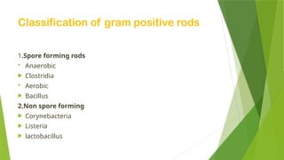
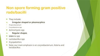
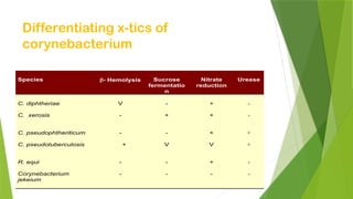
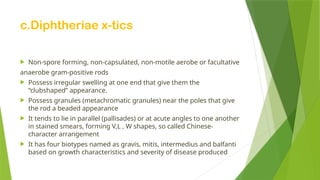
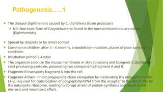
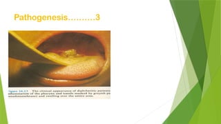
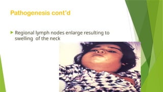
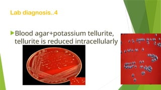
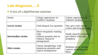
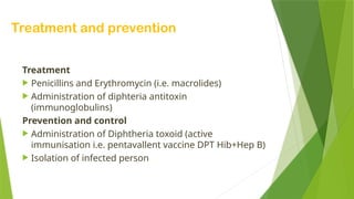
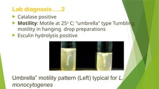
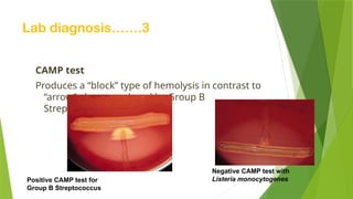
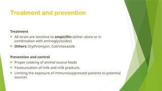
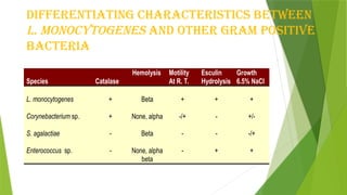
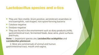
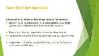
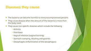
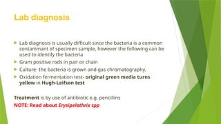
Gram positive
rods/bacilli
Presented by
HANABA COLLINS GODWIN AND EJANG SARAH
LECTURER;ANGOL DENISH CALMAX
Illustration of rods/bacilli
Classification of gram positive rods
1.Spore forming rods
 Anaerobic
 Clostridia
 Aerobic
 Bacillus
2.Non spore forming
 Corynebacteria
 Listeria
 lactobacillus
overview
Non spore forming gram positive
rods/bacilli
 They include;
A. Irregular shaped or pleomorphics
 Propionibacterium
 Corynebacteria spp
 Actinomyces spp
A. Regular shapes
 Listeria spp
 Lactobacillus spp
 Erysipelothirx
 Note ;our main emphasis is on corynebacterium, listeria and
lactobacillus
Corynebacteria spp
 Corynebacterium diphtheriae is the most significant human
pathogen.
 It Causes diphtheria
 Other species
I. C. Xerosis
II. C. Pseudodiphtheriticum
III. C. Pseudotuberculosis
IV. C. Jekeium
V. C. ulcerans
Differentiating x-tics of
corynebacterium
Species  Hemolysis Sucrose
fermentatio
n
Nitrate
reduction
Urease
C. diphtheriae
C. xerosis
V
-
-
+
+
+
-
-
C. pseudophtheriticum
C. pseudotuberculosis
-
+
-
V
+
V
+
+
R. equi
Corynebacterium
jekeium
-
-
-
-
+
-
-
-
c.Diphtheriae x-tics
 Non-spore forming, non-capsulated, non-motile aerobe or facultative
anaerobe gram-positive rods
 Possess irregular swelling at one end that give them the
“clubshaped” appearance.
 Possess granules (metachromatic granules) near the poles that give
the rod a beaded appearance
 It tends to lie in parallel (pallisades) or at acute angles to one another
in stained smears, forming V,L , W shapes, so called Chinese-
character arrangement
 It has four biotypes named as gravis, mitis, intermedius and balfanti
based on growth characteristics and severity of disease produced
Pathogenesis…..1
 The disease Diphtheria is caused by C. diphtheria (toxin producer)
 NB: Non toxic form of Corynebacteria found in the normal microbiota are called
(Diphtheroids)
 Spread by droplets or by direct contact
 Common in children after 3 – 6 months, crowded communities, places of poor sanitary
condition.
 Incubation period 2-4 days
 The organism colonize the mucus membrane or skin abrasions and toxigenic C.diphteriae
start producing exotoxin, possessing two components,fragment A and B.
 Fragment B transports fragment A into the cell
 Fragment A then inhibit polypeptide chain elongation by inactivating the elongation factor
EF-2, required for translocation of polypeptidyl-tRNA from the acceptor to the donor site on
the eukaryotic ribosome, leading to abrupt arrest of protein synthesis and result in cell
necrosis and neurotoxic effect.
Pathogenesis…….2
 Diphteria toxin causes respiratory tract epithelial destruction resulting in
formation of necrotic epithelium with pseudomembrane formation over the
tonsils, pharynx, and larynx.
 Distant toxic damage includes parenchymal degeneration and necrosis in heart
muscle, liver, kidney, adrenal glands and peripheral and cranial nerves.
 Wound/skin diphteria occurs chiefly in the tropics and forms membrane-covered
wound that fails to heal. Systemic effect is negligible.
 It clinically manifests with fever, sorethroat, suffocation (due to obstruction by
upper respiratory tract pseudomembrane formation), arrhythemia, and difficulty
of vision, swallowing and paralysis of upper and lower extremities
Pathogenesis……….3
Pathogenesis cont’d
 Regional lymph nodes enlarge resulting to
swelling of the neck
Clinical features of
diphtheria………….1
Diphtheria signs and symptoms usually begin two to five days after a person becomes
infected
It clinically manifests with:
 Fever and chills
 Sore throat
 Suffocation (due to obstruction by upper respiratory tract pseudomembrane
formation covering your throat and tonsils)
 Arrhythmia (abnormality of the heart's rhythm. It may beat too slowly, too quickly, or
irregularly)
 Difficulty breathing or rapid breathing
 Difficulty of vision, and swallowing
 Paralysis of upper and lower extremities
 Swollen glands (enlarged lymph nodes) in your neck
Clinical features of diphtheria’’’’’2
Skin infection
 A second type of diphtheria can affect the skin,
causing the typical pain, redness and swelling
associated with other bacterial skin infections.
 Ulcers covered by a gray membrane also may
develop in cutaneous diphtheria.
Laboratory diagnosis………1
 Specimen: Swabs from the nose, throat, or suspected lesion
 Smears: Beaded rods in typical arrangement when stained with alkaline
methylene blue or gram’s stain
 Culture: Small, granular, and gray, with irregular edges with small zone of
hemolysis on blood agar
 PCR to detect tox gene
Note: Selective media are necessary for isolation from clinical specimens.
The Selective media
1. Loeffler’s serum media: Grows rapidly with in 8 hrs. after inoculation and
show typical appearance
2. Blood tellurite agar: Produce characteristic grey-black colonies due to their
ability to reduce potassium tellurite to tellurium
Lab diagnosis ……2
 Throat or nose swab for gram stain,
 Purple (Gram-positive) rods
 Arrange in palisades: “L-V” shape;
“Chinese characters
 Pleomorphic: “club-ends”
Lab diagnosis……….3
Lab diagnosis..4
Blood agar+potassium tellurite,
tellurite is reduced intracellularly
Lab diagnosis….5
 X-tics of c.diphtheriae colonies
Strain Colony appearance in
Loeffler’s medium
Colony appearance in
Tellurite medium
Gravis strains Club-shaped, few granules Flat, grey with raised
center and irregular edge
Intermedius strains
Short irregularly staining
rods
without granules but in
Chinese character
arrangement
Small, smooth colonies;
grey-black with paler
periphery
Mitis strains
Classic morphology with
numerous granules and
typical arrangement
Medium-sized, circular
convex, glistening and
black
Lab diagnosis.,,..6
 Demonstration of toxin proton(elek’s test)
 A strip of filter paper impregnated with
diphtheria antitoxin is placed on the
surface of serum agar.
- A strain of C. diphtheriae streaked at right
angels to the filter paper.
- Incubate the plate at 37C for 1 to 2 days (24
to 48 hrs)
 Results:
 Toxigenic strains produce exotoxin, which
diffuses and results into precipitation lines
(1 to 4) radiating from intersection of the
strip and the growth of organism
Treatment and prevention
Treatment
 Penicillins and Erythromycin (i.e. macrolides)
 Administration of diphteria antitoxin
(immunoglobulins)
Prevention and control
 Administration of Diphtheria toxoid (active
immunisation i.e. pentavallent vaccine DPT Hib+Hep B)
 Isolation of infected person
Listeria spp
 Most important species is listeria
monocytogenes
Listeria monocytogenes(general x-tics)
 Gram-positive, non–sporulating rod
 Intracellular bacteria
 Facultative anaerobe
 Catalase positive
 Motile at 250
C by use of peritrichous flagella
 Widely present in plants, soil and surface water
 Zoonotic pathogen of domestic animals
 Possesses the antigenic structure Listreriolysin (hemolysin)
 Esculin hydrolysis positive
 Ferment variety of carbohydrate with no production of gas
 Human exposure is limited; direct or indirect
 Transient colonization occurs without disease
Virulence factors
 A number of adhesin protein (e.g. flagellin) that facilitate bacteria
binding to host cell.
 Internalin A (cell surface protein) that interact with receptor on
epithelial cell promoting phagocytosis in to the epithelial wall of the
cell.
 Iron is also an important virulence factor.
 Listerolysin O enzyme which enable them to lysis phagocytic cell
hence entry in to cell cytoplasm
Pathogenesis and clinical importance
 Distributed worldwide in animals, plants and soil
 Transmission to human by contact with animals or their feces,
unpasteurized milk, contaminated vegetables. Endogenously from
gastrointestinal tract.
 Adults infection (listeriosis)
 Bacteremia and meningitis in the compromised/elderly
 Mild flu-like syndrome in pregnant women could be fatal to fetus
 Abortion
 Neonatal listeriosis (Granulomatous infantisepticum)
 Neonatal sepsis: Early onset usually from intrauterine transmission results in; high
mortality rate
 Neonatal meningitis: late onset with lower mortality rate
Lab diagnosis ………1
Identification
 Gram stain: Gram Positive non–spore-forming
coccobacilli
 Culture:
-Blood and CSF samples on blood agar
-Colonies: Small, gray colonies
with a narrow zone of beta hemolysis
Lab diagnosis…..2
 Catalase positive
 Motility: Motile at 25o
C; "umbrella" type Tumbling
motility in hanging drop preparations
 Esculin hydrolysis positive
Umbrella” motility pattern (Left) typical for L.
monocytogenes
Lab diagnosis…….3
CAMP test
Produces a “block” type of hemolysis in contrast to
“arrow”-shape produced by Group B
Streptococcus
Negative CAMP test with
Listeria monocytogenes
Positive CAMP test for
Group B Streptococcus
Treatment and prevention
Treatment
 All strain are sensitive to ampicillin (either alone or in
combination with aminoglycosides)
 Others: Erythromycin, Cotrimoxazole
Prevention and control
 Proper cooking of animal source foods
 Pasteurization of milk and milk products
 Limiting the exposure of immunosuppressed patients to potential
sources
Differentiating Characteristics between
L. monocytogenes and Other Gram Positive
Bacteria
Species Catalase
Hemolysis Motility
At R. T.
Esculin
Hydrolysis
Growth
6.5% NaCl
L. monocytogenes
Corynebacterium sp.
S. agalactiae
Enterococcus sp.
+
+
-
-
Beta
None, alpha
Beta
None, alpha
beta
+
-/+
-
-
+
-
-
+
+
+/-
-/+
+
Lactobacillus species
Lactobacillus species and x-tics
 They are: Non-motile, Gram-positive, aerotolerant anaerobes or
microaerophilic, rod-shaped, non-spore-forming bacteria
 Catalase negative
 Oxidase negative
 They are found in the environment such as diary products (milk),
gastrointestinal tract, fermented foods, bear, wine, plant surfaces
and fruits.
Note: 1. Important species are; Lactobacillus acidophilus and
Lactobacillus plantarum
2. Most are commensals of animal and human
gastrointestinal tract, mouth and vagina.
Benefit of lactobacillus
Lactobacillus acidophilus has Some benefit for humans
 Yoghurt keep health balance of good bacteria i.e. protect
vaginal and intestinal lining from harmful bacteria
 They are probiotics help the body to absorb nutrients
 Produce vit K (help in blood coagulation)and Lactase enzyme
 Help to treat diarrhoea especially those caused by lactose
intolerance in children.
Diseases they cause
 The bacteria can become harmful to immunocompromised persons
 They cause disease when the amount of the bacteria is more than
the body need.
 They cause non-specific diseases which include the following
 Arthritis
 Diarrhoea
 Vaginal infection (vaginal burning)
 Stomach cramping, bloating and gastritis
 Oesophagitis (inflammation of the oesophagus)
Lab diagnosis
 Lab diagnosis is usually difficult since the bacteria is a common
contaminant of specimen sample, however the following can be
used to identify the bacteria
 Gram positive rods in pair or chain
 Culture- the bacteria is grown and gas chromatography.
 Oxidation fermentation test- original green media turns
yellow in Hugh-Leifson test
Treatment is by use of antibiotic e.g. pencillins
NOTE: Read about Erysipelothrix spp
Thank you
4 listening
Any qtns

hanaba presentation.pptxfghhgfgghhhhgfghjj

  • 1.
    Gram positive rods/bacilli Presented by HANABACOLLINS GODWIN AND EJANG SARAH LECTURER;ANGOL DENISH CALMAX
  • 2.
  • 3.
    Classification of grampositive rods 1.Spore forming rods  Anaerobic  Clostridia  Aerobic  Bacillus 2.Non spore forming  Corynebacteria  Listeria  lactobacillus
  • 4.
  • 5.
    Non spore forminggram positive rods/bacilli  They include; A. Irregular shaped or pleomorphics  Propionibacterium  Corynebacteria spp  Actinomyces spp A. Regular shapes  Listeria spp  Lactobacillus spp  Erysipelothirx  Note ;our main emphasis is on corynebacterium, listeria and lactobacillus
  • 6.
    Corynebacteria spp  Corynebacteriumdiphtheriae is the most significant human pathogen.  It Causes diphtheria  Other species I. C. Xerosis II. C. Pseudodiphtheriticum III. C. Pseudotuberculosis IV. C. Jekeium V. C. ulcerans
  • 7.
    Differentiating x-tics of corynebacterium Species Hemolysis Sucrose fermentatio n Nitrate reduction Urease C. diphtheriae C. xerosis V - - + + + - - C. pseudophtheriticum C. pseudotuberculosis - + - V + V + + R. equi Corynebacterium jekeium - - - - + - - -
  • 8.
    c.Diphtheriae x-tics  Non-sporeforming, non-capsulated, non-motile aerobe or facultative anaerobe gram-positive rods  Possess irregular swelling at one end that give them the “clubshaped” appearance.  Possess granules (metachromatic granules) near the poles that give the rod a beaded appearance  It tends to lie in parallel (pallisades) or at acute angles to one another in stained smears, forming V,L , W shapes, so called Chinese- character arrangement  It has four biotypes named as gravis, mitis, intermedius and balfanti based on growth characteristics and severity of disease produced
  • 9.
    Pathogenesis…..1  The diseaseDiphtheria is caused by C. diphtheria (toxin producer)  NB: Non toxic form of Corynebacteria found in the normal microbiota are called (Diphtheroids)  Spread by droplets or by direct contact  Common in children after 3 – 6 months, crowded communities, places of poor sanitary condition.  Incubation period 2-4 days  The organism colonize the mucus membrane or skin abrasions and toxigenic C.diphteriae start producing exotoxin, possessing two components,fragment A and B.  Fragment B transports fragment A into the cell  Fragment A then inhibit polypeptide chain elongation by inactivating the elongation factor EF-2, required for translocation of polypeptidyl-tRNA from the acceptor to the donor site on the eukaryotic ribosome, leading to abrupt arrest of protein synthesis and result in cell necrosis and neurotoxic effect.
  • 10.
    Pathogenesis…….2  Diphteria toxincauses respiratory tract epithelial destruction resulting in formation of necrotic epithelium with pseudomembrane formation over the tonsils, pharynx, and larynx.  Distant toxic damage includes parenchymal degeneration and necrosis in heart muscle, liver, kidney, adrenal glands and peripheral and cranial nerves.  Wound/skin diphteria occurs chiefly in the tropics and forms membrane-covered wound that fails to heal. Systemic effect is negligible.  It clinically manifests with fever, sorethroat, suffocation (due to obstruction by upper respiratory tract pseudomembrane formation), arrhythemia, and difficulty of vision, swallowing and paralysis of upper and lower extremities
  • 11.
  • 12.
    Pathogenesis cont’d  Regionallymph nodes enlarge resulting to swelling of the neck
  • 13.
    Clinical features of diphtheria………….1 Diphtheriasigns and symptoms usually begin two to five days after a person becomes infected It clinically manifests with:  Fever and chills  Sore throat  Suffocation (due to obstruction by upper respiratory tract pseudomembrane formation covering your throat and tonsils)  Arrhythmia (abnormality of the heart's rhythm. It may beat too slowly, too quickly, or irregularly)  Difficulty breathing or rapid breathing  Difficulty of vision, and swallowing  Paralysis of upper and lower extremities  Swollen glands (enlarged lymph nodes) in your neck
  • 14.
    Clinical features ofdiphtheria’’’’’2 Skin infection  A second type of diphtheria can affect the skin, causing the typical pain, redness and swelling associated with other bacterial skin infections.  Ulcers covered by a gray membrane also may develop in cutaneous diphtheria.
  • 15.
    Laboratory diagnosis………1  Specimen:Swabs from the nose, throat, or suspected lesion  Smears: Beaded rods in typical arrangement when stained with alkaline methylene blue or gram’s stain  Culture: Small, granular, and gray, with irregular edges with small zone of hemolysis on blood agar  PCR to detect tox gene Note: Selective media are necessary for isolation from clinical specimens. The Selective media 1. Loeffler’s serum media: Grows rapidly with in 8 hrs. after inoculation and show typical appearance 2. Blood tellurite agar: Produce characteristic grey-black colonies due to their ability to reduce potassium tellurite to tellurium
  • 16.
    Lab diagnosis ……2 Throat or nose swab for gram stain,  Purple (Gram-positive) rods  Arrange in palisades: “L-V” shape; “Chinese characters  Pleomorphic: “club-ends”
  • 17.
  • 18.
    Lab diagnosis..4 Blood agar+potassiumtellurite, tellurite is reduced intracellularly
  • 19.
    Lab diagnosis….5  X-ticsof c.diphtheriae colonies Strain Colony appearance in Loeffler’s medium Colony appearance in Tellurite medium Gravis strains Club-shaped, few granules Flat, grey with raised center and irregular edge Intermedius strains Short irregularly staining rods without granules but in Chinese character arrangement Small, smooth colonies; grey-black with paler periphery Mitis strains Classic morphology with numerous granules and typical arrangement Medium-sized, circular convex, glistening and black
  • 20.
    Lab diagnosis.,,..6  Demonstrationof toxin proton(elek’s test)  A strip of filter paper impregnated with diphtheria antitoxin is placed on the surface of serum agar. - A strain of C. diphtheriae streaked at right angels to the filter paper. - Incubate the plate at 37C for 1 to 2 days (24 to 48 hrs)  Results:  Toxigenic strains produce exotoxin, which diffuses and results into precipitation lines (1 to 4) radiating from intersection of the strip and the growth of organism
  • 21.
    Treatment and prevention Treatment Penicillins and Erythromycin (i.e. macrolides)  Administration of diphteria antitoxin (immunoglobulins) Prevention and control  Administration of Diphtheria toxoid (active immunisation i.e. pentavallent vaccine DPT Hib+Hep B)  Isolation of infected person
  • 22.
    Listeria spp  Mostimportant species is listeria monocytogenes
  • 23.
    Listeria monocytogenes(general x-tics) Gram-positive, non–sporulating rod  Intracellular bacteria  Facultative anaerobe  Catalase positive  Motile at 250 C by use of peritrichous flagella  Widely present in plants, soil and surface water  Zoonotic pathogen of domestic animals  Possesses the antigenic structure Listreriolysin (hemolysin)  Esculin hydrolysis positive  Ferment variety of carbohydrate with no production of gas  Human exposure is limited; direct or indirect  Transient colonization occurs without disease
  • 24.
    Virulence factors  Anumber of adhesin protein (e.g. flagellin) that facilitate bacteria binding to host cell.  Internalin A (cell surface protein) that interact with receptor on epithelial cell promoting phagocytosis in to the epithelial wall of the cell.  Iron is also an important virulence factor.  Listerolysin O enzyme which enable them to lysis phagocytic cell hence entry in to cell cytoplasm
  • 25.
    Pathogenesis and clinicalimportance  Distributed worldwide in animals, plants and soil  Transmission to human by contact with animals or their feces, unpasteurized milk, contaminated vegetables. Endogenously from gastrointestinal tract.  Adults infection (listeriosis)  Bacteremia and meningitis in the compromised/elderly  Mild flu-like syndrome in pregnant women could be fatal to fetus  Abortion  Neonatal listeriosis (Granulomatous infantisepticum)  Neonatal sepsis: Early onset usually from intrauterine transmission results in; high mortality rate  Neonatal meningitis: late onset with lower mortality rate
  • 26.
    Lab diagnosis ………1 Identification Gram stain: Gram Positive non–spore-forming coccobacilli  Culture: -Blood and CSF samples on blood agar -Colonies: Small, gray colonies with a narrow zone of beta hemolysis
  • 27.
    Lab diagnosis…..2  Catalasepositive  Motility: Motile at 25o C; "umbrella" type Tumbling motility in hanging drop preparations  Esculin hydrolysis positive Umbrella” motility pattern (Left) typical for L. monocytogenes
  • 28.
    Lab diagnosis…….3 CAMP test Producesa “block” type of hemolysis in contrast to “arrow”-shape produced by Group B Streptococcus Negative CAMP test with Listeria monocytogenes Positive CAMP test for Group B Streptococcus
  • 29.
    Treatment and prevention Treatment All strain are sensitive to ampicillin (either alone or in combination with aminoglycosides)  Others: Erythromycin, Cotrimoxazole Prevention and control  Proper cooking of animal source foods  Pasteurization of milk and milk products  Limiting the exposure of immunosuppressed patients to potential sources
  • 30.
    Differentiating Characteristics between L.monocytogenes and Other Gram Positive Bacteria Species Catalase Hemolysis Motility At R. T. Esculin Hydrolysis Growth 6.5% NaCl L. monocytogenes Corynebacterium sp. S. agalactiae Enterococcus sp. + + - - Beta None, alpha Beta None, alpha beta + -/+ - - + - - + + +/- -/+ +
  • 31.
  • 32.
    Lactobacillus species andx-tics  They are: Non-motile, Gram-positive, aerotolerant anaerobes or microaerophilic, rod-shaped, non-spore-forming bacteria  Catalase negative  Oxidase negative  They are found in the environment such as diary products (milk), gastrointestinal tract, fermented foods, bear, wine, plant surfaces and fruits. Note: 1. Important species are; Lactobacillus acidophilus and Lactobacillus plantarum 2. Most are commensals of animal and human gastrointestinal tract, mouth and vagina.
  • 33.
    Benefit of lactobacillus Lactobacillusacidophilus has Some benefit for humans  Yoghurt keep health balance of good bacteria i.e. protect vaginal and intestinal lining from harmful bacteria  They are probiotics help the body to absorb nutrients  Produce vit K (help in blood coagulation)and Lactase enzyme  Help to treat diarrhoea especially those caused by lactose intolerance in children.
  • 34.
    Diseases they cause The bacteria can become harmful to immunocompromised persons  They cause disease when the amount of the bacteria is more than the body need.  They cause non-specific diseases which include the following  Arthritis  Diarrhoea  Vaginal infection (vaginal burning)  Stomach cramping, bloating and gastritis  Oesophagitis (inflammation of the oesophagus)
  • 35.
    Lab diagnosis  Labdiagnosis is usually difficult since the bacteria is a common contaminant of specimen sample, however the following can be used to identify the bacteria  Gram positive rods in pair or chain  Culture- the bacteria is grown and gas chromatography.  Oxidation fermentation test- original green media turns yellow in Hugh-Leifson test Treatment is by use of antibiotic e.g. pencillins NOTE: Read about Erysipelothrix spp
  • 36.